1 · Introduction to Anatomy & Anatomical Terminology
Instructional Objectives
- Compare and contrast the various anatomical imaginary planes of the body.
- Compare and contrast the anatomical position.
- Compare and contrast the movements of the body.
- Compare and contrast the cavities of the body.
- Recall anatomical terms.
- Describe the organs located in the various body cavities.
- Recall the levels of the structural organization of the body.
- Compare and contrast abdominal regions and quadrants.
1.1 · Objective 1 — Anatomical planes
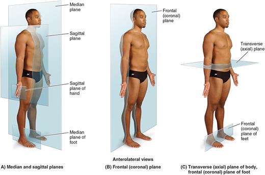
Three standard imaginary planes section the body for imaging and dissection. A sagittal plane divides the body into left and right parts — a midsagittal (median) plane splits it into equal halves, while any off-center sagittal cut is parasagittal. A frontal (coronal) plane divides the body into anterior and posterior parts. A transverse (horizontal / axial / cross-sectional) plane divides the body into superior and inferior parts. An oblique plane cuts at any angle between these.
1.2 · Objective 2 — Anatomical position
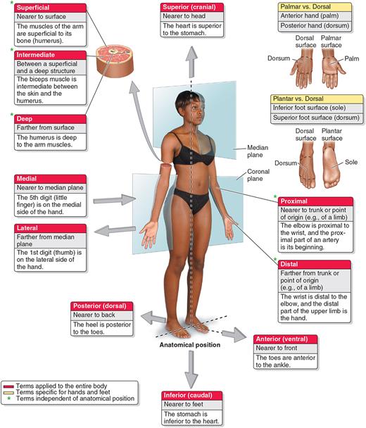
All anatomical descriptions assume the body is in the anatomical position: standing upright, feet flat and directed forward, arms at the sides, palms facing forward (forearms supinated), with the head and eyes facing forward. This fixed reference frame means directional terms mean the same thing regardless of the body’s actual orientation. Contrast it with supine (lying face up) and prone (lying face down).
1.3 · Objective 3 — Movements of the body
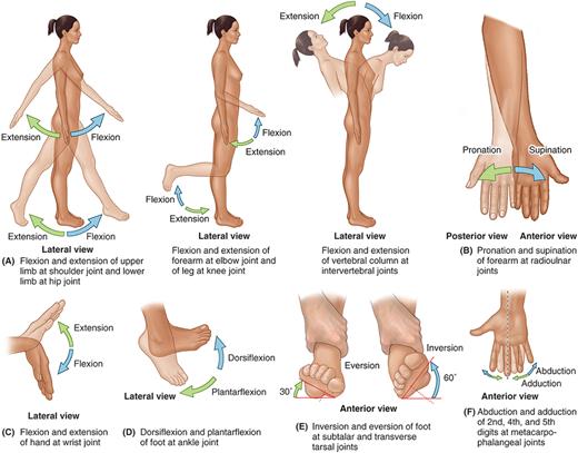
Terms of movement describe joint actions: flexion (decreasing a joint angle) vs. extension (increasing it); abduction (moving away from midline) vs. adduction (moving toward it) and circumduction (a cone-shaped combination); medial vs. lateral rotation; forearm pronation (palm down) vs. supination (palm up); foot inversion (sole toward midline) vs. eversion (sole away) and dorsiflexion/plantarflexion; and elevation/depression, protraction/retraction, and opposition for structures like the scapula, mandible, and thumb.
1.4 · Objective 4 — Cavities of the body
Internal organs sit within two major cavities. The dorsal cavity subdivides into the cranial cavity and the continuous vertebral (spinal) canal. The ventral cavity subdivides into the thoracic cavity — itself split into paired pleural cavities and a central mediastinum (containing the heart within its own pericardial cavity) — and the abdominopelvic cavity (abdominal + pelvic portions, with no wall between them).
Each cavity is lined by a serous membrane: a parietal layer lines the wall and a visceral layer covers the organ, with a film of serous fluid between them reducing friction during breathing, heartbeat, and peristalsis. The three serous membranes are the pleura (lungs), pericardium (heart), and peritoneum (abdominal organs).
1.5 · Objective 5 — Anatomical terms
Terms of relationship describe position along the body’s axes: anterior/ventral (front) vs. posterior/dorsal (back); superior/cranial (toward head) vs. inferior/caudal (toward feet). Terms of laterality & depth add: medial (toward midline) vs. lateral (away); proximal (nearer the trunk/attachment) vs. distal (farther); superficial (nearer the surface) vs. deep; and ipsilateral (same side) vs. contralateral (opposite side). Remember that textbook descriptions represent the typical body plan — anatomical variation (extra bones, accessory muscles, unusual vessel branching) is common and clinically important.
1.6 · Objective 6 — Organs of the body cavities
| Cavity | Principal organs |
|---|---|
| Cranial cavity | Brain |
| Vertebral (spinal) canal | Spinal cord |
| Pleural cavities | Lungs |
| Mediastinum / pericardial cavity | Heart, great vessels, trachea, esophagus, thymus |
| Abdominal cavity | Stomach, small and large intestine, liver, gallbladder, spleen, pancreas, kidneys |
| Pelvic cavity | Urinary bladder, rectum, internal reproductive organs |
1.7 · Objective 7 — Levels of structural organization
The body is organized from broad to fine: chemical → cellular → tissue → organ → organ system → organism, each level building on the one below. Anatomy is also studied by approach: gross (macroscopic) vs. microscopic; and systemic anatomy (by functional system — integumentary, musculoskeletal, nervous, circulatory, respiratory, digestive, urinary, reproductive, endocrine) vs. regional anatomy (all structures within one area, e.g. the thorax, studied together).
1.8 · Objective 8 — Abdominal regions & quadrants
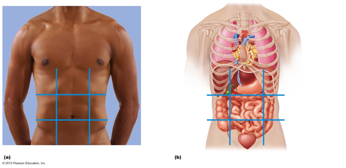
Two schemes localize abdominal findings. The nine regions (two horizontal, two vertical lines) are, top row: right hypochondriac, epigastric, left hypochondriac; middle row: right lumbar, umbilical, left lumbar; bottom row: right iliac (inguinal), hypogastric (pubic), left iliac (inguinal). The simpler four quadrants (one horizontal + one vertical line through the umbilicus) are the right upper (RUQ), left upper (LUQ), right lower (RLQ), and left lower (LLQ) quadrants — e.g., the appendix is RLQ and the spleen is LUQ.
2 · Histology
Instructional Objectives
- Compare and contrast the structure of the types of tissue found in the human body.
- Compare and contrast the locations of the tissue types.
- Differentiate the subcategories of tissue types.
- Identify the various cells of the tissue types.
2.1 · Objective 1 — Structure of the tissue types
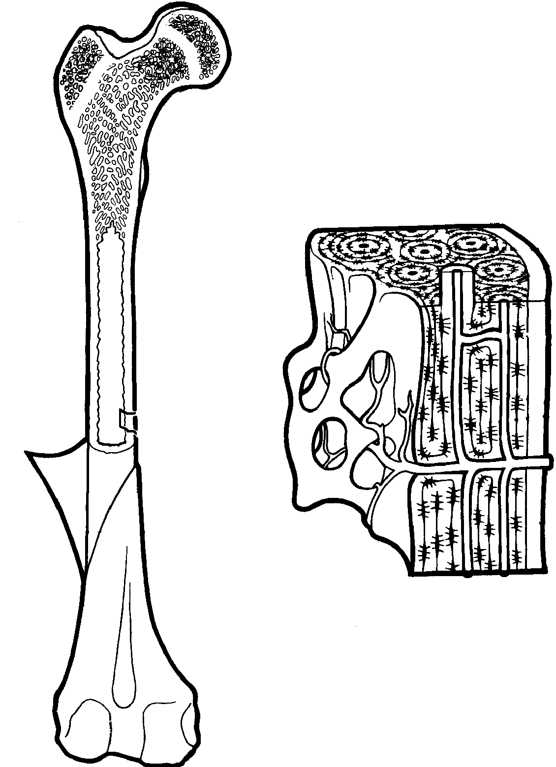
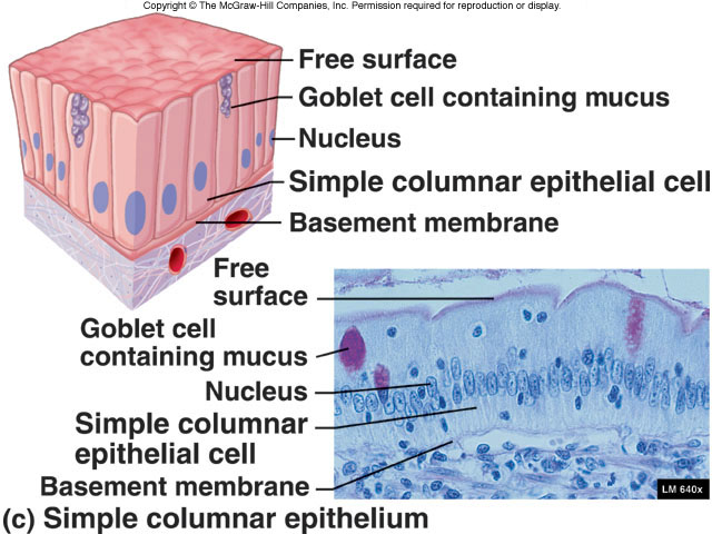
All body structures are built from four primary tissue types, distinguished by their structure. Epithelial tissue is sheets of tightly packed cells with very little extracellular matrix, resting on a basement membrane, avascular. Connective tissue is the opposite — relatively few cells scattered in an abundant extracellular matrix of ground substance plus fibers (collagen for tensile strength, elastic for stretch/recoil, reticular for a supportive mesh). Muscle tissue is elongated contractile cells (striated in skeletal/cardiac, non-striated in smooth). Nervous tissue is excitable neurons plus supporting neuroglia. In the supportive connective tissues, compact bone is organized into cylindrical osteons (Haversian systems) — concentric lamellae around a central Haversian canal — while spongy bone is a lattice of trabeculae; cartilage is a firm gel matrix with no blood supply.
2.2 · Objective 2 — Locations of the tissue types
Epithelial tissue covers body surfaces (skin), lines cavities, vessels, and ducts, and forms glands. Connective tissue is the most widespread — it binds, supports, and packs structures everywhere (deep to skin, around organs, in tendons/ligaments, and as bone, cartilage, and blood). Muscle tissue: skeletal muscle attaches to the skeleton, cardiac muscle forms the heart wall, and smooth muscle lines hollow organs and blood vessels. Nervous tissue makes up the brain, spinal cord, and peripheral nerves. Among supportive tissues, hyaline cartilage covers articular surfaces and forms respiratory rings, fibrocartilage is in intervertebral discs and knee menisci, and elastic cartilage is in the external ear and epiglottis.
2.3 · Objective 3 — Subcategories of the tissue types
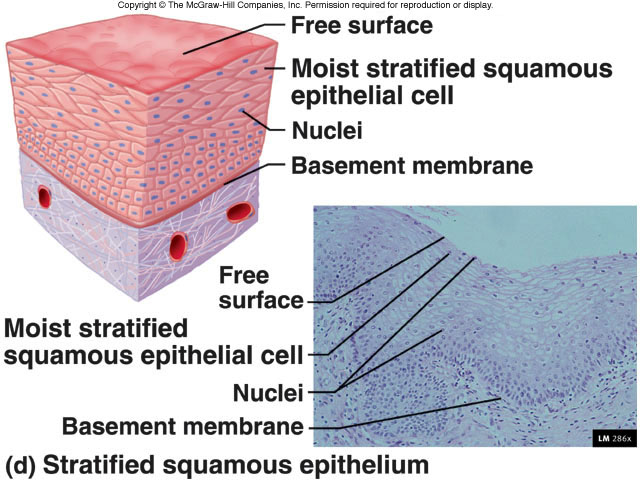
Epithelium is classified on two axes: cell shape (squamous/flat, cuboidal, columnar) and layering (simple = one layer, stratified = many, pseudostratified = one layer appearing layered), plus special transitional epithelium (stretches, lines the bladder). Connective tissue subdivides into loose (areolar, adipose, reticular), dense (regular in tendons/ligaments, irregular in dermis/capsules, elastic in large arteries), supportive (cartilage and bone), and fluid (blood). Muscle has three subtypes: skeletal (striated, voluntary, multinucleate), cardiac (striated, involuntary, branching with intercalated discs), and smooth (non-striated, involuntary). Cartilage has three subtypes (hyaline, fibrocartilage — highest tensile strength, elastic), and bone occurs as compact and spongy.
2.4 · Objective 4 — Cells of the tissue types
Each tissue has characteristic cells. Connective tissue cells include fibroblasts (produce fibers and ground substance), adipocytes (store fat), and defensive cells (macrophages, mast cells). Cartilage is maintained by chondrocytes sitting in cavities called lacunae. Bone contains osteoblasts (build matrix), osteocytes (mature cells in lacunae, connected by canaliculi), and osteoclasts (resorb bone). Muscle is made of muscle fibers (myocytes), and nervous tissue of neurons (signal-conducting) and neuroglia (support). All of these are built from cells sharing the same core organelles — nucleus, mitochondria, endoplasmic reticulum, and Golgi apparatus.
Long bones lengthen at the epiphyseal (growth) plate, a hyaline-cartilage layer between the diaphysis and epiphysis with four zones — resting, proliferative, hypertrophic, and calcification — that ossifies at maturity, leaving the epiphyseal line.
3 · Neuroanatomy: Brain and ANS
Instructional Objectives
- Describe the anatomical divisions of the brain.
- Compare and contrast the anatomy of cerebrum-cerebellum-brain stem.
- Define the structures of the reticular system and the limbic system.
- Describe the autonomic system: sympathetic, parasympathetic, and enteric nervous systems in anatomy and innervations.
3.1 · Objective 1 — Anatomical divisions of the brain
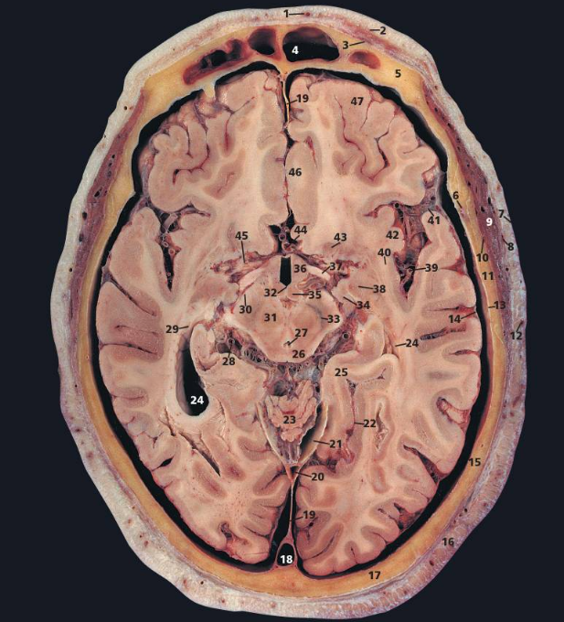
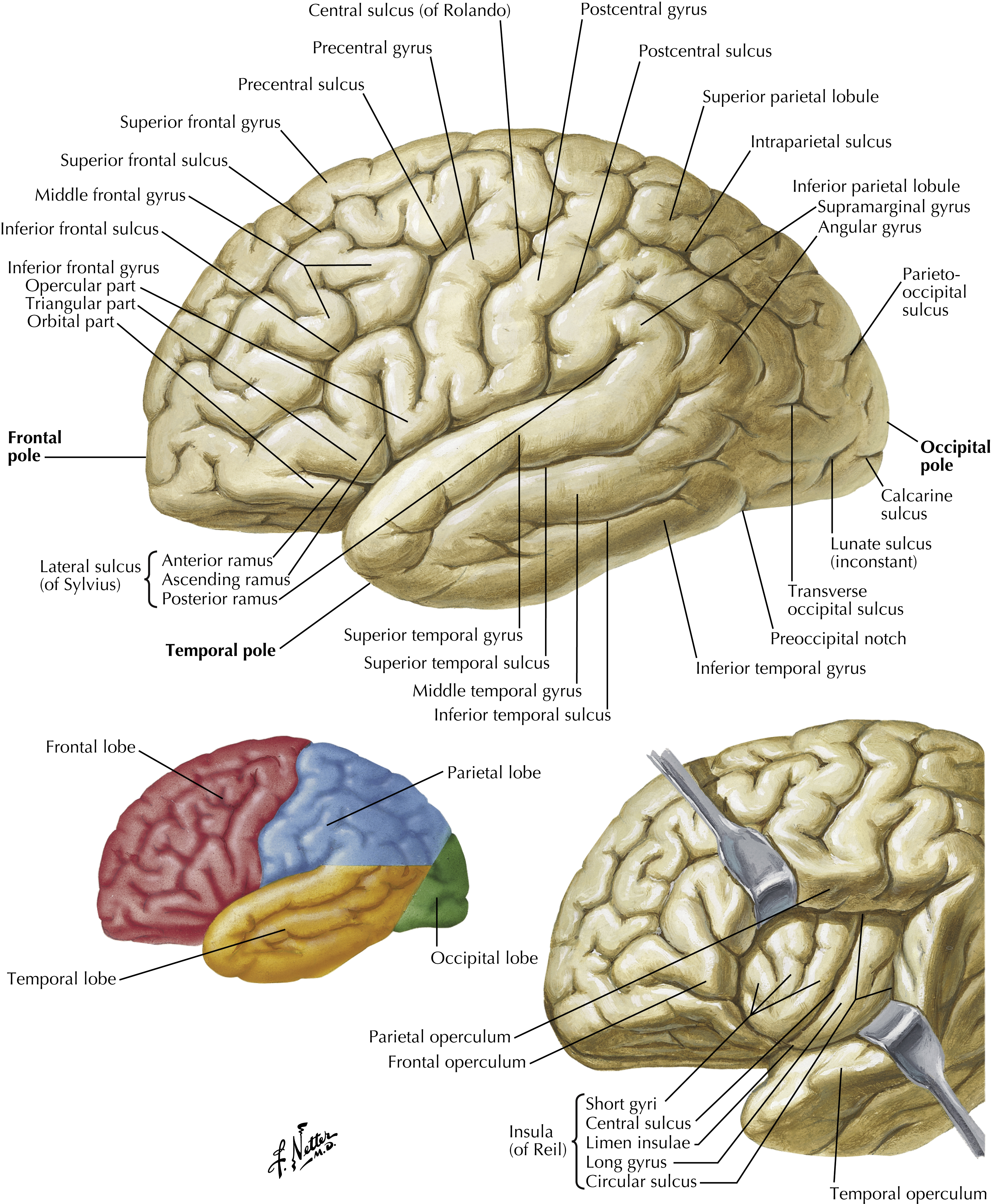
The brain has four major anatomical divisions. The cerebrum is the largest — paired hemispheres joined by the corpus callosum, with a folded surface of gyri (ridges) and sulci (grooves). The central and lateral sulci partition each hemisphere into four lobes: frontal (voluntary motor, executive function, personality), parietal (somatosensory, spatial awareness), temporal (auditory, memory, language), and occipital (vision). The diencephalon sits below and contains the thalamus, hypothalamus, and epithalamus. The brainstem connects the brain to the spinal cord as the midbrain, pons, and medulla oblongata. The cerebellum lies beneath the occipital lobe. Throughout the brain, gray matter (neuron cell bodies) forms the outer cortex and deep nuclei, while white matter (myelinated axons) fills the interior.
3.2 · Objective 2 — Cerebrum vs. cerebellum vs. brainstem
The cerebrum is the seat of conscious thought: its outer cerebral cortex (gray matter) handles perception, voluntary movement, and higher cognition, while deep gray-matter basal nuclei (caudate, putamen, globus pallidus) smooth and modulate voluntary movement. Its diencephalon relays and regulates — the thalamus forwards nearly all sensory input (except smell) to the cortex, the hypothalamus is the master homeostatic regulator (temperature, hunger, thirst, circadian rhythm, pituitary control), and the epithalamus houses the melatonin-secreting pineal gland. The cerebellum does not initiate movement; it coordinates fine motor control, posture, and balance by comparing intended to actual movement. The brainstem is the vital through-route and reflex center: the midbrain carries descending motor fibers (cerebral peduncles) and visual/auditory reflex centers (superior/inferior colliculi), the pons relays between cerebrum and cerebellum and helps drive respiration, and the medulla oblongata houses cardiovascular and respiratory control centers and the decussation of the pyramids where most motor fibers cross.
3.3 · Objective 3 — Reticular system and limbic system
The reticular system (reticular formation) is a diffuse net of neurons threaded through the core of the brainstem (midbrain, pons, and medulla). Its ascending component, the reticular activating system (RAS), projects to the cortex to maintain consciousness, wakefulness, and arousal and to filter repetitive/unimportant sensory input; damage can produce coma. It also overlaps the brainstem's autonomic centers (cardiovascular, respiratory) and helps regulate motor tone and sleep-wake cycles. The limbic system is a ring of structures spanning the cerebrum and diencephalon that governs emotion and memory: the hippocampus (consolidation of new memories), the amygdala (emotional processing, especially fear), the cingulate gyrus (links emotion to behavior), plus the fornix and mammillary bodies (memory circuitry) and olfactory input.
3.4 · Objective 4 — Autonomic nervous system
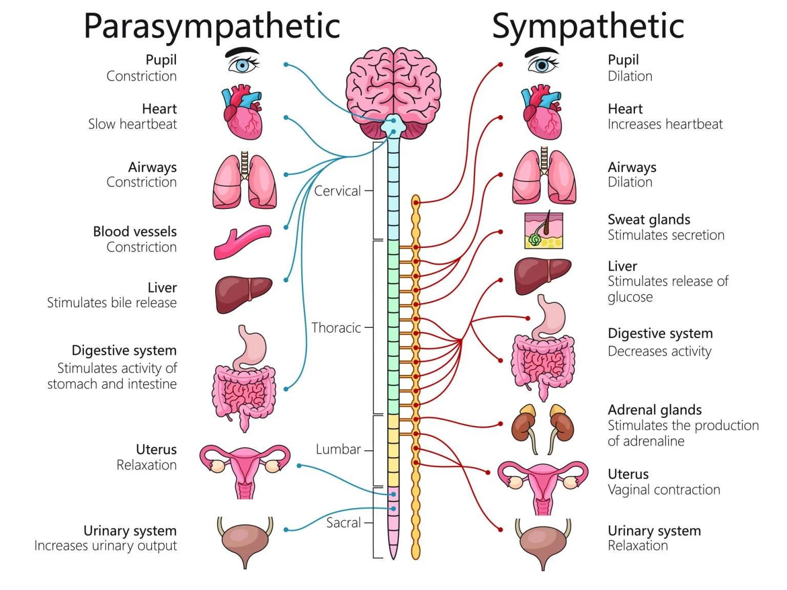
The autonomic nervous system (ANS) controls involuntary effectors — smooth muscle, cardiac muscle, and glands — through a two-neuron chain: a preganglionic neuron (cell body in the CNS) synapses on a postganglionic neuron (cell body in a peripheral ganglion) that reaches the target. The sympathetic division ("fight or flight") has a thoracolumbar outflow (T1–L2), with short preganglionic fibers synapsing in the paravertebral sympathetic chain ganglia and long postganglionic fibers to targets — enabling rapid, widespread activation. The parasympathetic division ("rest and digest") has a craniosacral outflow (cranial nerve nuclei III, VII, IX, X and S2–S4), with long preganglionic fibers synapsing on short postganglionic fibers in ganglia within or near the target organ — producing localized effects. The enteric nervous system governs the GI tract semi-independently through the myenteric plexus (between muscle layers, controls motility) and submucosal plexus (secretion and local blood flow); sympathetic input inhibits GI activity while parasympathetic (vagal) input stimulates it.
Supplementary — Meninges, ventricles, CSF & blood-brain barrier
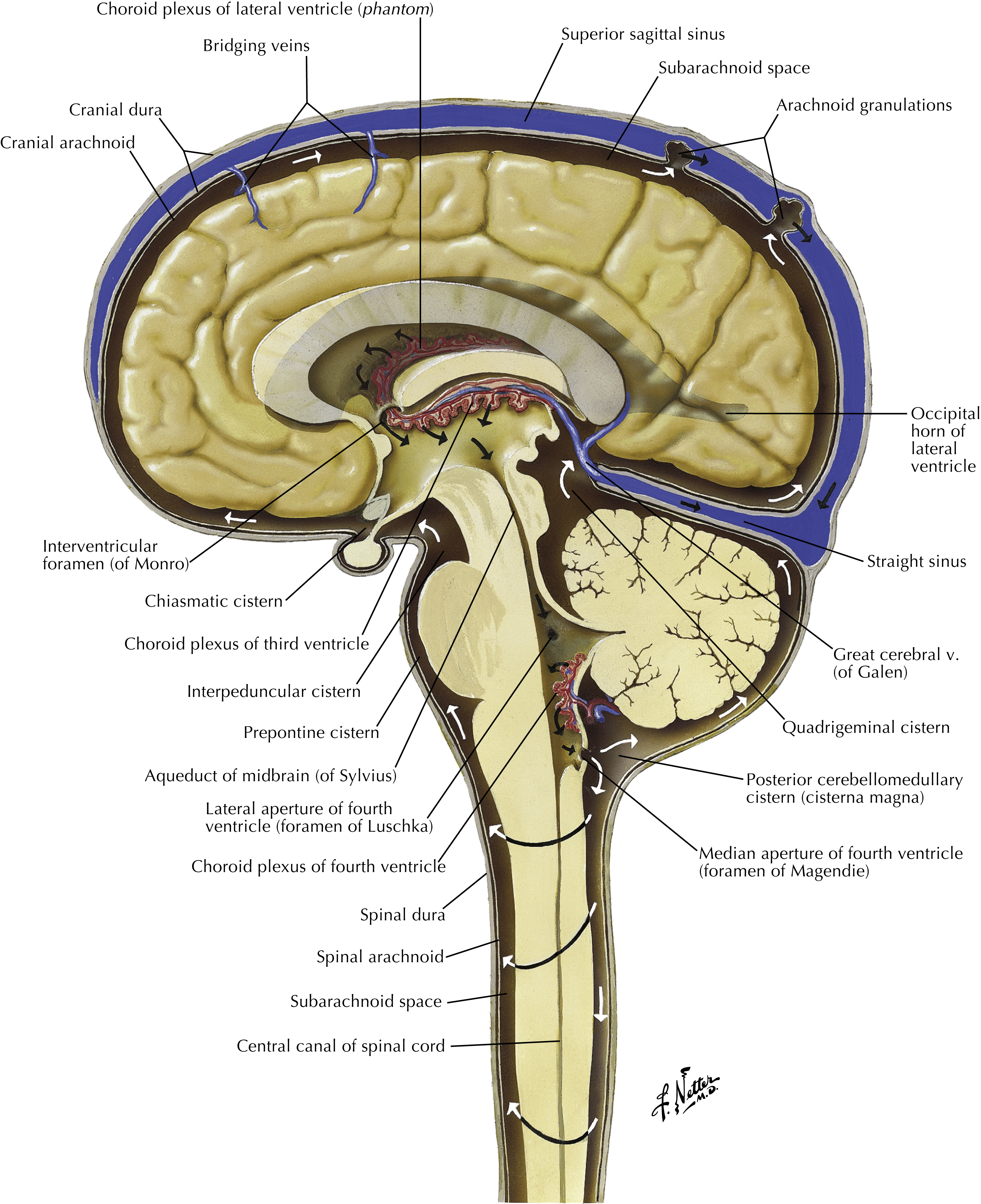
Not a listed objective for this topic, but core brain anatomy worth knowing: three cranial meninges wrap the brain — the tough dura mater (folds into the falx cerebri and tentorium cerebelli; its layers separate to form the dural venous sinuses), the web-like arachnoid mater (over the CSF-filled subarachnoid space), and the delicate pia mater adherent to the brain. Cerebrospinal fluid (CSF), made by the choroid plexus, flows from the paired lateral ventricles → interventricular foramina → third ventricle → cerebral aqueduct → fourth ventricle → subarachnoid space, and is reabsorbed via arachnoid granulations. The blood-brain barrier — capillary tight junctions reinforced by astrocyte end-feet — blocks most large/hydrophilic molecules while letting small lipid-soluble molecules cross.
4 · Neuroanatomy: Nervous Tissue & Cerebral Blood Flow
Instructional Objectives
- Describe the cells that make up nervous tissue.
- Describe the structure and components of a neuron.
- Compare and contrast the classification of a neuron.
- Compare and contrast the vasculature of the brain.
- Describe the arterial blood flow to the brain.
- Describe the venous drainage of the brain.
4.1 · Objective 1 — Cells of nervous tissue
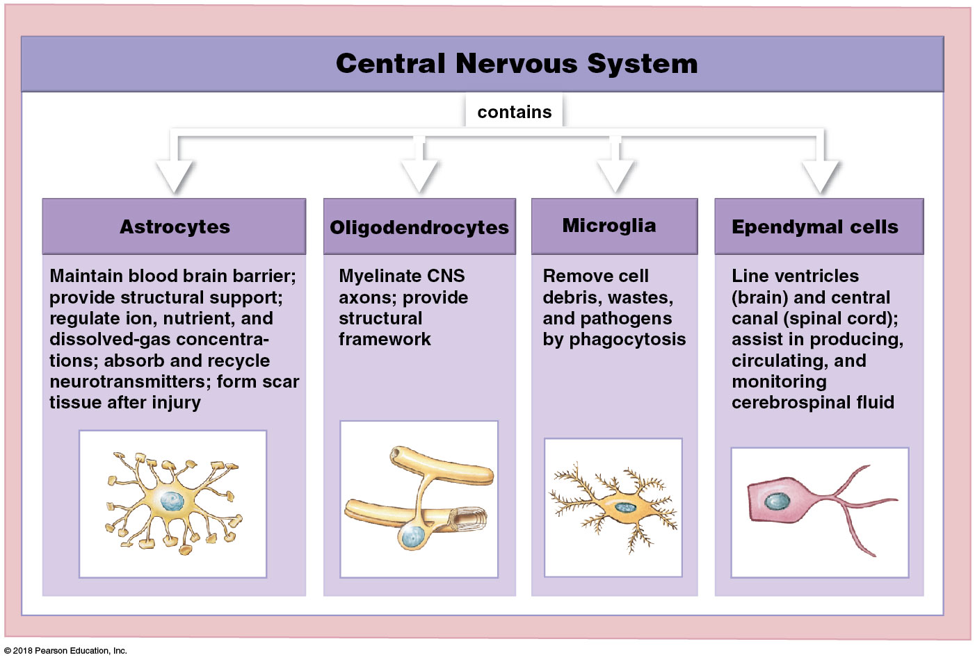
Nervous tissue is built from two cell classes: signal-conducting neurons and supporting neuroglia. Neuroglia differ between the two divisions. In the central nervous system (CNS): astrocytes (most abundant — support neurons, help form the blood-brain barrier, regulate the extracellular environment), oligodendrocytes (myelinate CNS axons; one cell wraps segments of several axons), microglia (resident immune cells clearing debris/pathogens), and ependymal cells (line the ventricles, help produce/circulate CSF). In the peripheral nervous system (PNS): Schwann cells (myelinate PNS axons; one cell wraps a single segment of one axon) and satellite cells (support neuron cell bodies in peripheral ganglia).
4.2 · Objective 2 — Structure and components of a neuron
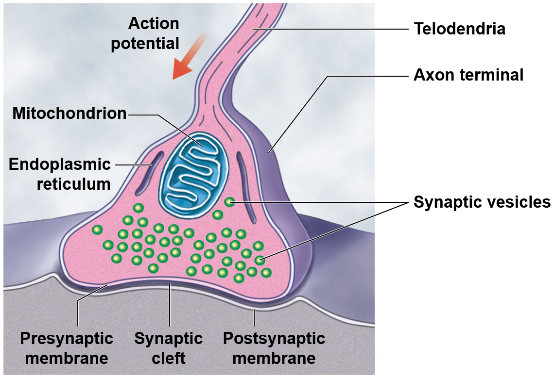
A neuron has a cell body (soma) containing the nucleus, branching dendrites that receive incoming signals, and a single axon arising from the axon hillock (the action-potential trigger zone) that conducts the outgoing signal to its axon terminals. Many axons are insulated by a myelin sheath, interrupted at regular gaps called nodes of Ranvier that let the impulse "jump" node to node — saltatory conduction — much faster than continuous conduction. At a synapse, the presynaptic terminal releases neurotransmitter into the synaptic cleft to excite or inhibit the next cell.
4.3 · Objective 3 — Classification of a neuron
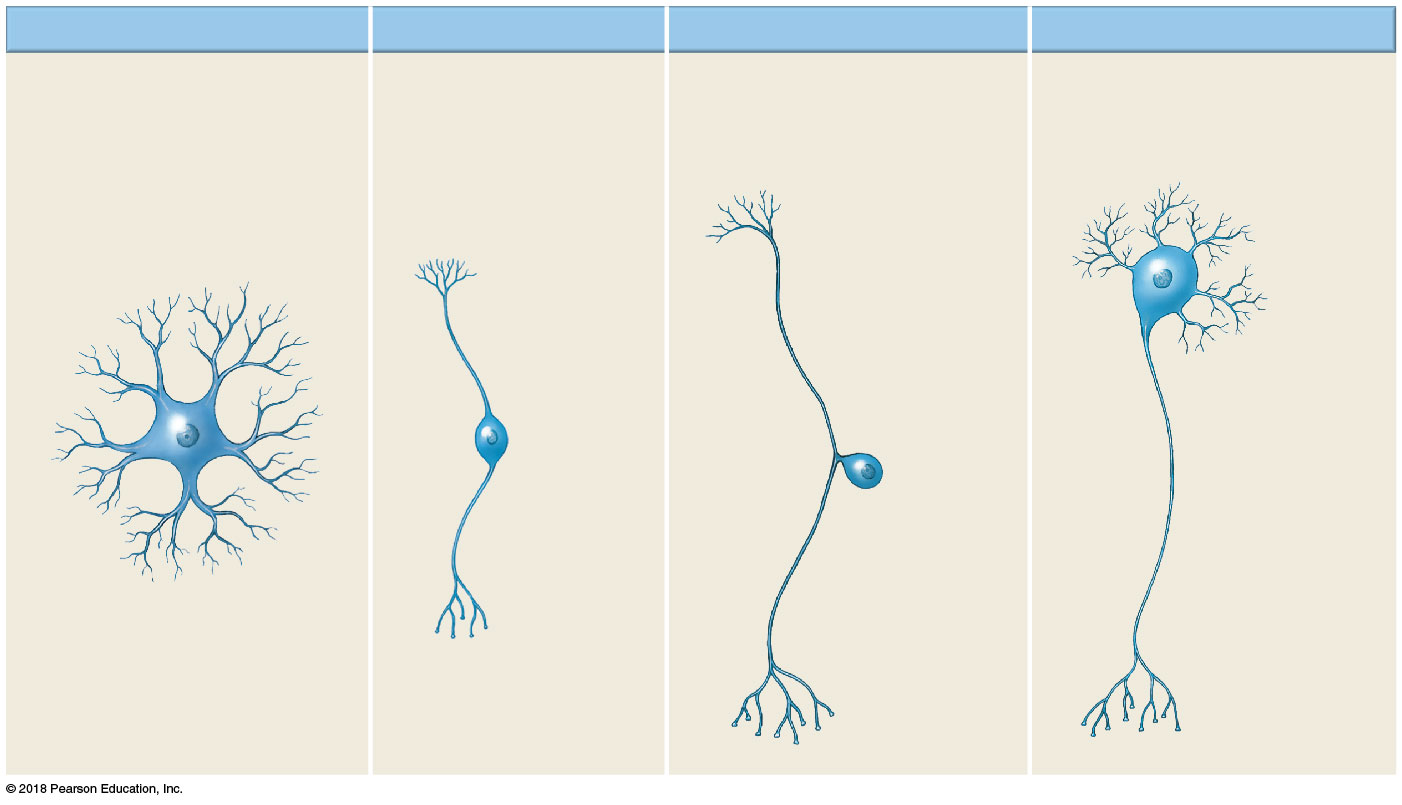
Neurons are classified two ways. Functionally: sensory (afferent) neurons carry signals toward the CNS, motor (efferent) neurons carry signals away to effectors, and interneurons lie entirely within the CNS integrating between the two. Structurally (by number of processes off the soma): multipolar (one axon, many dendrites — most CNS and motor neurons), bipolar (one axon, one dendrite — special senses such as the retina), and unipolar (pseudounipolar) (a single process splits into peripheral and central branches — typical sensory neurons with cell bodies in dorsal root ganglia).
4.4 · Objective 4 — Vasculature of the brain
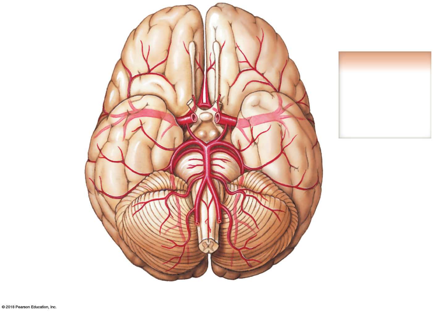
The brain is fed by two paired arterial systems that together form a protective loop. The internal carotid arteries supply the anterior circulation (most of the cerebrum), while the paired vertebral arteries ascend through the transverse foramina and merge into the single basilar artery to supply the posterior circulation (brainstem, cerebellum, occipital lobes). The two systems are joined at the brain's base by the cerebral arterial circle (Circle of Willis), so that if one feeding vessel narrows gradually, flow can be redirected around the ring to preserve perfusion — the key contrast with the single-route venous drainage below.
4.5 · Objective 5 — Arterial blood flow to the brain
Tracing the flow through the Circle of Willis: each internal carotid gives rise to an anterior cerebral artery (the two joined across the midline by the short anterior communicating artery) and a middle cerebral artery (the largest branch, supplying most of the lateral cerebral surface). The basilar artery terminates as the paired posterior cerebral arteries, each linked forward to its internal carotid by a posterior communicating artery — completing the ring.
4.6 · Objective 6 — Venous drainage of the brain
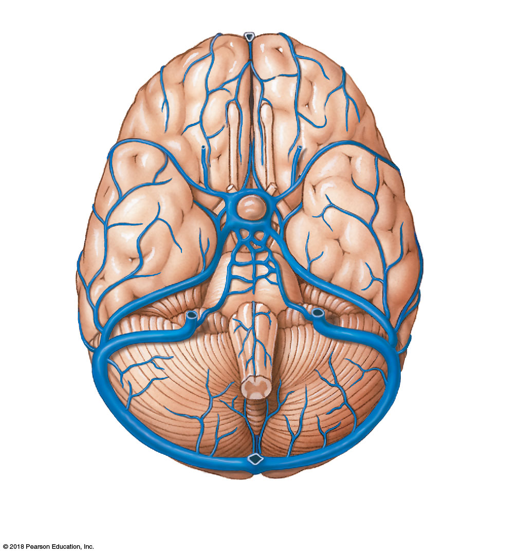
Venous drainage follows a different route: superficial and deep cerebral veins empty into the dural venous sinuses — endothelium-lined channels between the two dura layers — including the superior sagittal sinus (atop the falx cerebri), inferior sagittal sinus, straight sinus, and the paired transverse and sigmoid sinuses, which drain into the internal jugular veins to return blood to the heart.
5 · Neuroanatomy: Spinal Cord and Sensory Receptors
Instructional Objectives
- Describe the anatomical divisions of the spinal cord.
- Compare and contrast the major efferent and afferent tracts of the spinal cord and their corresponding nuclei.
- Describe the spinocerebellar tracts.
- Describe the anatomical arrangement of the white and gray matter of the spinal cord.
- Identify the blood supply, venous drainage, and lymphatic drainage of the spinal cord.
- Discuss neural anatomical communication within the body.
- Compare and contrast the sensory receptors anatomically.
5.1 · Objective 1 — Anatomical divisions of the spinal cord
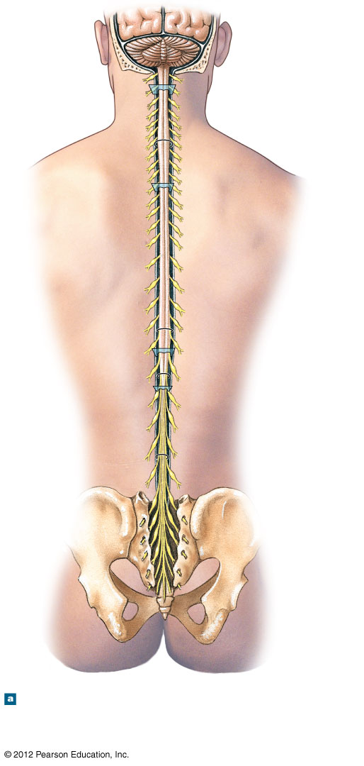
The spinal cord runs from the foramen magnum to about the L1–L2 vertebral level, giving off 31 pairs of spinal nerves across its cervical, thoracic, lumbar, sacral, and coccygeal regions. It is thicker at two points that innervate the limbs: the cervical enlargement (upper limb) and the lumbosacral enlargement (lower limb). Inferiorly it tapers into the conus medullaris; below that, the fibrous filum terminale anchors the cord to the coccyx, surrounded by the descending lumbar and sacral nerve roots that form the cauda equina ("horse's tail").
5.2 · Objective 2 — Efferent and afferent tracts and their nuclei
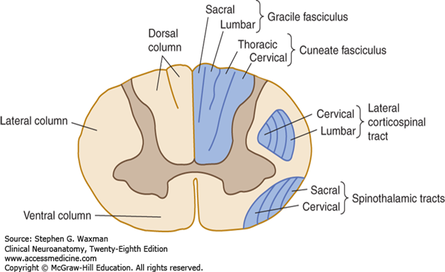
Afferent (ascending, sensory) tracts carry information up the cord. The dorsal columns (fasciculus gracilis and fasciculus cuneatus) carry fine touch, vibration, and conscious proprioception, staying ipsilateral until they synapse in the nucleus gracilis and nucleus cuneatus of the medulla. The spinothalamic tracts carry pain, temperature, and crude touch, crossing to the opposite side near their level of entry and relaying in the thalamus. Efferent (descending, motor) tracts carry commands down: the corticospinal tracts deliver voluntary motor signals from the cortex — the larger lateral corticospinal tract (already crossed at the pyramidal decussation) controls limb muscles, and the smaller anterior corticospinal tract (crossing at its level of exit) controls axial/trunk muscles.
5.3 · Objective 3 — Spinocerebellar tracts
The spinocerebellar tracts are ascending afferent tracts that carry unconscious proprioception — information about muscle length, tension, and joint position — from the trunk and limbs directly to the cerebellum, which uses it to coordinate movement, posture, and balance. They travel in the lateral funiculus and, unlike the conscious pathways, do not reach the cerebral cortex, so the information never enters conscious awareness.
5.4 · Objective 4 — Arrangement of gray and white matter
In cross-section the gray matter forms a central butterfly/H-shape: dorsal (posterior) horns receive incoming sensory input, ventral (anterior) horns hold motor neuron cell bodies, and — only at T1–L2 — small lateral horns contain the intermediolateral gray horn with sympathetic preganglionic cell bodies. Surrounding it, white matter is arranged into funiculi (columns) — dorsal, lateral, and ventral — that carry the ascending and descending tracts. Sympathetic fibers exit and rejoin the spinal nerve through the rami communicantes: a white ramus (myelinated preganglionic fibers to a chain ganglion) and a gray ramus (unmyelinated postganglionic fibers rejoining the nerve).
5.5 · Objective 5 — Blood supply, venous and lymphatic drainage
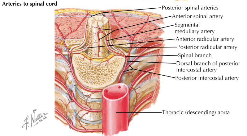
The cord is supplied by a single anterior spinal artery and paired posterior spinal arteries, reinforced at intervals by segmental radicular arteries — the largest, the artery of Adamkiewicz, is clinically important in aortic surgery. Corresponding anterior and posterior spinal veins drain into the epidural venous plexus and segmental veins, and lymphatic drainage runs alongside the vasculature toward regional nodes.
5.6 · Objective 6 — Neural anatomical communication
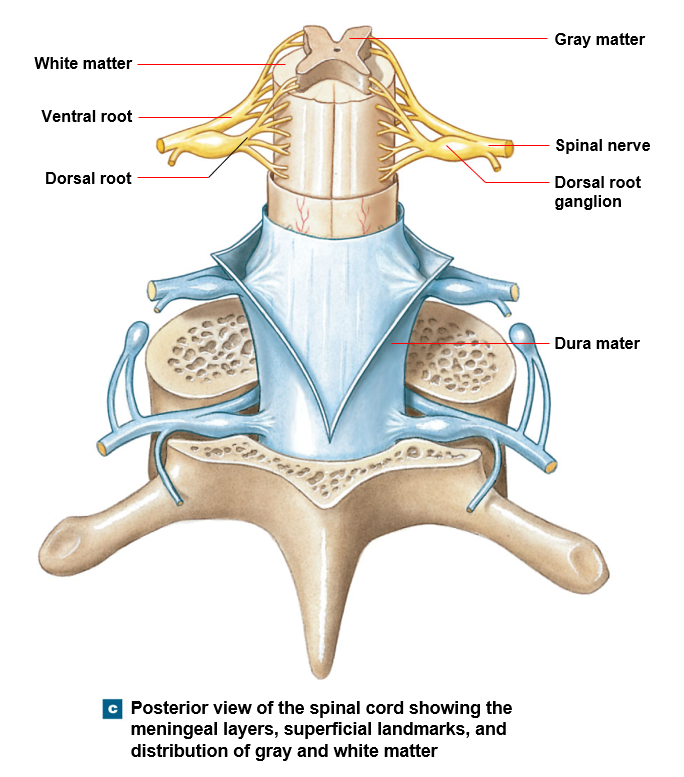
The cord communicates with the body through spinal nerves, each formed by a dorsal (posterior) root carrying sensory fibers in (cell bodies in the dorsal root ganglion) and a ventral (anterior) root carrying motor fibers out. The simplest circuit is the reflex arc: a receptor detects a stimulus → an afferent neuron conducts to the cord → an integration center (often an interneuron) processes it → an efferent neuron drives an effector — producing a rapid, often involuntary response before the signal reaches the brain. Each spinal nerve supplies a defined skin area (dermatome) and muscle group (myotome), giving the communication a predictable anatomical map.
5.7 · Objective 7 — Sensory receptors compared anatomically
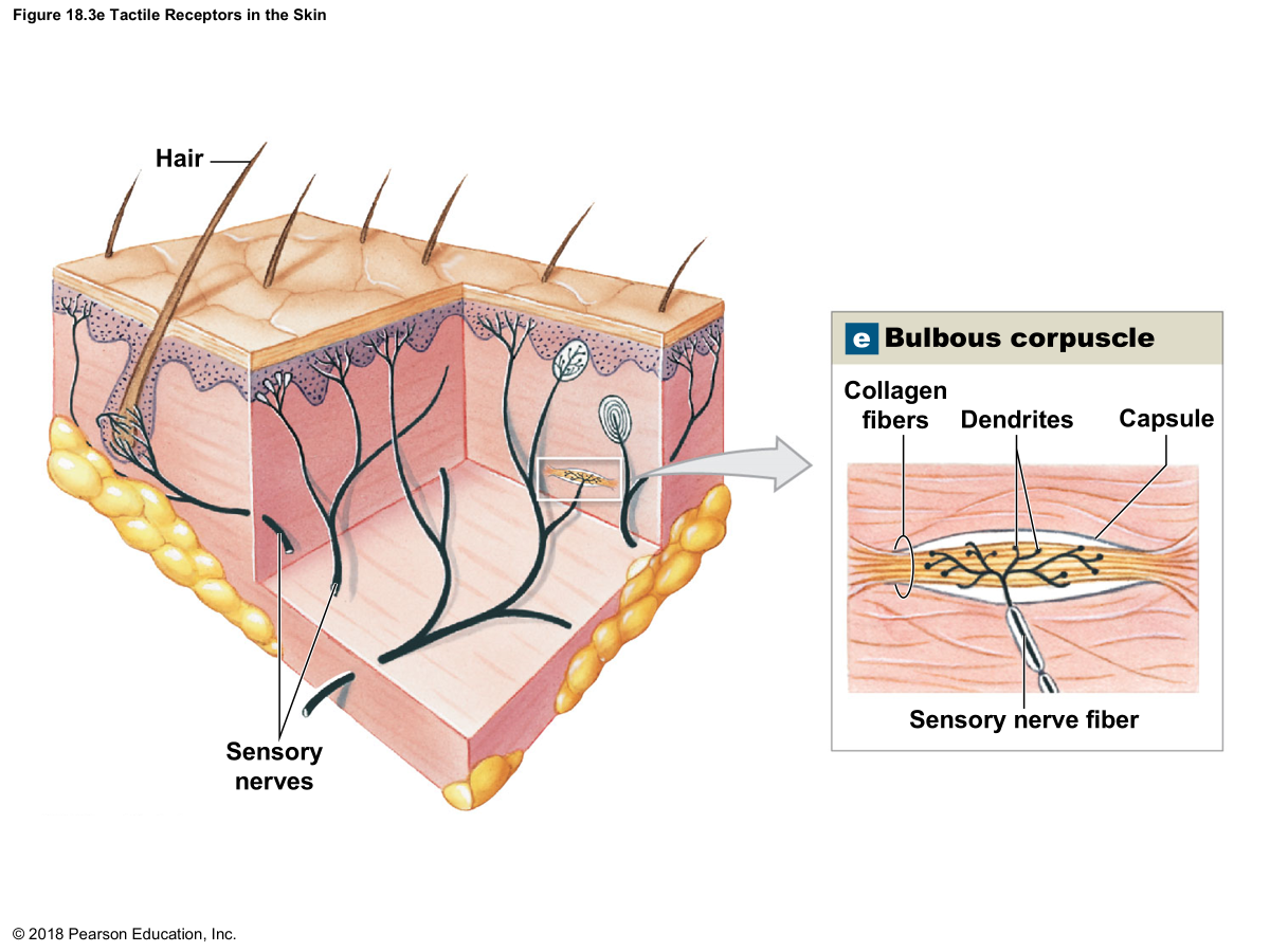
The general senses (touch, pressure, temperature, pain, proprioception) use receptors distributed throughout the body, classified by the stimulus they detect. Mechanoreceptors respond to physical deformation: Meissner corpuscles (light touch/fine discrimination, abundant in fingertips), Pacinian corpuscles (deep pressure/vibration, deep in the dermis), Merkel discs (sustained light touch/pressure), and Ruffini endings (skin stretch). Thermoreceptors are free nerve endings tuned to warming or cooling. Nociceptors are mostly free nerve endings that detect tissue damage as pain. Chemoreceptors detect chemical changes — internally monitoring blood gases/pH (carotid and aortic bodies), and underlying the special senses of smell and taste. Which receptor fires and where it sits together set the sensation's quality and location (the "labeled line" principle), with conscious interpretation in the primary somatosensory cortex of the parietal lobe.
6 · Neuroanatomy: Peripheral and Cranial Nerves
Instructional Objectives
- Describe the anatomical divisions and organization of the cranial nerves.
- Describe the organization of the peripheral nervous system.
- Discuss the location and courses of the 12 cranial nerves.
6.1 · Objective 1 — Divisions and organization of the cranial nerves
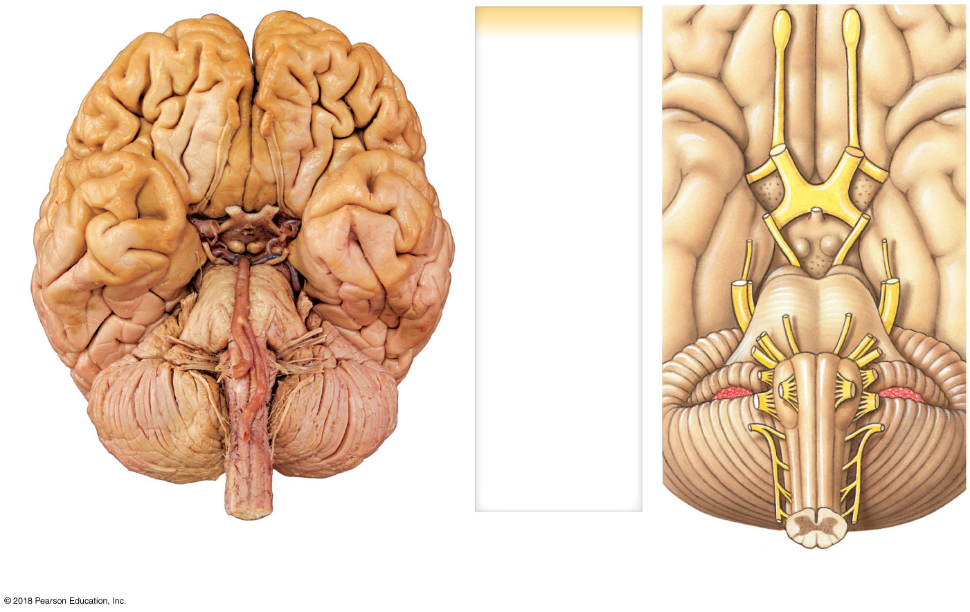
The twelve pairs of cranial nerves are part of the peripheral nervous system but arise directly from the brain rather than the spinal cord, and they emerge in rostral-to-caudal sequence: CN I (olfactory) from the cerebrum and CN II (optic) from the diencephalon, CN III–IV from the midbrain, CN V–VIII from the pons, and CN IX–XII from the medulla. Functionally they organize into three groups: purely sensory (I, II, VIII), purely motor (III, IV, VI, XI, XII), and mixed sensory-and-motor (V, VII, IX, X). A distinct subset — III, VII, IX, and X — also carries parasympathetic fibers, forming the cranial half of the craniosacral parasympathetic outflow.
6.2 · Objective 2 — Organization of the peripheral nervous system
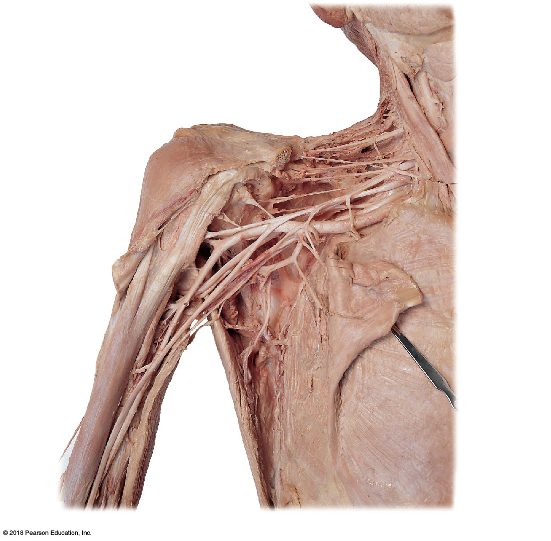
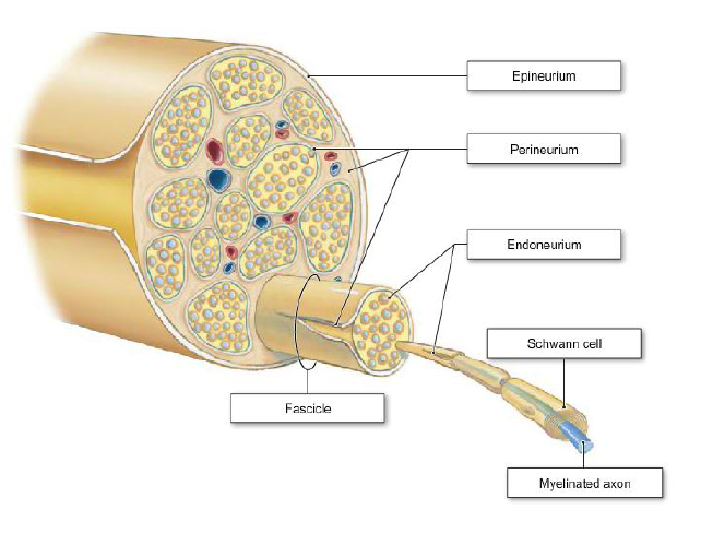
The peripheral nervous system (PNS) is everything outside the brain and spinal cord. It is split into a sensory (afferent) division carrying information toward the CNS and a motor (efferent) division carrying commands out — the motor division further dividing into the somatic (voluntary control of skeletal muscle) and autonomic (involuntary control of smooth/cardiac muscle and glands) systems. Structurally the PNS carries 31 pairs of spinal nerves (8 cervical, 12 thoracic, 5 lumbar, 5 sacral, 1 coccygeal), each formed by a sensory dorsal root (cell bodies in the dorsal root ganglion) and a motor ventral root. After exiting, each nerve splits into a dorsal ramus (deep back muscles/skin) and a larger ventral ramus (anterolateral body wall and limbs). Ventral rami interweave into four major plexuses: the cervical (C1–C4, incl. the phrenic nerve to the diaphragm), the brachial (C5–T1 → median, ulnar, radial, musculocutaneous, axillary nerves), the lumbar (L1–L4 → femoral, obturator), and the sacral (L4–S4 → sciatic, the largest nerve) — the latter two often grouped as the lumbosacral plexus.
6.3 · Objective 3 — Location and courses of the 12 cranial nerves
Each cranial nerve exits the cranium through a specific foramen to reach its target. Two carry notable clinical weight: CN III (oculomotor) routes parasympathetic fibers through the ciliary ganglion to constrict the pupil and accommodate the lens, on top of moving four of the six extra-ocular muscles and the eyelid; and CN IX (glossopharyngeal) gives a carotid sinus/body branch carrying baroreceptor and chemoreceptor input for reflex control of blood pressure and breathing.
| Nerve | Foramen | Principal function |
|---|---|---|
| I — Olfactory | Cribriform plate (ethmoid) | Smell |
| II — Optic | Optic canal | Vision |
| III — Oculomotor | Superior orbital fissure | Most eye movements, eyelid elevation, pupillary constriction & lens accommodation (parasympathetic) |
| IV — Trochlear | Superior orbital fissure | Superior oblique (eye movement) |
| V — Trigeminal | V1 & V2: superior orbital fissure/foramen rotundum; V3: foramen ovale | Facial sensation; muscles of mastication (V3 only) |
| VI — Abducens | Superior orbital fissure | Lateral rectus (eye abduction) |
| VII — Facial | Internal acoustic meatus → stylomastoid foramen | Facial expression; taste (anterior 2/3 tongue); lacrimal/salivary glands |
| VIII — Vestibulocochlear | Internal acoustic meatus | Hearing & balance |
| IX — Glossopharyngeal | Jugular foramen | Taste/sensation (posterior tongue, pharynx); stylopharyngeus; carotid body/sinus reflex input |
| X — Vagus | Jugular foramen | Parasympathetic supply to thoracic/abdominal viscera; pharyngeal/laryngeal muscles |
| XI — Accessory | Jugular foramen | Sternocleidomastoid & trapezius |
| XII — Hypoglossal | Hypoglossal canal | Tongue muscles |