1 · Gastrointestinal System I & II
Prof. Hugh G. Rappa, MD
Instructional Objectives
- Describe the regions and quadrants of the abdomen and corresponding viscera.
- Compare and contrast the parietal and visceral peritoneum and the relationship to abdominal organs.
- Describe the various cell types, tissue, and organs of the gastrointestinal tract.
- Describe the anatomy of the stomach including regions and the relationship to other abdominal viscera.
- Describe the anatomical features of the spleen and list its anatomical relationships.
- Compare and contrast the parts of the small intestines and its relationship with other abdominal organs.
- Compare and contrast the parts of the large intestines and its relationship with other abdominal organs.
- Describe the anatomy of the pancreas and its relationships with other abdominal organs.
- Identify the lobes of the liver and describe their anatomical relations within the abdominal cavity.
- Identify the anatomy of the portal vein and portal venous drainage and its relationship with other abdominal organs.
- Describe the anatomy of the gall bladder and biliary tree and its relationship with other abdominal organs.
- Describe the origins course and major branches of the abdominal aorta that supply the gastrointestinal tract.
- Describe the origins course and major tributaries of the inferior vena cava that supply the gastrointestinal tract.
- Define the anatomy of abdominal lymph drainage.
1.1 · Objective 1 — Describe the regions and quadrants of the abdomen and corresponding viscera
The abdomen is mapped two ways so you can localize organs and pain precisely.
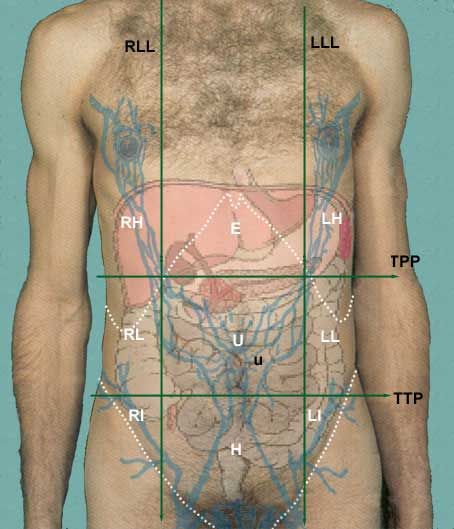
Four quadrants (divided by a vertical median plane and a horizontal transumbilical plane):
- Right Upper Quadrant (RUQ): liver (right lobe), gallbladder, right kidney, hepatic flexure, part of duodenum & pancreas head.
- Left Upper Quadrant (LUQ): stomach, spleen, left lobe of liver, tail of pancreas, left kidney, splenic flexure.
- Right Lower Quadrant (RLQ): cecum, appendix, terminal ileum, right ovary/tube.
- Left Lower Quadrant (LLQ): sigmoid colon, descending colon, left ovary/tube.
Nine regions (two vertical midclavicular planes + subcostal and transtubercular planes) give finer localization: right/left hypochondriac flanking the central epigastric; right/left lumbar (lateral) flanking the central umbilical; right/left inguinal (iliac) flanking the central hypogastric (pubic).
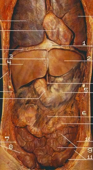
1.2 · Objective 2 — Compare and contrast the parietal and visceral peritoneum and the relationship to abdominal organs
The peritoneum is a serous membrane lining the abdominopelvic cavity. It has two continuous layers:
- Parietal peritoneum — lines the abdominal/pelvic walls; somatically innervated (sharp, well-localized pain).
- Visceral peritoneum — covers the organs; autonomically innervated (dull, poorly localized, referred pain).
Between the layers is the peritoneal cavity, a potential space with a thin film of serous fluid. It is divided into a greater sac and a smaller lesser sac (omental bursa), which communicate through the omental (epiploic) foramen of Winslow.
Organ relationships to the peritoneum:
- Intraperitoneal — nearly surrounded by visceral peritoneum and suspended by a mesentery: stomach, spleen, liver, jejunum, ileum, transverse & sigmoid colon, cecum, appendix.
- Retroperitoneal (primary) — behind the peritoneum from the start: kidneys, adrenal glands, ureters, abdominal aorta, IVC.
- Secondarily retroperitoneal — began intraperitoneal, then fused to the wall: most of the duodenum, pancreas, ascending & descending colon.
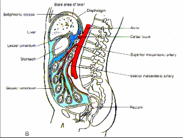
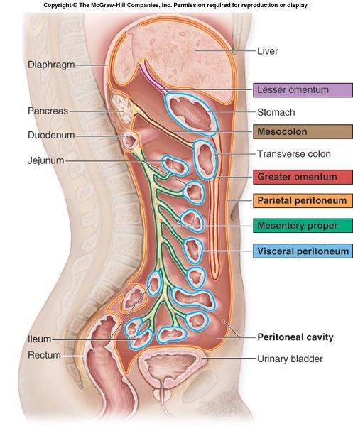
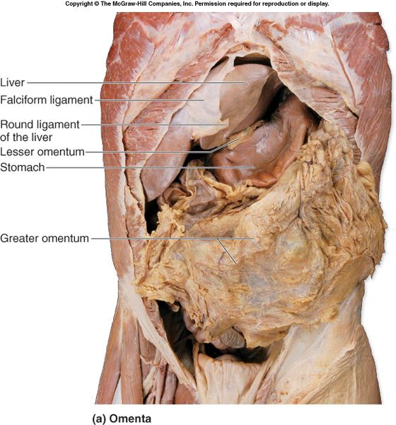
Mesenteries & omenta are double folds of peritoneum that suspend organs and carry vessels, nerves, and lymphatics:
- Greater omentum — hangs from the greater curvature of the stomach, drapes over the transverse colon and small bowel ("fatty apron").
- Lesser omentum — from the porta hepatis and lesser curvature; contains the hepatogastric and hepatoduodenal ligaments (the latter carries the portal triad).
- Mesentery proper — root runs from the duodenojejunal flexure to the right iliac fossa; suspends jejunum and ileum.
1.3 · Objective 3 — Describe the various cell types, tissue, and organs of the gastrointestinal tract
The GI tract has two categories of organs:
- Digestive organs (the tube / alimentary canal): oral cavity → pharynx → esophagus → stomach → small intestine → large intestine → anus. A continuous ~30 ft (9–10 m) tube; smooth muscle in the wall provides motility.
- Accessory digestive organs: teeth, tongue, salivary glands, liver, gallbladder, pancreas — connected by ducts, they assist digestion but don't form the tube.
The four tunics (layers) of the GI wall, from lumen outward:
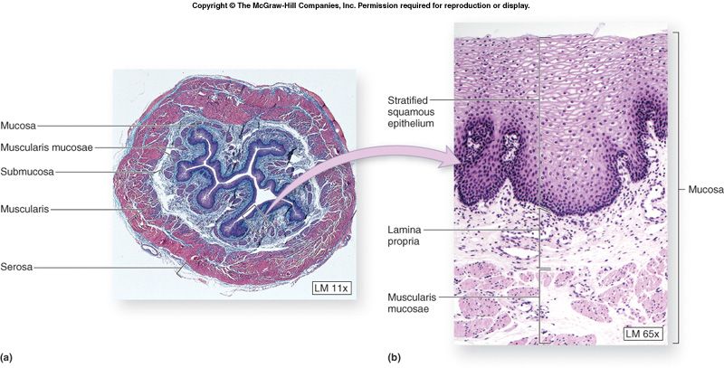
- Mucosa — epithelium + lamina propria + muscularis mucosae. Epithelium varies by function: nonkeratinized stratified squamous (mouth, esophagus, anal canal — protection) vs. simple columnar (stomach → large intestine — secretion/absorption).
- Submucosa — dense irregular CT with vessels, glands, and the submucosal (Meissner) plexus.
- Muscularis externa — inner circular + outer longitudinal smooth muscle; the myenteric (Auerbach) plexus sits between them.
- Serosa (visceral peritoneum) where intraperitoneal, or adventitia where retroperitoneal/fixed.
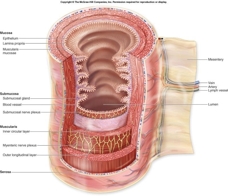
Enteric nervous system (ENS) — the gut's intrinsic innervation via the two intramural plexuses above; coordinates smooth-muscle contraction and secretion. Extrinsic control is primarily parasympathetic.
Surface-area amplifiers: rugae (temporary longitudinal folds that flatten as the stomach fills) and plicae circulares (permanent transverse folds of the small intestine).
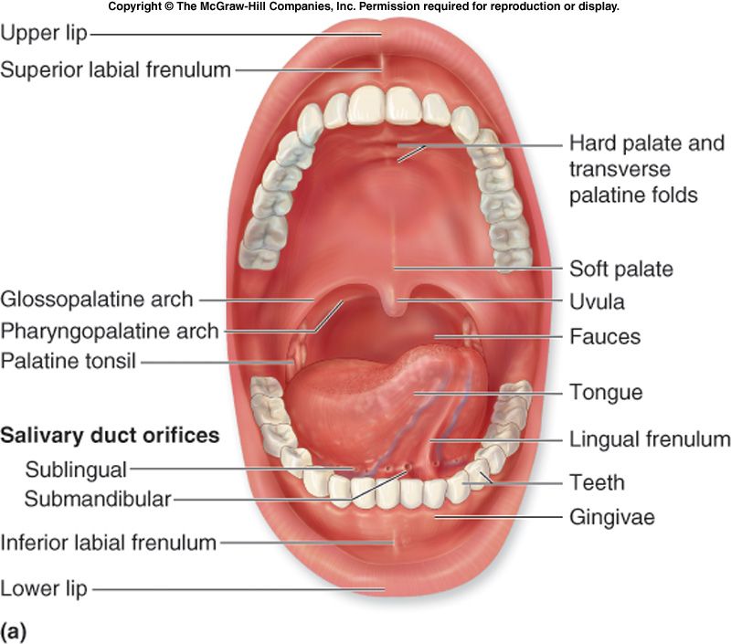
Salivary glands (three pairs): parotid (largest, ~25–30% of saliva, Stensen's duct, related to facial nerve), submandibular (most saliva, ~60–70%, Wharton's duct), sublingual (smallest, ~3–5%).
Esophagus histology: nonkeratinized stratified squamous mucosa; submucosa with mucous glands; muscularis that is skeletal muscle superiorly, mixed in the middle, and smooth muscle inferiorly; adventitia (no serosa in the thorax). Upper esophageal sphincter = skeletal (cricopharyngeus); lower (cardiac) sphincter = physiologic, aided by the diaphragm.
1.4 · Objective 4 — Describe the anatomy of the stomach including regions and the relationship to other abdominal viscera
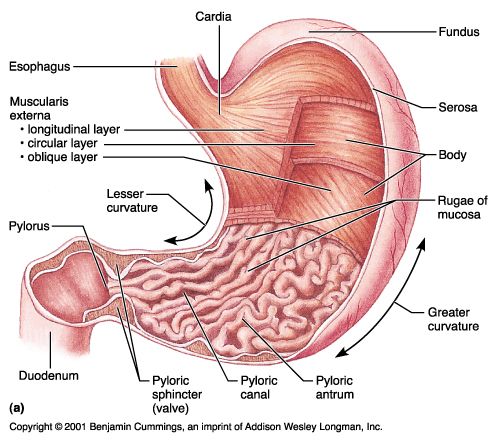
The stomach is an intraperitoneal, J-shaped organ in the LUQ/epigastric region, continuous with the esophagus at the cardia and the duodenum at the pylorus.
Regions:
- Cardia — surrounds the esophageal opening.
- Fundus — dome, the most superior region, under the left diaphragm.
- Body — largest central portion.
- Pyloric region — pyloric antrum → pyloric canal → pyloric sphincter (into the duodenum).
Curvatures: the lesser curvature (right/superior, attaches lesser omentum) and the greater curvature (left/inferior, attaches greater omentum).
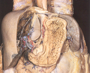
Relationships: anterior — left lobe of liver, diaphragm, anterior abdominal wall; posterior — the lesser sac (omental bursa), then pancreas, spleen, left kidney & adrenal, splenic artery (the "stomach bed"). The spleen lies to its left, the liver to its right.
Interior: rugae (folds), a gastric canal along the lesser curvature, and gastric pits leading into gastric glands.
1.5 · Objective 5 — Describe the anatomical features of the spleen and list its anatomical relationships
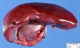
The spleen is the largest lymphatic organ — intraperitoneal, in the LUQ, lying against ribs 9–11 along its long axis.
- White pulp — lymphatic tissue (immune surveillance).
- Red pulp — filters blood, removes old RBCs; a site of hematopoiesis in the fetus.
Relationships & ligaments: connected to the stomach by the gastrosplenic ligament (carries short gastric vessels) and to the left kidney by the splenorenal ligament (carries the splenic vessels and pancreatic tail). Its hilum contacts the tail of the pancreas.
1.6 · Objective 6 — Compare and contrast the parts of the small intestines and its relationship with other abdominal organs
The small intestine finishes chemical digestion and absorbs most nutrients (~6 m total, coiled from pylorus to the ileocecal valve). Three parts:
1. Duodenum (~25 cm, C-shaped, mostly retroperitoneal) — 4 parts:
- Superior (1st): the duodenal cap/ampulla is intraperitoneal; the rest is retroperitoneal.
- Descending (2nd): receives the major duodenal papilla (bile + main pancreatic duct via ampulla of Vater) and minor papilla.
- Horizontal/inferior (3rd): crossed anteriorly by the SMA/SMV.
- Ascending (4th): ends at the duodenojejunal flexure, suspended by the ligament of Treitz.
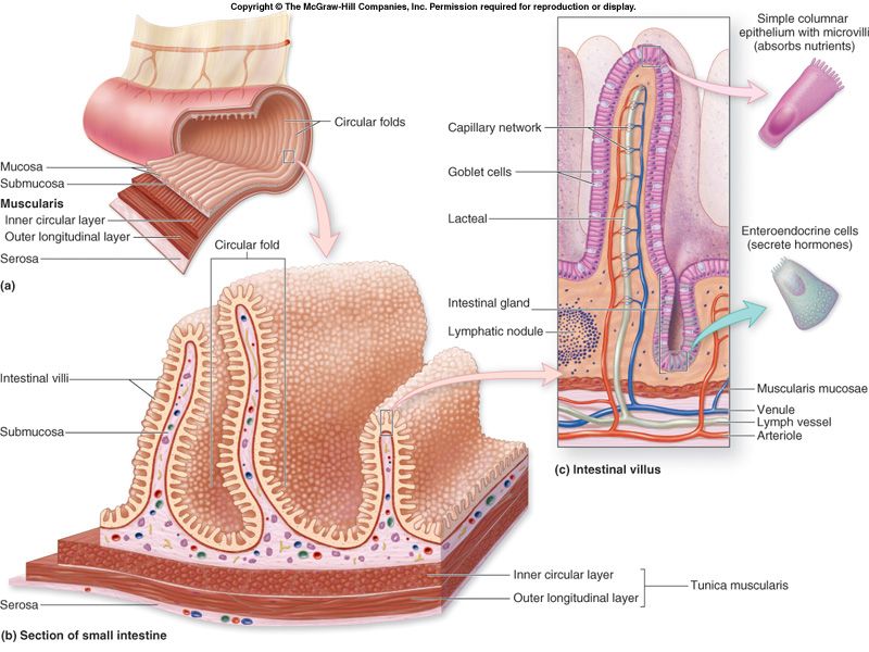
2. Jejunum (proximal ~40%, mostly LUQ) — larger diameter, thicker wall, tall plicae circulares, more vascular (longer vasa recta, fewer arcades). Primary site of chemical digestion and absorption.
3. Ileum (distal ~60%, RLQ) — thinner wall, shorter plicae, prominent Peyer's patches (lymphoid follicles); terminates at the ileocecal valve.
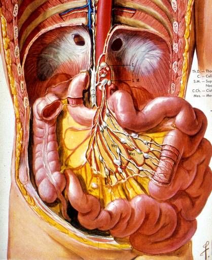
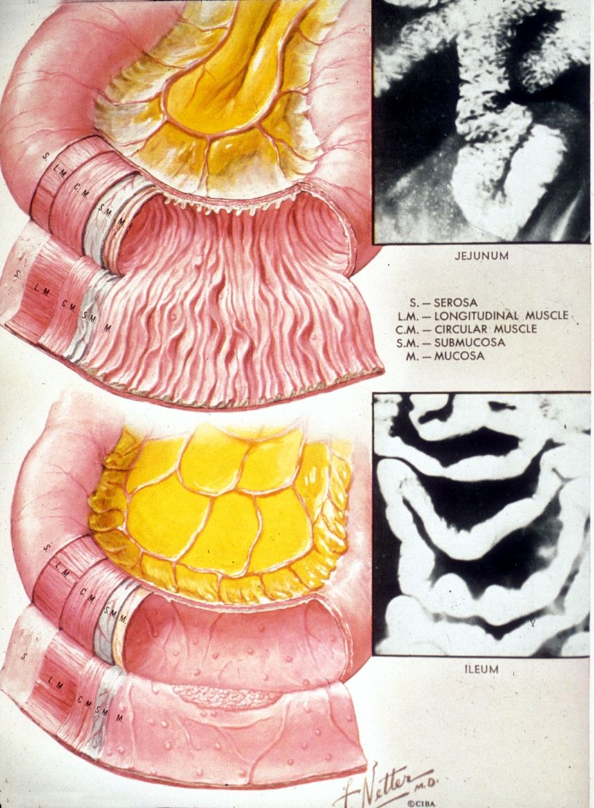
Jejunum and ileum are intraperitoneal, suspended by the mesentery proper.
1.7 · Objective 7 — Compare and contrast the parts of the large intestines and its relationship with other abdominal organs
Horseshoe-shaped, ~1.5 m long, it frames the small intestine, absorbs water & electrolytes, and stores feces. Segments: cecum → colon → rectum → anal canal.
Distinguishing features: teniae coli (3 longitudinal muscle bands), haustra (sacculations), and omental (epiploic) appendices (fat tags). Colon lacks villi, has abundant goblet cells, and does <10% of absorption.
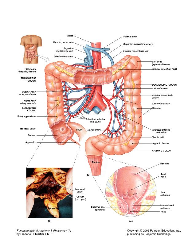
- Cecum & appendix (RLQ, intraperitoneal) — blind pouch below the ileocecal valve; the appendix is a lymphoid-rich tube fixed by the mesoappendix.
- Ascending colon — retroperitoneal; ileocecal valve → right colic (hepatic) flexure.
- Transverse colon — intraperitoneal, most mobile, longest; slung by the transverse mesocolon between the right and left colic flexures.
- Descending colon — retroperitoneal, narrowest; left colic (splenic) flexure → sigmoid.
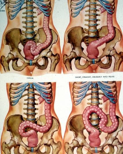
- Sigmoid colon — intraperitoneal, S-shaped, extends into pelvis; stores feces.
- Rectum — retroperitoneal, last ~15 cm, expandable store.
- Anal canal — internal anal sphincter (smooth, involuntary) + external anal sphincter (skeletal, voluntary).
1.8 · Objective 8 — Describe the anatomy of the pancreas and its relationships with other abdominal organs
The pancreas is a retroperitoneal (secondarily) mixed exocrine/endocrine gland lying across the posterior abdomen at ~L1–L2, running from the duodenal C-loop to the splenic hilum.
Regions: head (in the duodenal concavity) with the uncinate process (hooks posterior to the SMA/SMV) → neck (anterior to SMV) → body (crosses the aorta/left kidney) → tail (reaches the splenic hilum, intraperitoneal within the splenorenal ligament).
Ducts: main pancreatic duct (of Wirsung) joins the common bile duct at the ampulla of Vater → major duodenal papilla; the accessory duct (of Santorini) drains to the minor papilla.
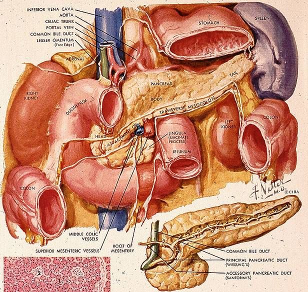
Key posterior relations of the body: left crus of the diaphragm, left adrenal, left renal vein, and splenic vein; the celiac trunk lies superior. The common bile duct passes behind the 1st part of the duodenum and through the head to the ampulla — why pancreatic head cancer causes obstructive jaundice.
1.9 · Objective 9 — Identify the lobes of the liver and describe their anatomical relations within the abdominal cavity
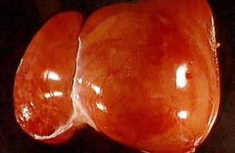
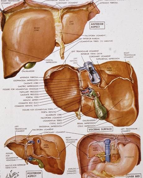
The liver is the largest gland and visceral organ, intraperitoneal in the RUQ, covered by Glisson's capsule (with a posterior bare area lacking peritoneum).
Four anatomical lobes:
- Right lobe — largest.
- Left lobe — separated from the right by the falciform ligament.
- Quadrate lobe — inferior, between the gallbladder and the ligamentum teres (functionally left).
- Caudate lobe — posterior, between the IVC and the ligamentum venosum (functionally independent).
Peritoneal ligaments reflect onto the liver from the diaphragm and surround the bare area: falciform, ligamentum teres (remnant of the umbilical vein), coronary, and right/left triangular ligaments.
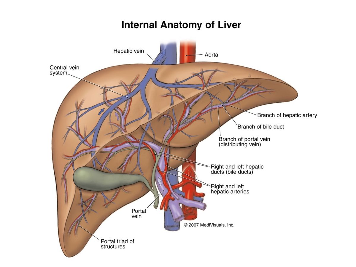
Triple blood supply: hepatic portal vein (~75% of flow — nutrient-rich blood for processing), hepatic artery (~25%, oxygen; supplies the ducts), and hepatic veins (outflow to the IVC → right atrium).
1.10 · Objective 10 — Identify the anatomy of the portal vein and portal venous drainage and its relationship with other abdominal organs
The hepatic portal vein carries nutrient-rich, deoxygenated blood from the GI tract, spleen, and pancreas to the liver for first-pass processing. It forms posterior to the neck of the pancreas by the union of the superior mesenteric vein (SMV) and the splenic vein.
- Splenic vein — drains the spleen; receives the inferior mesenteric vein (IMV) and short gastric/left gastroepiploic veins.
- SMV — drains the small intestine, cecum, ascending & transverse colon.
- Left & right gastric veins — drain to the portal vein directly.
The portal vein ascends in the hepatoduodenal ligament as part of the portal triad (portal vein posteriorly, hepatic artery and bile duct anteriorly), then divides into right and left branches at the porta hepatis.
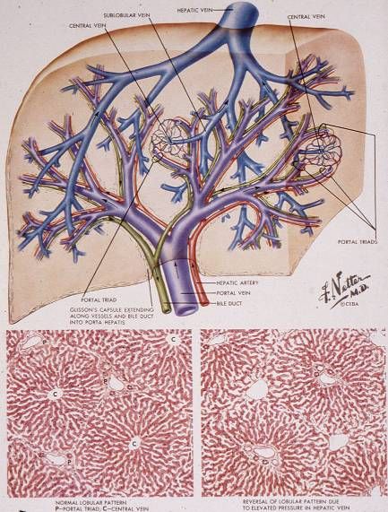
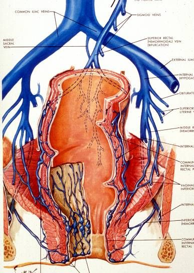
Portosystemic anastomoses (clinically vital in portal hypertension): gastroesophageal (→ esophageal varices), rectal (→ hemorrhoids), paraumbilical (→ caput medusae), and retroperitoneal.
1.11 · Objective 11 — Describe the anatomy of the gall bladder and biliary tree and its relationship with other abdominal organs
The gallbladder stores and concentrates bile. It sits on the visceral surface of the liver between the right and quadrate lobes. Parts: fundus (projects at the tip of the 9th costal cartilage), body, and neck (→ cystic duct, with the spiral valve of Heister).
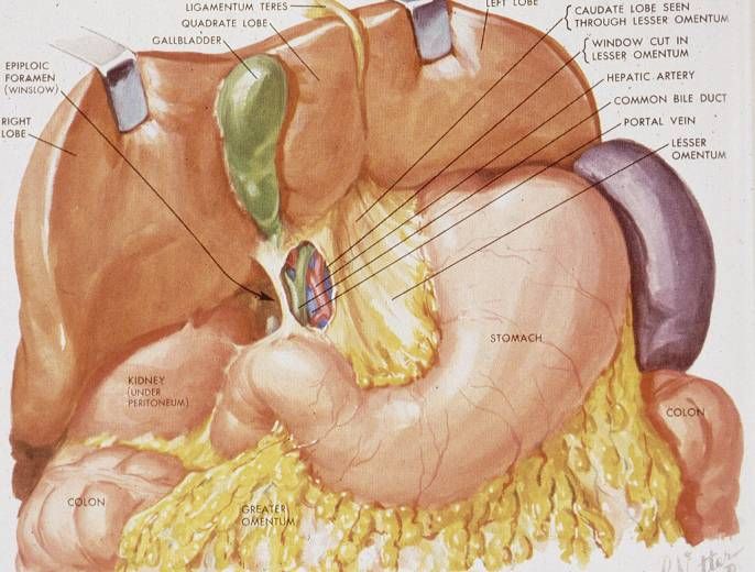
The biliary tree (bile flow):
- Right + left hepatic ducts → common hepatic duct.
- Common hepatic duct + cystic duct (from gallbladder) → common bile duct (CBD).
- CBD descends behind the 1st part of the duodenum, through the pancreatic head, and joins the main pancreatic duct at the ampulla of Vater → major duodenal papilla, guarded by the sphincter of Oddi.
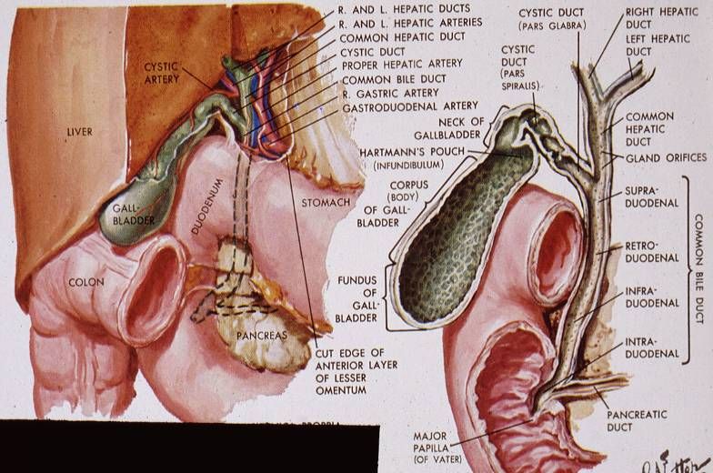
Cystohepatic triangle of Calot (landmark for the cystic artery in cholecystectomy): bounded by the cystic duct, common hepatic duct, and inferior liver edge.
1.12 · Objective 12 — Describe the origins course and major branches of the abdominal aorta that supply the gastrointestinal tract
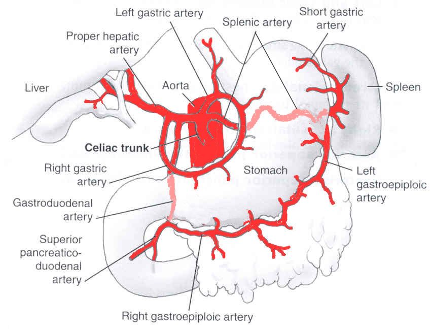
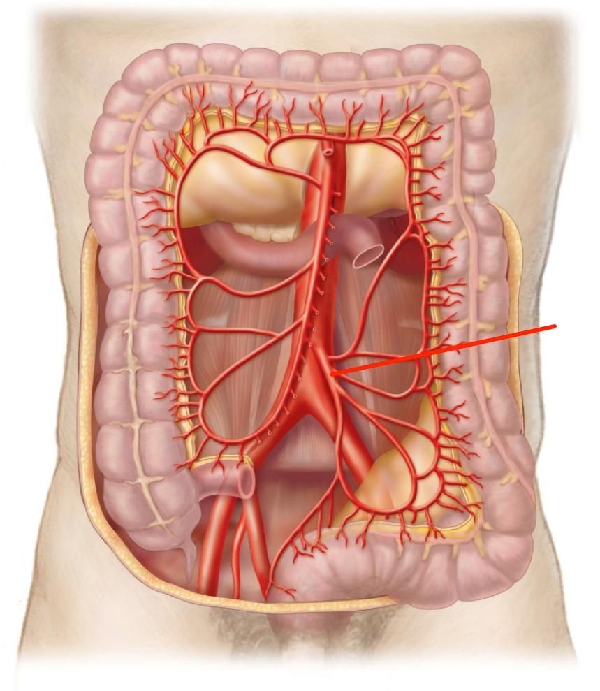
The abdominal aorta enters at the aortic hiatus (T12) between the diaphragmatic crura and descends to L4, where it bifurcates into the common iliac arteries. Three unpaired anterior branches supply the GI tract (following embryonic gut divisions):
1. Celiac trunk (T12) — foregut. Three branches:
- Left gastric artery — lesser curvature + esophageal branches.
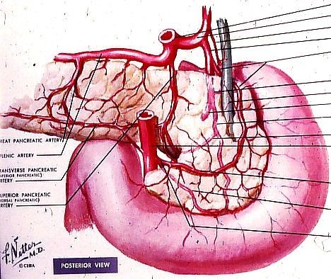
- Splenic artery — runs along the superior pancreas to the spleen; gives pancreatic branches, short gastrics, left gastroepiploic.
- Common hepatic artery → proper hepatic (→ right/left hepatic, cystic, right gastric) and gastroduodenal (→ right gastroepiploic + superior pancreaticoduodenal).
2. Superior mesenteric artery (SMA, L1) — midgut: distal duodenum → proximal 2/3 of the transverse colon (jejunal/ileal branches, ileocolic, right colic, middle colic).
3. Inferior mesenteric artery (IMA, L3) — hindgut: distal 1/3 transverse colon → upper rectum (left colic, sigmoid, superior rectal).
1.13 · Objective 13 — Describe the origins course and major tributaries of the inferior vena cava that supply the gastrointestinal tract
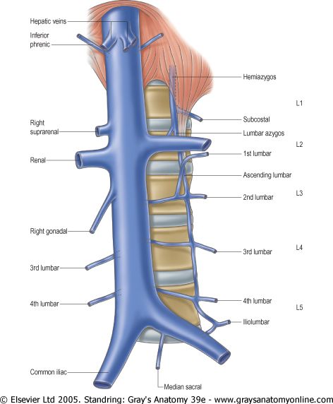
The IVC is the largest vein, retroperitoneal to the right of the aorta. It forms at L5 from the union of the two common iliac veins, ascends, and pierces the diaphragm at the caval hiatus (T8) to enter the right atrium.
Tributaries (ascending order):
- Common iliac veins (origin).
- Lumbar veins.
- Right gonadal (testicular/ovarian) vein — the left gonadal vein instead drains into the left renal vein.
- Renal veins — the left renal vein also receives the left suprarenal and left gonadal veins and crosses anterior to the aorta, under the SMA.
- Right suprarenal vein — direct to IVC (left drains to left renal vein).
- Inferior phrenic veins.
- Hepatic veins — just before the diaphragm.
Note on the GI tract: GI venous blood does not drain directly to the IVC — it goes through the portal system → liver → hepatic veins → IVC. The IVC directly drains the "systemic" abdominal organs (kidneys, adrenals, gonads, diaphragm, body wall).
1.14 · Objective 14 — Define the anatomy of abdominal lymph drainage
Abdominal lymph follows the arteries in reverse, draining through prevertebral (pre-aortic and lateral aortic/lumbar) nodes up to the cisterna chyli.
Pre-aortic nodes (named for the three unpaired GI arteries) drain the GI tract:
- Celiac nodes — foregut organs (stomach, liver, spleen, pancreas, proximal duodenum).
- Superior mesenteric nodes — midgut (distal duodenum → proximal 2/3 transverse colon).
- Inferior mesenteric nodes — hindgut (distal transverse colon → upper rectum).
Lateral aortic (lumbar) nodes drain the paired/retroperitoneal structures (kidneys, adrenals, gonads, posterior wall).
Convergence: intestinal + lumbar lymph trunks → cisterna chyli (at ~L1–L2, behind the aorta) → thoracic duct → drains into the junction of the left subclavian and internal jugular veins (left venous angle).
2 · Endocrine System
Prof. Hugh G. Rappa, MD
Instructional Objectives
- Describe the organs of the endocrine system.
- Compare and contrast the histology of the various organs of the endocrine system.
- Describe the anatomical relationships of each of the endocrine organs and surrounding anatomical structures and their blood supplies.
- Compare and contrast hormones and other factors involved in calcium metabolism.
2.1 · Objective 1 — Describe the organs of the endocrine system
The endocrine system secretes hormones directly into the blood (ductless). Primary endocrine organs: pituitary, hypothalamus, pineal, thyroid, parathyroids, thymus, adrenals (suprarenals), pancreatic islets, and gonads (ovaries/testes).
Organs with secondary endocrine tissue: heart (ANP/BNP), kidney (erythropoietin, calcitriol, renin), adipose (leptin, resistin), and the digestive tract (gut hormones).
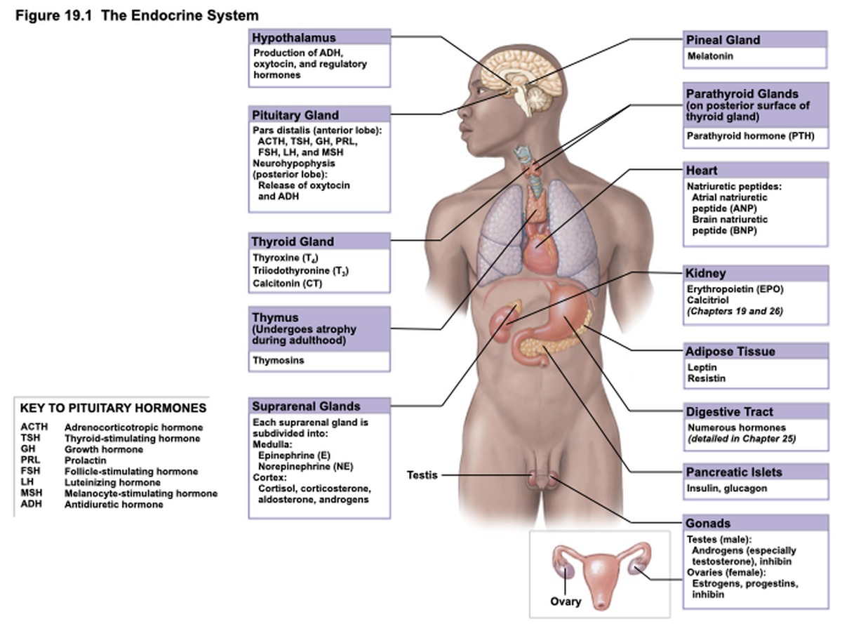
This lecture focuses on the thyroid, parathyroids, pituitary, pancreas, and adrenal glands.
2.2 · Objective 2 — Compare and contrast the histology of the various organs of the endocrine system
Anterior pituitary (adenohypophysis) — pars distalis: cords of cells with sinusoids. Two staining classes:
- Chromophobes (~50%) — pale, no granules.
- Chromophils (~50%): acidophils (~10%, reddish — GH & prolactin) and basophils (~40%, bluish — TSH, ACTH, FSH, LH).
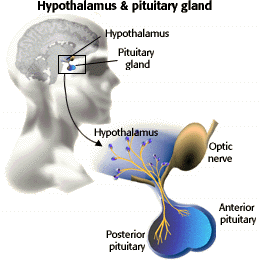
Posterior pituitary (neurohypophysis) — pars nervosa: unmyelinated axons, pituicytes (glia), and Herring bodies (stored ADH/oxytocin). No true secretory cells — it releases hypothalamic hormones.
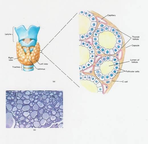
Thyroid: spherical follicles lined by simple cuboidal follicular cells around a central colloid (stores thyroglobulin). Active follicles have taller epithelium. Parafollicular (C) cells lie between follicles and secrete calcitonin (larger, paler-staining).
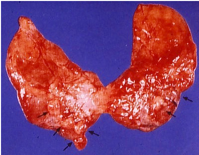
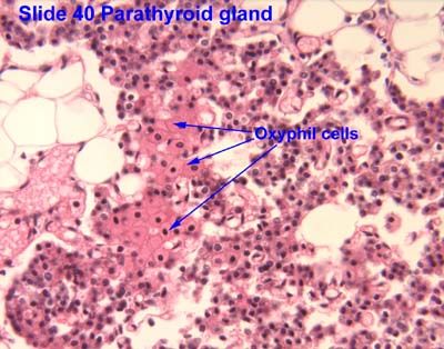
Parathyroid: cords of chief (principal) cells (secrete PTH; slightly eosinophilic) and larger, very eosinophilic oxyphil cells (function unclear); adipocytes increase with age.
Endocrine pancreas — islets of Langerhans (~1% of pancreas): α cells (glucagon), β cells (insulin), δ cells (somatostatin), F/PP cells (pancreatic polypeptide). Surrounded by exocrine acini.
Adrenal cortex — three zones (superficial→deep): zona glomerulosa (mineralocorticoids — aldosterone), zona fasciculata (glucocorticoids — cortisol; radial columns of lipid-laden cells), zona reticularis (androgens; meshwork). Adrenal medulla — chromaffin cells (modified sympathetic neurons) secrete epinephrine/norepinephrine; contains a central vein.
2.3 · Objective 3 — Describe the anatomical relationships of each of the endocrine organs and surrounding anatomical structures and their blood supplies
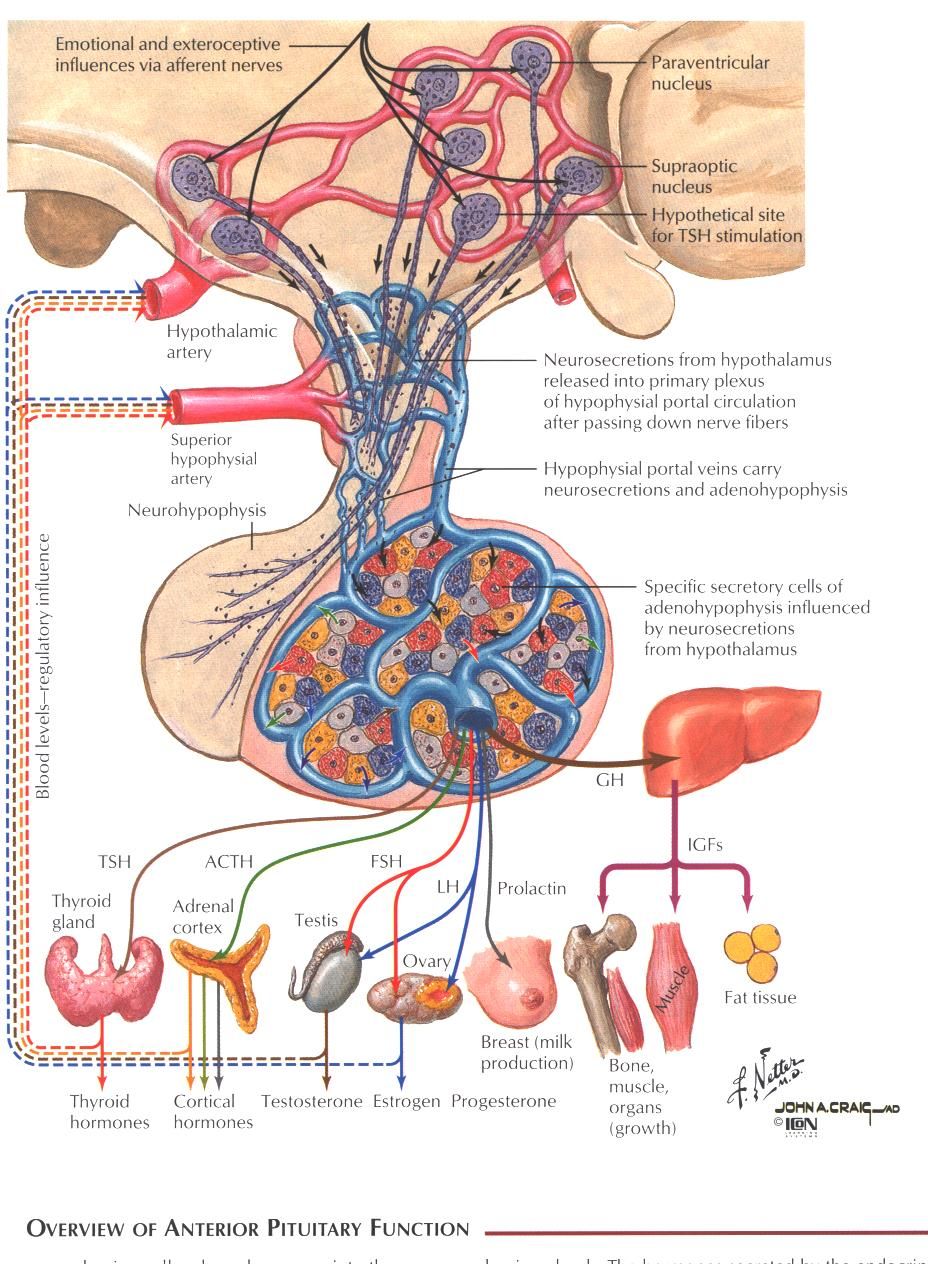
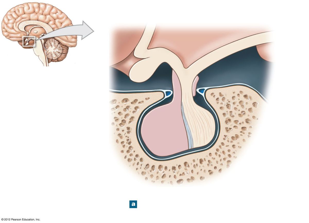
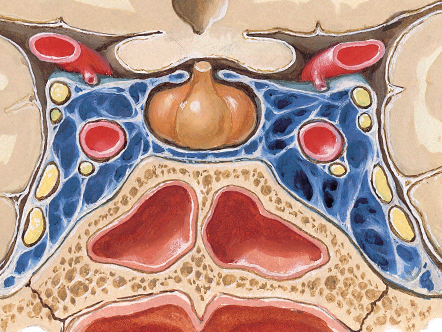
Pituitary gland (hypophysis): sits in the hypophyseal fossa of the sella turcica (sphenoid), attached to the hypothalamus by the infundibulum. Superiorly the diaphragma sellae separates it from the optic chiasm; laterally lie the cavernous sinuses; inferiorly the sphenoid air sinus; posteriorly the dorsum sellae.
- Anterior lobe receives portal blood via the hypophyseal portal system (superior hypophyseal artery → capillary plexus in the infundibulum → portal veins → adenohypophysis).
- Posterior lobe receives arterial blood (inferior hypophyseal artery).
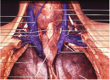
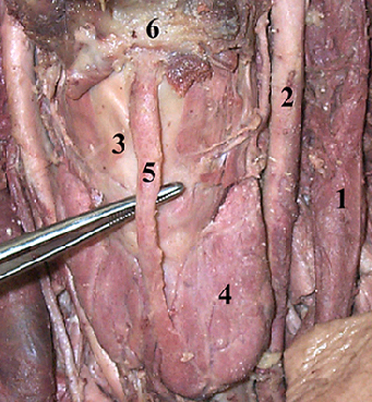
Thyroid gland: in the anterior neck at C5–T1, two lobes joined by an isthmus (over tracheal rings 2–3); enclosed in pretracheal fascia (moves with swallowing). Medial relations: larynx, trachea, pharynx, esophagus, external & recurrent laryngeal nerves. Posterolateral: carotid sheath.
Arterial supply: superior thyroid artery (from external carotid) and inferior thyroid artery (from thyrocervical trunk); occasional thyroidea ima. Venous: superior & middle thyroid veins → IJV; inferior thyroid vein → brachiocephalic.
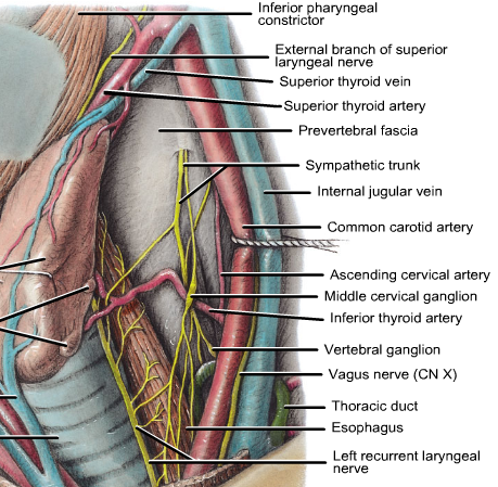
Parathyroid glands: usually four, on the posterior surface of the thyroid lobes (between the lobe and its capsule). Superior pair are more constant; inferior pair are variable. Blood supply: mainly the inferior thyroid artery.
Pancreas: retroperitoneal, epigastric/LUQ at T12–L3. Head in the duodenal C-loop, body crosses the left kidney, tail reaches the splenic hilum. Highly vascular (fenestrated capillaries); supplied by the pancreaticoduodenal arteries (from GDA & SMA) and pancreatic branches of the splenic artery; venous drainage → portal system.
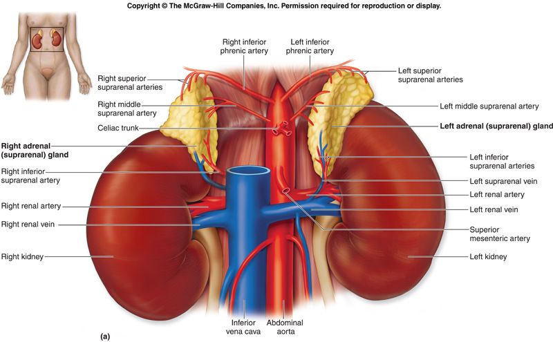
Adrenal (suprarenal) glands: retroperitoneal, capping the superior pole of each kidney within the renal fascia. Right = pyramidal (relates to liver & IVC); Left = semilunar (relates to stomach, pancreas, spleen). Triple arterial supply: superior suprarenal (from inferior phrenic), middle suprarenal (from aorta), inferior suprarenal (from renal artery). Venous: right suprarenal vein → IVC; left suprarenal vein → left renal vein.
2.4 · Objective 4 — Compare and contrast hormones and other factors involved in calcium metabolism
Three hormones regulate serum calcium; two raise it, one lowers it.
| Hormone | Source | Effect on Ca²⁺ | Actions |
|---|---|---|---|
| Parathyroid hormone (PTH) | Chief cells of parathyroid | ↑ (raises) | ↑ bone resorption (osteoclasts), ↑ renal Ca²⁺ reabsorption & phosphate excretion, ↑ activation of vitamin D (calcitriol) in the kidney. |
| Calcitriol (active vitamin D) | Kidney (activates vit D) | ↑ (raises) | ↑ intestinal absorption of Ca²⁺ and phosphate; assists PTH on bone. |
| Calcitonin | Parafollicular (C) cells of thyroid | ↓ (lowers) | ↓ osteoclast activity (inhibits bone resorption); minor role in adult humans. |
The PTH loop: low serum Ca²⁺ → parathyroid chief cells release PTH → bone, kidney, and (via calcitriol) gut all raise Ca²⁺ → normal Ca²⁺ feeds back to shut off PTH.
Relevant anatomy: the parathyroids (posterior thyroid) sense calcium directly; the thyroid C cells supply calcitonin; the kidney performs the final hydroxylation of vitamin D to calcitriol and executes PTH's renal effects.
3 · Male Reproductive System
Prof. Hugh E. Griffenkranz, MPAS, PA-C
Instructional Objectives
- Recall the embryologic development of the male reproductive tract.
- Identify the major muscles that make up the pelvic floor.
- Identify the major blood supply to the pelvis and male reproductive organs.
- Identify the nerves that innervate the pelvic floor and male reproductive organs.
- Describe the organs of the male reproductive system and their relationship to other anatomical features.
- Describe the pathway for semen from origin to ejaculation.
- Describe the parts of the urethra in relationship to the male reproduction system.
3.1 · Objective 1 — Recall the embryologic development of the male reproductive tract
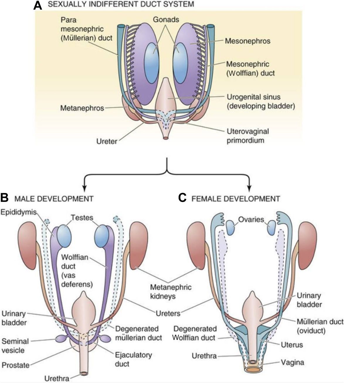
The male tract develops under the influence of the SRY gene (testis-determining factor) and testicular hormones.
- Testes form in the posterior abdomen near the developing kidneys.
- Sertoli cells secrete anti-Müllerian hormone (AMH) → regression of the paramesonephric (Müllerian) ducts.
- Leydig cells secrete testosterone → the mesonephric (Wolffian) ducts persist and form the epididymis, ductus deferens, seminal vesicles, and ejaculatory duct.
- DHT (from testosterone) drives development of the prostate, penis, and scrotum from the urogenital sinus and external genitalia.
Descent of the testes: the gubernaculum testis anchors the testis; as the body grows, the gubernaculum doesn't lengthen, so the testis is drawn inferiorly through the inguinal canal into the scrotum (usually by birth). The ductus deferens, vessels, lymphatics, and nerves stay bundled as the spermatic cord.
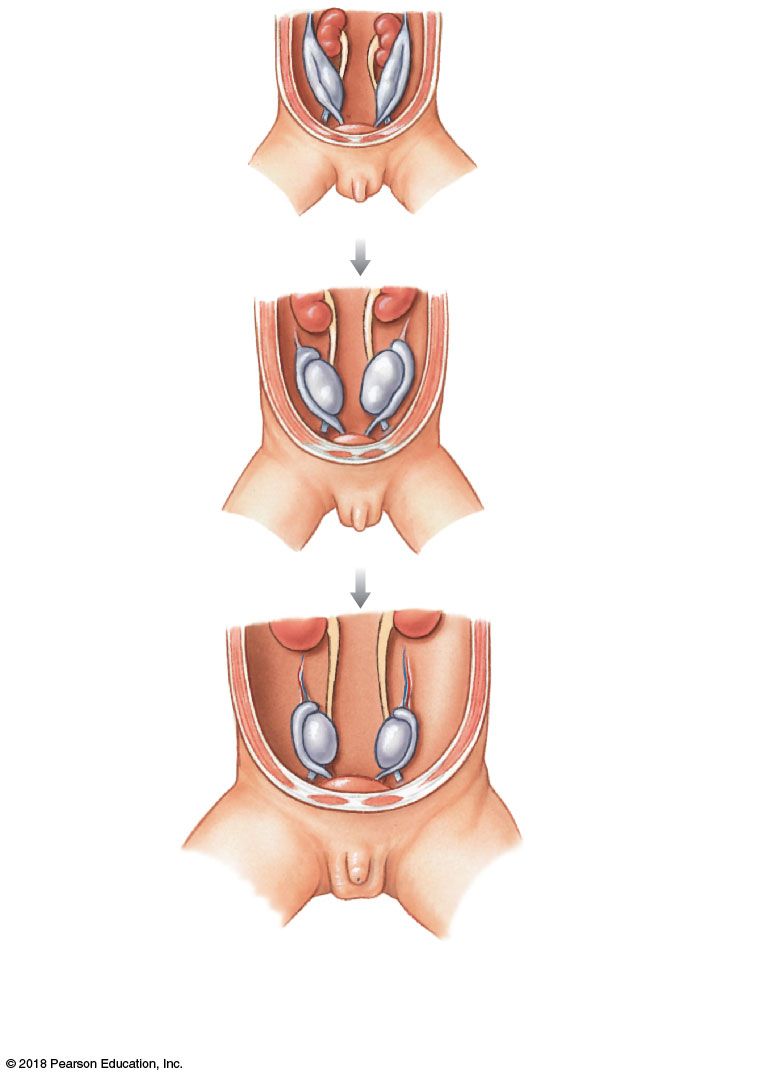
Why descend? Sperm development needs ~2 °F below core temperature — hence the extra-abdominal scrotal location.
3.2 · Objective 2 — Identify the major muscles that make up the pelvic floor
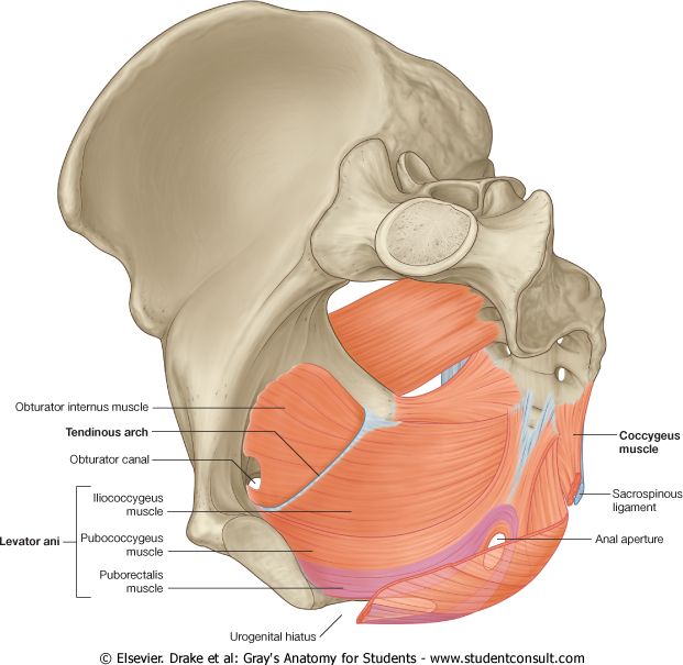
The pelvic floor (pelvic diaphragm) is formed mainly by the levator ani and coccygeus muscles, supporting the pelvic viscera and sealing the pelvic outlet.
- Levator ani — three parts: puborectalis (sling around the anorectal junction, maintains fecal continence), pubococcygeus, and iliococcygeus.
- Coccygeus (ischiococcygeus) — posterior part, from ischial spine to coccyx/sacrum.
Perineal muscles relevant to the male tract include the bulbospongiosus and ischiocavernosus (contribute to erection and ejaculation) and the external urethral sphincter in the deep perineal pouch.
3.3 · Objective 3 — Identify the major blood supply to the pelvis and male reproductive organs
The pelvis is supplied chiefly by the internal iliac artery, with the gonads supplied from the abdominal aorta.
- Testicular arteries — arise directly from the abdominal aorta (below the renal arteries), reflecting the testes' abdominal origin; travel in the spermatic cord.
- Internal pudendal artery (from internal iliac) — main supply to the perineum and external genitalia; gives the deep and dorsal arteries of the penis and the artery to the bulb.
- Scrotum — anterior via external pudendal (from femoral), posterior via internal pudendal branches.
- Ductus deferens — deferential artery (from the inferior vesical/internal iliac).
Venous drainage: the pampiniform plexus surrounds the testicular artery (counter-current heat exchange) and coalesces into the testicular vein: right → IVC, left → left renal vein (why left-sided varicoceles are more common).
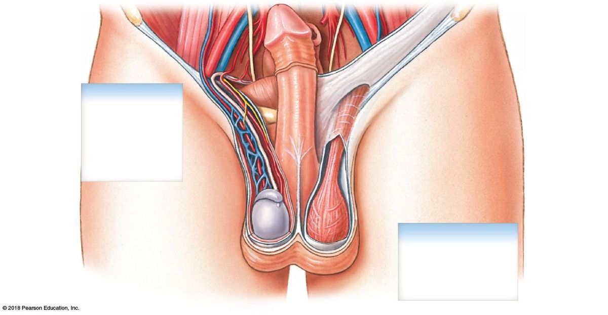
3.4 · Objective 4 — Identify the nerves that innervate the pelvic floor and male reproductive organs
- Pudendal nerve (S2–S4) — the main somatic nerve of the perineum: motor to the pelvic floor/perineal muscles and external urethral sphincter; sensory to the external genitalia (via the dorsal nerve of the penis).
- Pelvic splanchnic nerves (S2–S4, parasympathetic) — drive erection ("point").
- Hypogastric / sympathetic (T11–L2) — drive emission and ejaculation ("shoot"); also close the internal urethral sphincter to prevent retrograde ejaculation.
- Ilioinguinal & genitofemoral nerves — sensory to the scrotum and anterior groin; the genital branch also supplies the cremaster muscle (cremasteric reflex).
3.5 · Objective 5 — Describe the organs of the male reproductive system and their relationship to other anatomical features
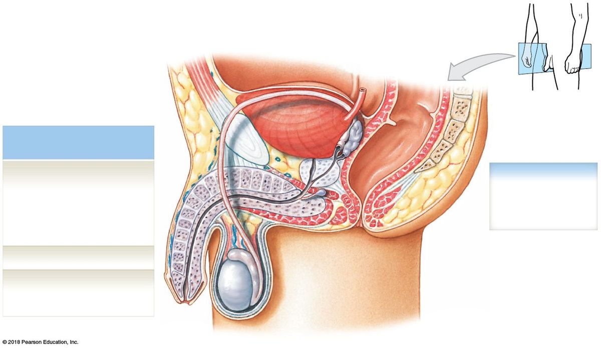
Principal structures: scrotum, testes, epididymis, ductus (vas) deferens, urethra, accessory glands (seminal vesicles, prostate, bulbourethral), and penis.
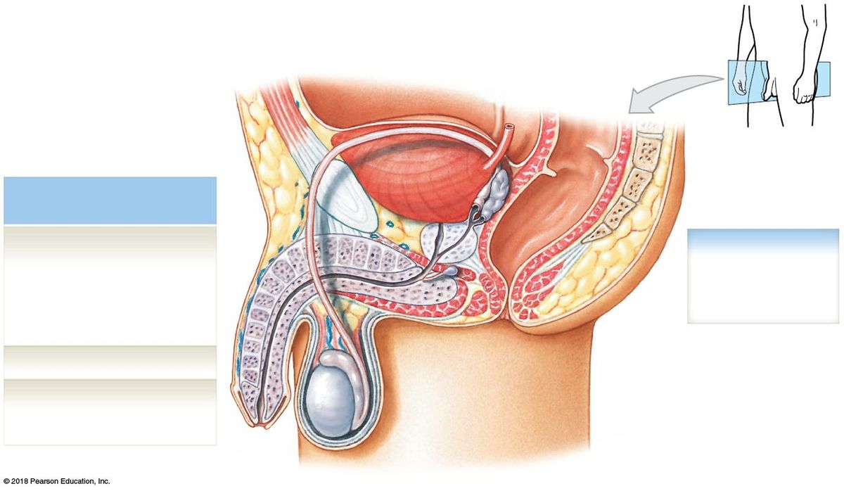
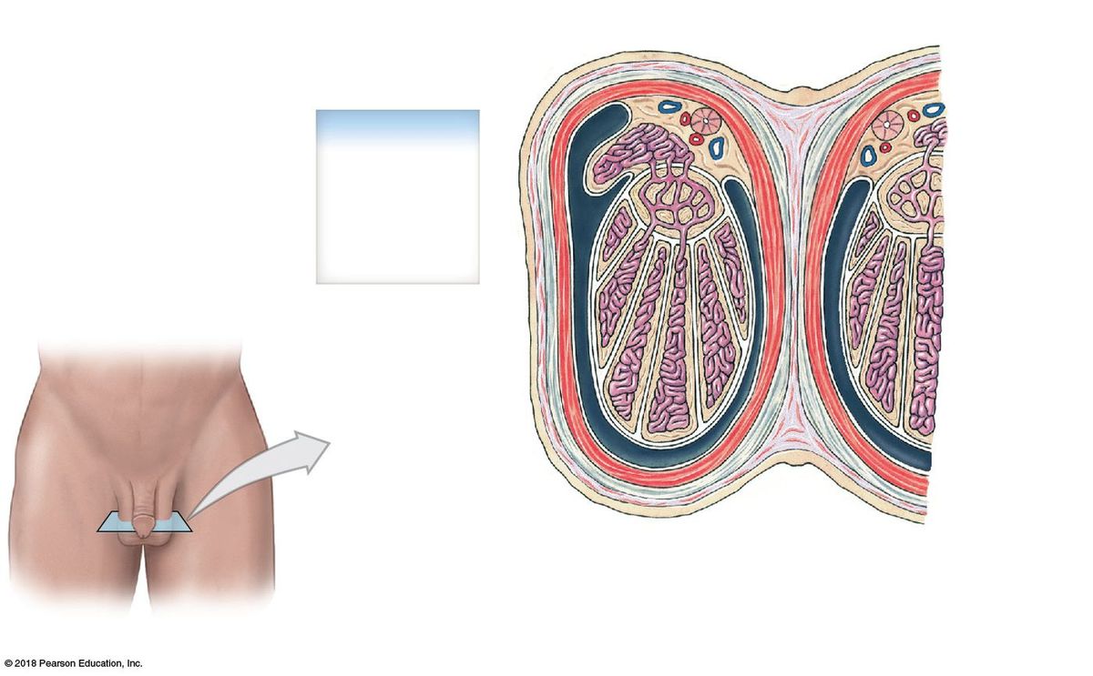
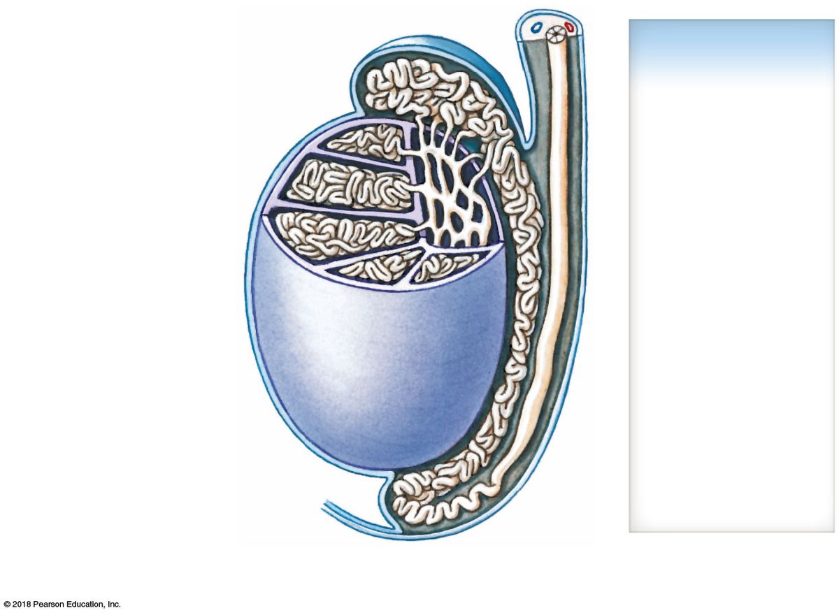
Testes: within the scrotum, each ~5×3×2.5 cm. Tunica albuginea (fibrous capsule) sends septa that divide the testis into lobules; septa converge at the mediastinum testis. Tunica vaginalis (serous, from peritoneum) surrounds it and reduces friction.
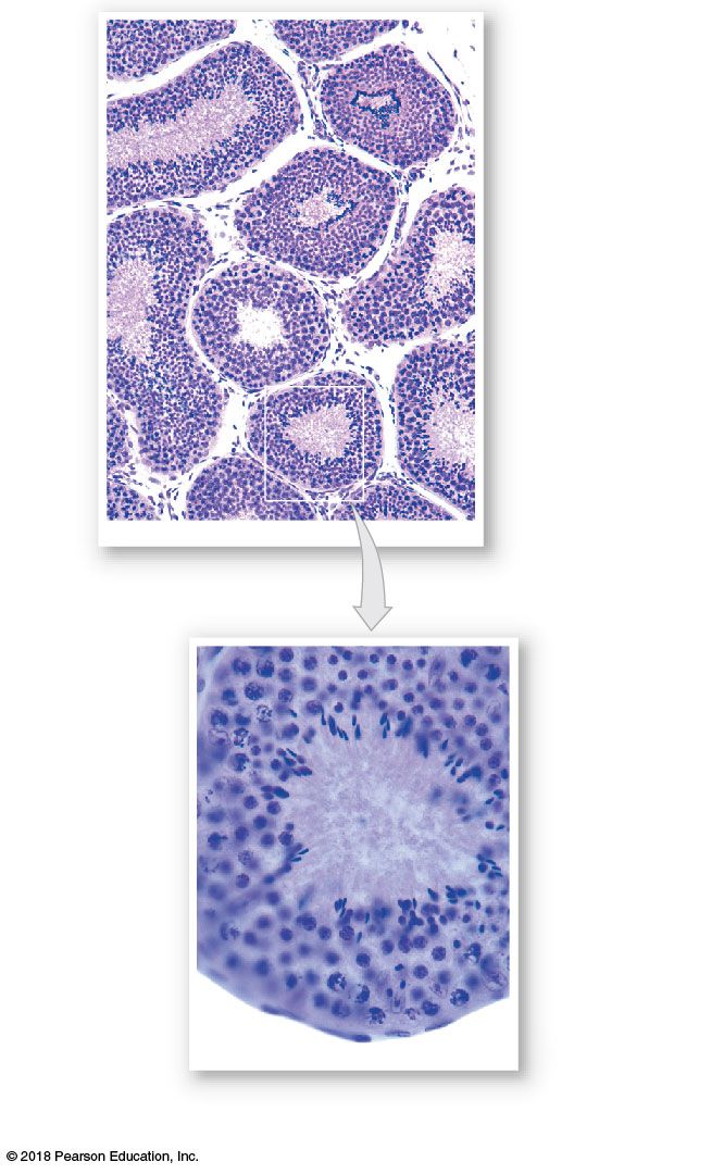
Histology: seminiferous tubules (site of spermatogenesis) contain developing germ cells and nurse (Sertoli) cells (blood–testis barrier, inhibin, ABP). Between tubules, interstitial (Leydig) cells make testosterone.
Epididymis (head, body, tail on the posterior testis) — matures and stores sperm; sperm gain motility here (~2 weeks transit). Its tail becomes the ductus deferens.
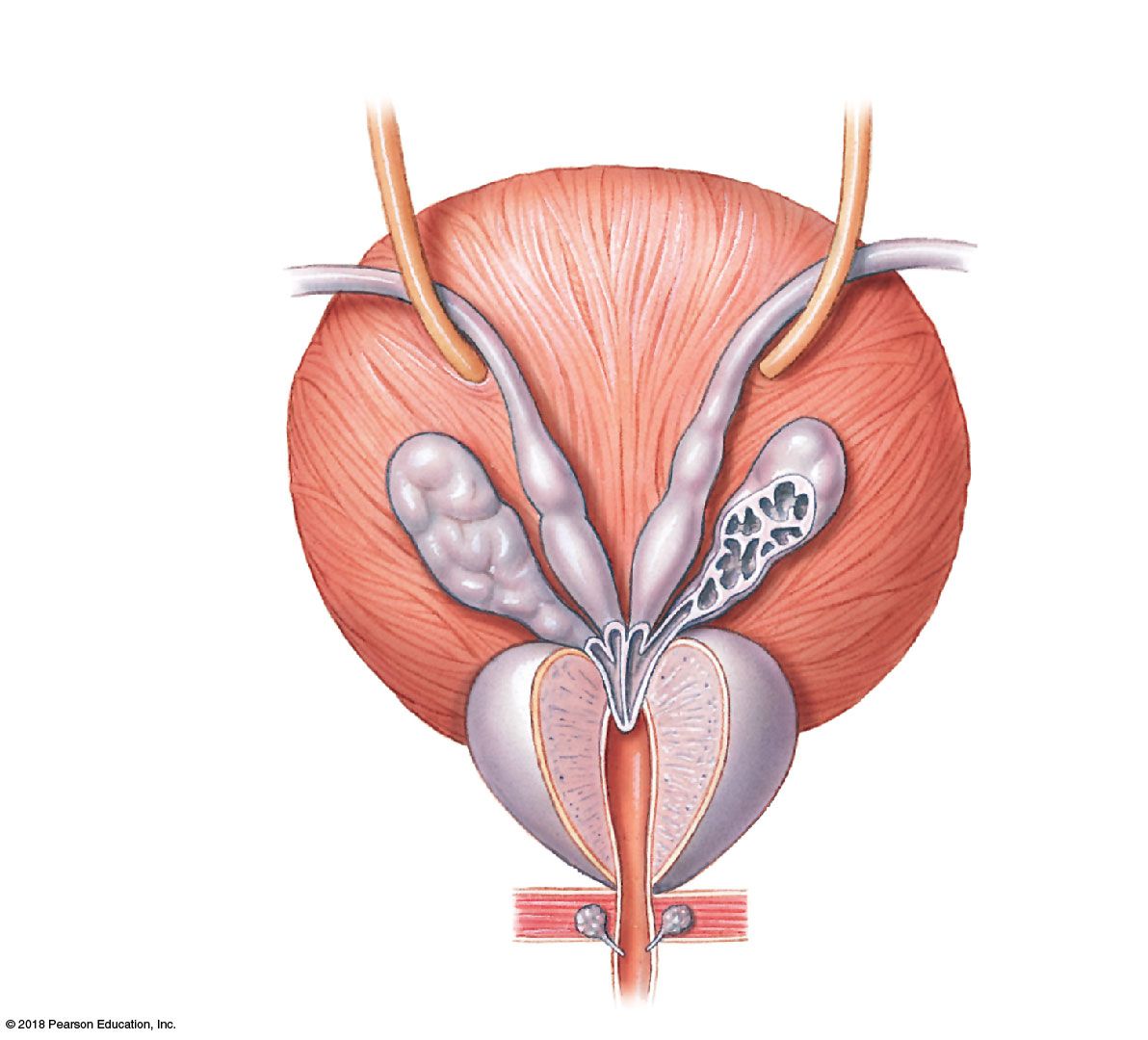
Accessory glands: seminal vesicles (60% of semen — fructose, prostaglandins, clotting factors), prostate (20–30% — weakly acidic, anti-coagulant enzymes, seminalplasmin), bulbourethral (Cowper's) glands (5% — alkaline mucus, neutralizes urethral acid, lubrication).
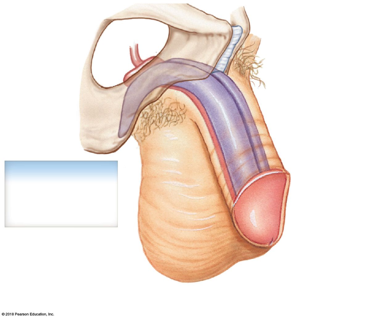
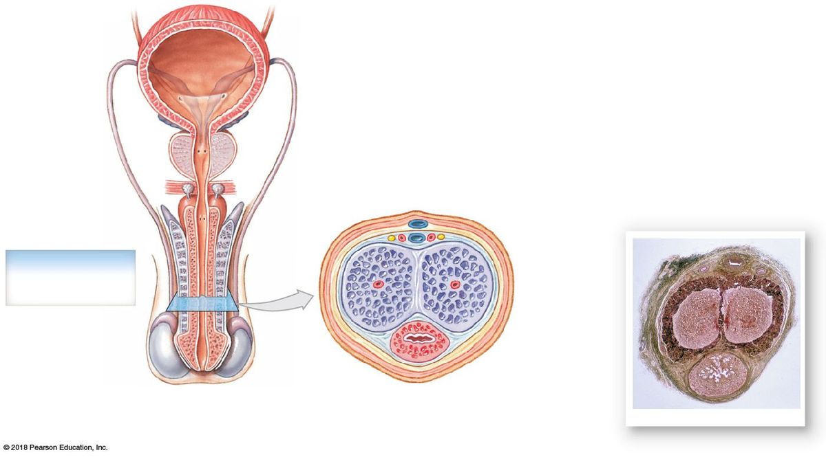
Penis: root, body (shaft), glans. Three erectile columns — two dorsal corpora cavernosa (each ends as a crus attached to the ischial ramus; contains the deep artery) and one ventral corpus spongiosum (surrounds the spongy urethra; expands as the bulb and the glans).
3.6 · Objective 6 — Describe the pathway for semen from origin to ejaculation
Follow one cell from production to exit:
Seminiferous tubules → straight tubules → rete testis → efferent ductules → epididymis (head→body→tail) → ductus (vas) deferens → ampulla of the ductus deferens → (joins seminal vesicle duct) → ejaculatory duct → prostatic urethra → membranous urethra → spongy (penile) urethra → external urethral orifice.
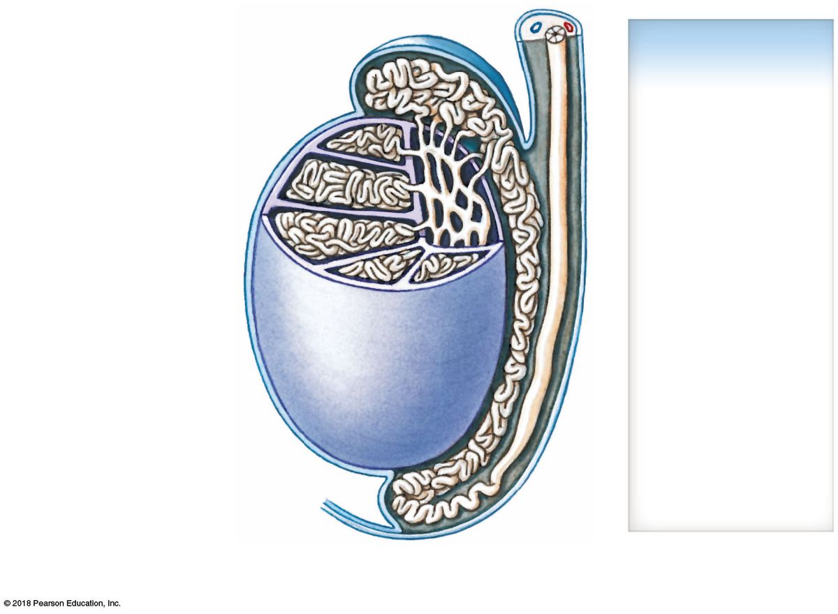
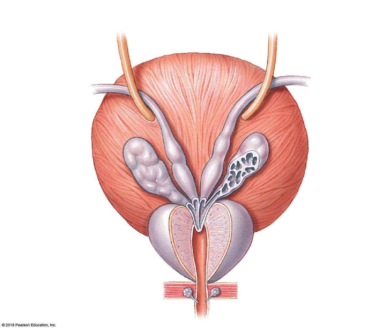
Physiology overlay: emission (sympathetic) mixes sperm with glandular secretions in the urethra; ejaculation is the forceful expulsion by rhythmic contractions of the bulbospongiosus and ischiocavernosus. Sperm complete capacitation (final functional maturation) only in the female tract.
3.7 · Objective 7 — Describe the parts of the urethra in relationship to the male reproduction system
The male urethra (~18–20 cm) is shared by the urinary and reproductive systems and has three regions:
- Prostatic urethra — passes through the prostate; receives the ejaculatory ducts and multiple prostatic ducts. This is where the reproductive and urinary tracts join.
- Membranous urethra — shortest; passes through the urogenital diaphragm / deep perineal pouch, surrounded by the external urethral sphincter (voluntary).
- Spongy (penile) urethra — longest; runs within the corpus spongiosum to the external urethral orifice; receives the bulbourethral gland ducts proximally.
4 · Female Reproductive System
Prof. Hugh G. Rappa, MD
Instructional Objectives
- Recall the embryologic development of all organs of the female reproductive tract.
- Describe the perineum, including the anterior and posterior triangles.
- Compare and contrast the various ligaments / peritoneal folds of the female reproductive tract.
- Identify the major muscles that make up the pelvic floor.
- Identify the major blood supply to the pelvis and each organ of the female reproductive.
- Identify the nerves that innervate the pelvic floor and female reproductive organs.
- Describe each of the organs of the female reproduction system and their relationship to other anatomical features.
- Describe the cell type of the organs of the female reproductive tract.
- Identify the peritoneal coverings of the female internal genitalia.
4.1 · Objective 1 — Recall the embryologic development of all organs of the female reproductive tract
In the absence of AMH and testosterone, the female pattern develops by default.
- Paramesonephric (Müllerian) ducts persist (no AMH to regress them) and form the major internal organs. They arise from coelomic epithelium lateral to the mesonephros (~week 6), grow caudally, and fuse in the midline into the uterovaginal primordium.
- Unfused cranial portions → fallopian (uterine) tubes.
- Fused caudal portions → uterus and cervix, and the upper ~1/3 of the vagina.
- Lower vagina → from the urogenital sinus.
- Mesonephric (Wolffian) ducts regress (no testosterone).
4.2 · Objective 2 — Describe the perineum, including the anterior and posterior triangles
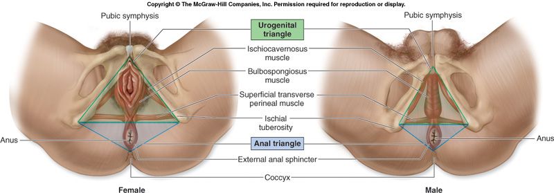
The perineum is the diamond-shaped region between the thighs, bounded anteriorly by the pubic symphysis, laterally by the ischial tuberosities, and posteriorly by the coccyx.
An imaginary line between the ischial tuberosities divides it into two triangles:
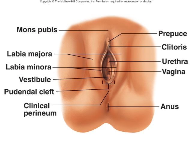
- Anterior (urogenital) triangle — contains the urethral and vaginal orifices and the external genitalia; contains the urogenital diaphragm.
- Posterior (anal) triangle — contains the anus and ischioanal fossae.
4.3 · Objective 3 — Compare and contrast the various ligaments / peritoneal folds of the female reproductive tract
Peritoneum drapes the pelvic organs, forming supporting folds and two clinically important pouches.
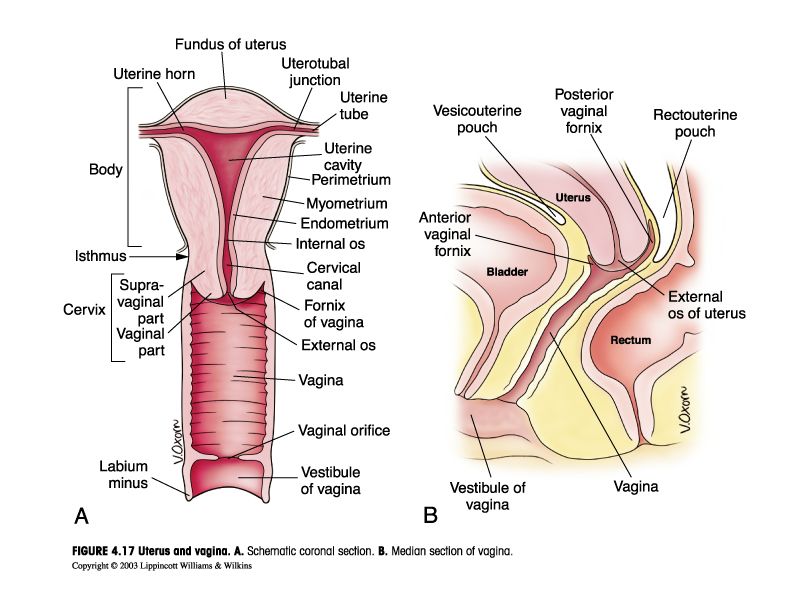
Peritoneal pouches: the vesicouterine pouch (between bladder and uterus) and the deeper rectouterine pouch (of Douglas) (between uterus and rectum — the lowest point of the female peritoneal cavity, where fluid collects).
Ligaments / folds:
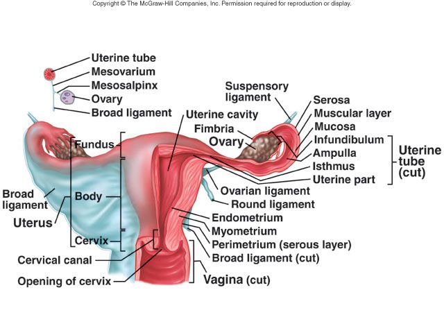
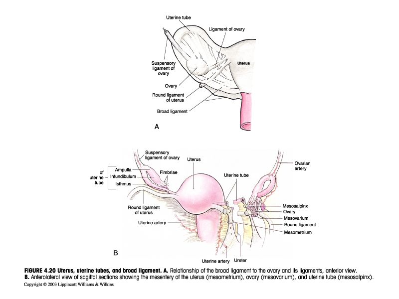
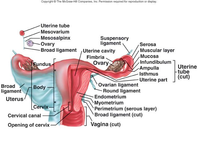
- Broad ligament — a peritoneal sheet draping the uterus, tubes, and ovaries; subdivided into mesovarium, mesosalpinx, and mesometrium.
- Mesovarium — attaches the ovary to the broad ligament.
- Ovarian ligament — ovary → uterus (superolateral body).
- Suspensory ligament of the ovary — ovary → pelvic (lateral) wall; carries the ovarian vessels.
- Round ligament — uterus → through the inguinal canal → labia majora (maintains anteversion; remnant of the gubernaculum).
- Cardinal (transverse cervical) ligaments — cervix/upper vagina → lateral pelvic wall; carry the uterine vessels (key uterine support).
- Uterosacral ligaments — cervix → sacrum.
4.4 · Objective 4 — Identify the major muscles that make up the pelvic floor
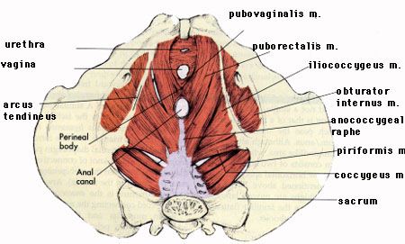
Same pelvic diaphragm as in the male: levator ani (puborectalis, pubococcygeus, iliococcygeus) + coccygeus. It supports the pelvic organs, seals the pelvic outlet, and helps continence and defecation.
In females, the levator ani also includes the pubovaginalis slip encircling the vagina. Perineal muscles around the vagina: bulbospongiosus, external urethral sphincter, and the urethrovaginal sphincter.
4.5 · Objective 5 — Identify the major blood supply to the pelvis and each organ of the female reproductive
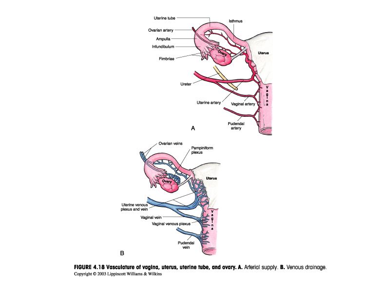
- Ovarian arteries — from the abdominal aorta (below the renals), travel in the suspensory ligament (reflecting ovarian origin near the kidneys).
- Uterine artery — from the internal iliac; runs in the cardinal ligament and crosses over the ureter ("water under the bridge"). Supplies uterus and anastomoses with ovarian and vaginal arteries.
- Vaginal artery — from the internal iliac (or uterine); supplies the vagina.
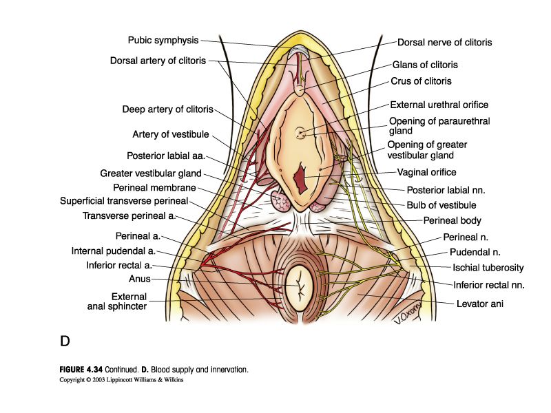
- Internal pudendal artery — from the internal iliac; supplies the perineum, clitoris, and external genitalia.
Venous drainage: the uterine/vaginal/pampiniform plexuses → internal iliac veins; ovarian veins mirror the male — right ovarian vein → IVC, left ovarian vein → left renal vein.
4.6 · Objective 6 — Identify the nerves that innervate the pelvic floor and female reproductive organs
- Pudendal nerve (S2–S4) — somatic supply to the perineum, external genitalia, and pelvic floor/perineal muscles (including the external urethral & anal sphincters). Stretched in childbirth → incontinence.
- Pelvic splanchnic nerves (S2–S4, parasympathetic) — to the pelvic organs; drive clitoral erection.
- Sympathetic (T10–L2, via hypogastric plexus) — uterine/ovarian; ovarian afferents ride with the ovarian vessels back to T10–T11 (why ovarian/periovulatory pain can refer to the umbilical/flank region).
Uterine/cervical pain above the pelvic pain line travels with sympathetics; below the pelvic pain line (e.g., lower vagina, perineum) travels with the pudendal nerve.
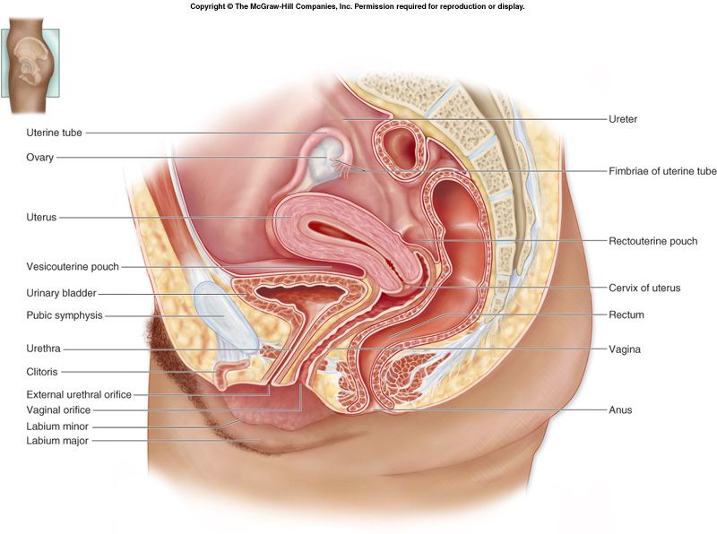
4.7 · Objective 7 — Describe each of the organs of the female reproduction system and their relationship to other anatomical features
Ovaries — the primary sex organs (produce oocytes + estrogen/progesterone). Lie against the lateral pelvic wall, posterior to the broad ligament; anchored by the ovarian and suspensory ligaments. Blood via the ovarian artery; right ovarian vein → IVC, left → left renal vein.
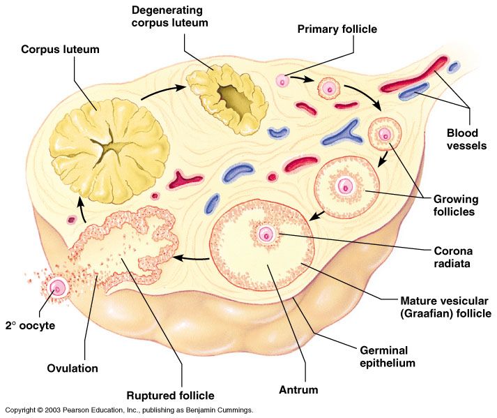
Uterine (fallopian) tubes — receive the ovulated oocyte and are the usual site of fertilization; lie in the free (superior) edge of the broad ligament. Parts: infundibulum (with fimbriae) → ampulla (widest; fertilization site) → isthmus → intramural/uterine part. Do not physically attach to the ovary.
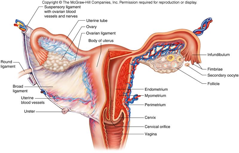
Uterus — organ of gestation, between the bladder (anterior) and rectum (posterior); typically anteverted (at the cervix–vagina junction) and anteflexed (at the body–cervix junction). Regions: fundus (above the tube entrances), body, isthmus, and cervix (with internal & external os). Supported by the levator ani, cardinal, uterosacral, round, and broad ligaments.
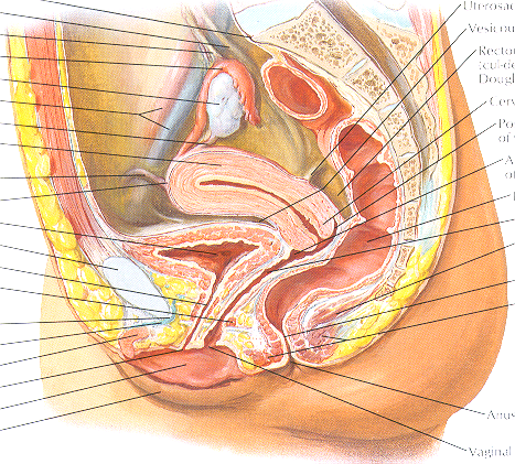
Vagina — ~10 cm fibromuscular tube from cervix to vestibule; posterior to the urethra/bladder, anterior to the rectum. The fornices surround the cervix; serves as birth canal, copulatory organ, and menstrual passage.
External genitalia (vulva): mons pubis, labia majora (homologous to the scrotum), labia minora, clitoris (erectile, homologous to the penis), vestibule (contains urethral & vaginal orifices), and greater vestibular (Bartholin) glands (homologous to bulbourethral glands).
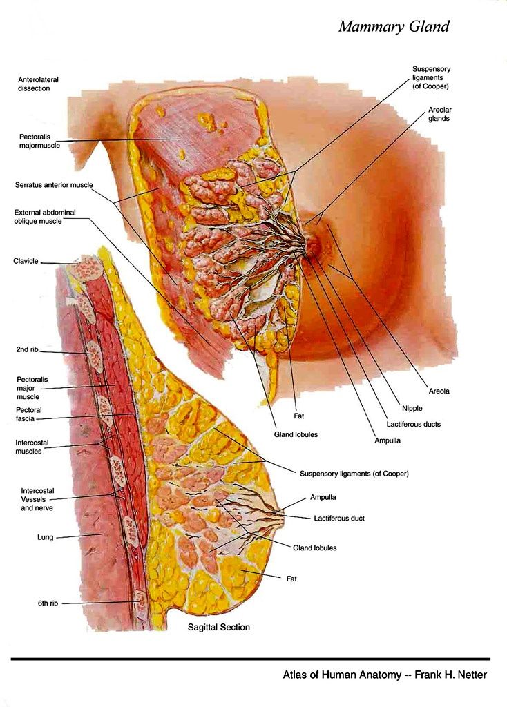
Breast — glandular lobes drained by lactiferous ducts to the nipple; supported by suspensory (Cooper's) ligaments; lymph drains mostly to the axillary nodes. Lies over pectoralis major/serratus anterior (ribs 2–6).
4.8 · Objective 8 — Describe the cell type of the organs of the female reproductive tract
| Organ | Epithelium / cell type |
|---|---|
| Ovary (surface) | Simple cuboidal ("germinal") epithelium over a tunica albuginea; cortex holds follicles (oocyte + follicular/granulosa cells). |
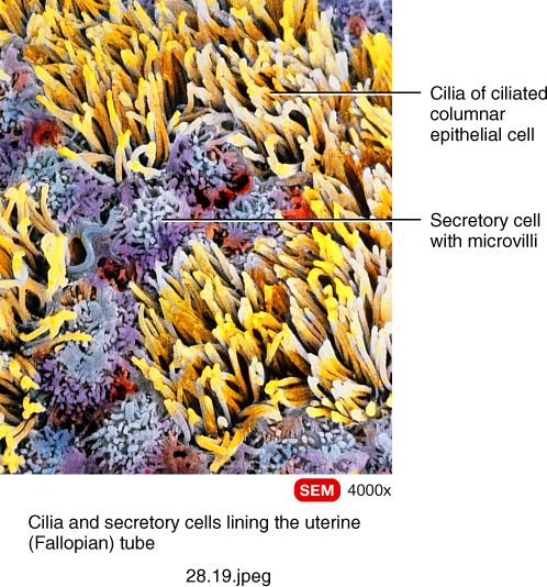
| Uterine tube | Simple columnar with ciliated and non-ciliated (secretory/peg) cells — cilia + peristalsis move the ovum toward the uterus. |
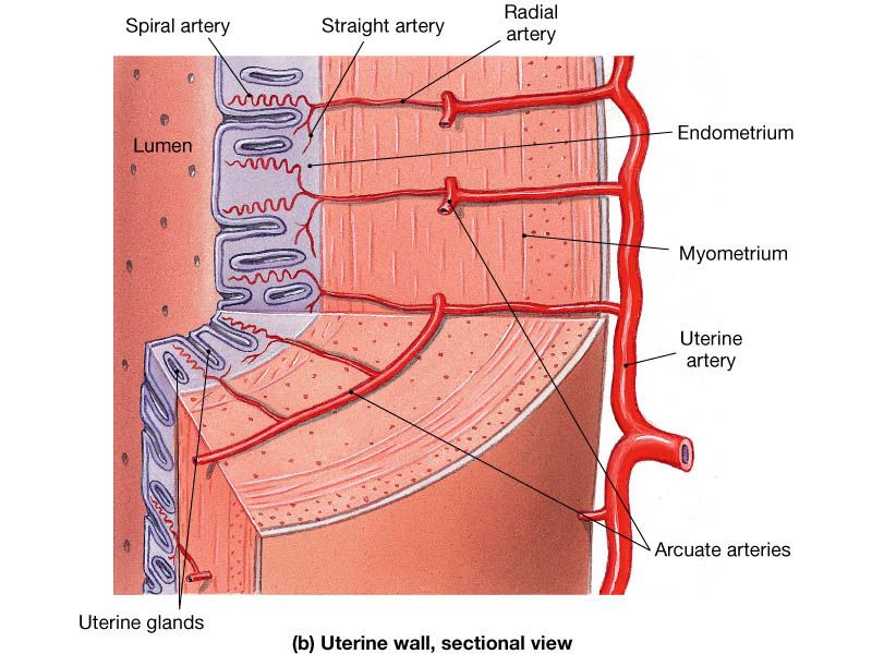
| Uterus — endometrium | Simple columnar epithelium with uterine glands; two layers — stratum functionalis (sheds at menstruation) and stratum basalis (regenerates it). |
| Uterus — myometrium | Thick smooth muscle (hypertrophies in pregnancy). |
| Uterus — perimetrium | Serosa (visceral peritoneum), continuous with the broad ligament. |
| Cervix / vagina | Nonkeratinized stratified squamous epithelium (protective, abrasion-resistant). The cervical canal is simple columnar; the transformation zone is clinically important. |
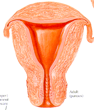
Endometrial cycle: the stratum functionalis builds each month under ovarian hormones; at menstruation the spiral arteries constrict, the functionalis self-digests and sloughs, and the basalis regenerates it.
4.9 · Objective 9 — Identify the peritoneal coverings of the female internal genitalia
The peritoneum reflects over the pelvic organs, giving each a characteristic covering:
- Uterus — the fundus and body are covered by peritoneum (perimetrium), continuous laterally with the broad ligament. The uterus is intraperitoneal.
- Uterine tubes — enclosed in the superior free margin of the broad ligament (mesosalpinx); largely peritoneal-covered; the fimbriated end opens into the peritoneal cavity.
- Ovaries — not covered by peritoneum on their surface (they have germinal epithelium instead); attached to the posterior broad ligament by the mesovarium. This is why the ovary opens the female peritoneal cavity to the exterior (tube → uterus → vagina).
- Peritoneal pouches — vesicouterine (anterior) and rectouterine of Douglas (posterior, the lowest point).
5 · Nephrology / Urinary System
Prof. Lauren Reynolds, MSPA, PA-C
Instructional Objectives
- Identify the organs that make up the urinary system.
- Describe the anatomy of the kidneys.
- Describe the blood flow to the kidney(s) and nephrons.
- Compare and contrast the various sections and parts of the nephron.
- Describe the anatomical relationships of each component of the urinary system and the surrounding anatomical structures.
- Compare and contrast the cell types of the urinary system.
5.1 · Objective 1 — Identify the organs that make up the urinary system
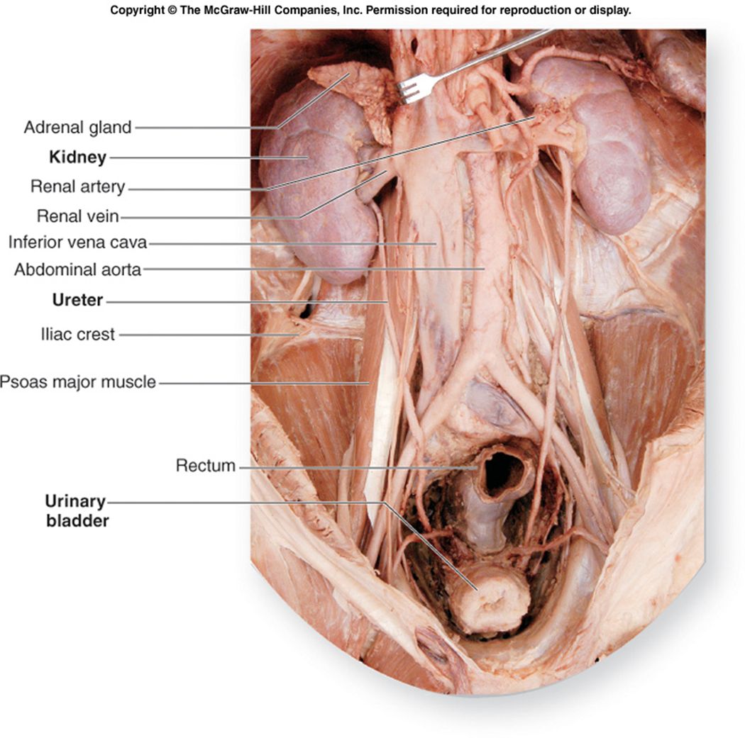
The urinary system = kidneys (primary organs) + the urinary tract (ureters, bladder, urethra).
- Kidneys — filter waste from the blood and convert filtrate into urine; also regulate BP, electrolytes, pH, and produce erythropoietin & calcitriol.
- Ureters — carry urine from kidney to bladder.
- Urinary bladder — stores urine.
- Urethra — conducts urine out of the body.
The act of urination is micturition.
5.2 · Objective 2 — Describe the anatomy of the kidneys
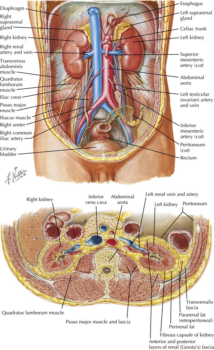
The kidneys are bean-shaped, retroperitoneal organs, ~11 cm long. Vertebral levels: right T12–L3 (lower, pushed down by the liver), left T10/11–L2.
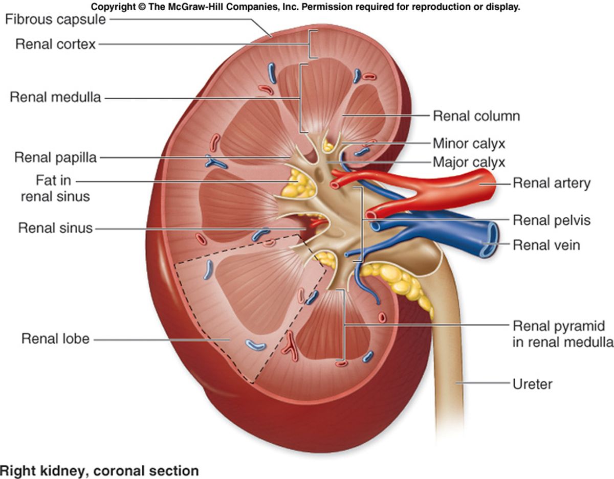
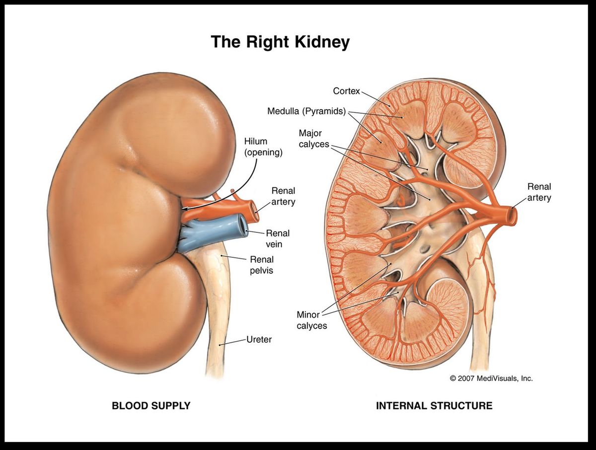
Hilum (concave medial border) transmits the NAVs and pelvis: renal artery, renal vein, renal pelvis, nerves, lymphatics. The renal sinus is the internal fat-filled space housing vessels, calyces, and the pelvis.
Coronal section (external → internal): renal cortex (outer) with renal columns dipping between pyramids; renal medulla arranged as 8–15 renal pyramids (base against cortex, apex = renal papilla); papillae drain into minor calyces → major calyces → renal pelvis → ureter.
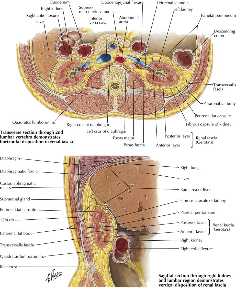
Surrounding layers (deep → superficial): fibrous (renal) capsule → perinephric (perirenal) fat → renal fascia → paranephric (pararenal) fat → peritoneum.
5.3 · Objective 3 — Describe the blood flow to the kidney(s) and nephrons
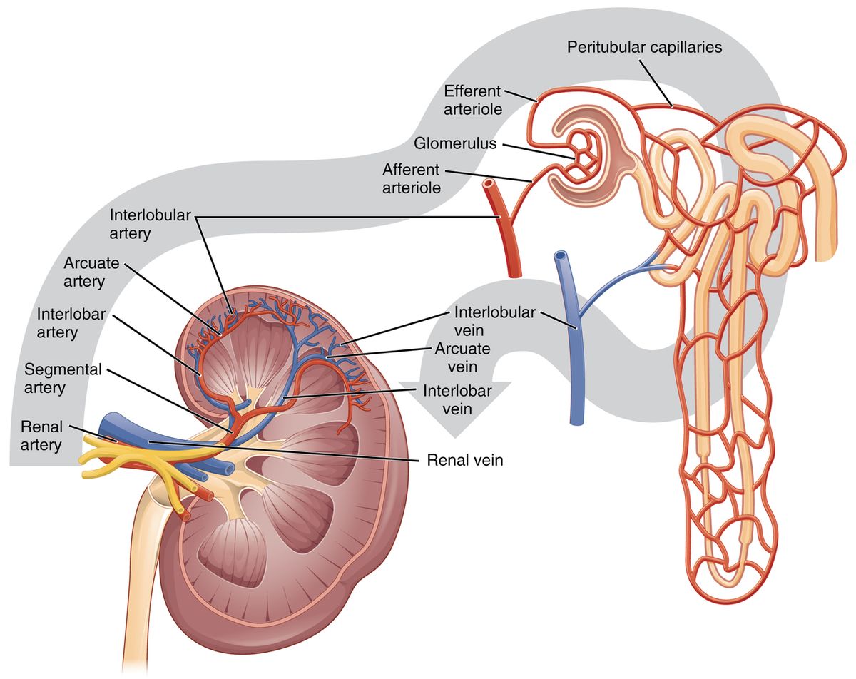
Arterial inflow (aorta → glomerulus):
Renal artery → segmental → interlobar (between pyramids) → arcuate (arching over pyramid bases) → interlobular (cortical radiate) → afferent arteriole → glomerulus.
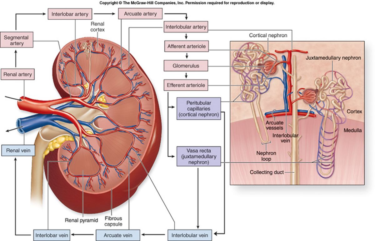
Plasma is filtered across the glomerulus into the glomerular (Bowman's) space. Then a second capillary bed forms — the kidney's unusual "two capillary beds in series":
Glomerulus → efferent arteriole → either peritubular capillaries (around cortical tubules) or vasa recta (long loops paralleling juxtamedullary nephron loops; concentrate urine).
Venous outflow (reverse of arterial, no "segmental"): vasa recta/peritubular → interlobular → arcuate → interlobar → renal vein → IVC.
5.4 · Objective 4 — Compare and contrast the various sections and parts of the nephron
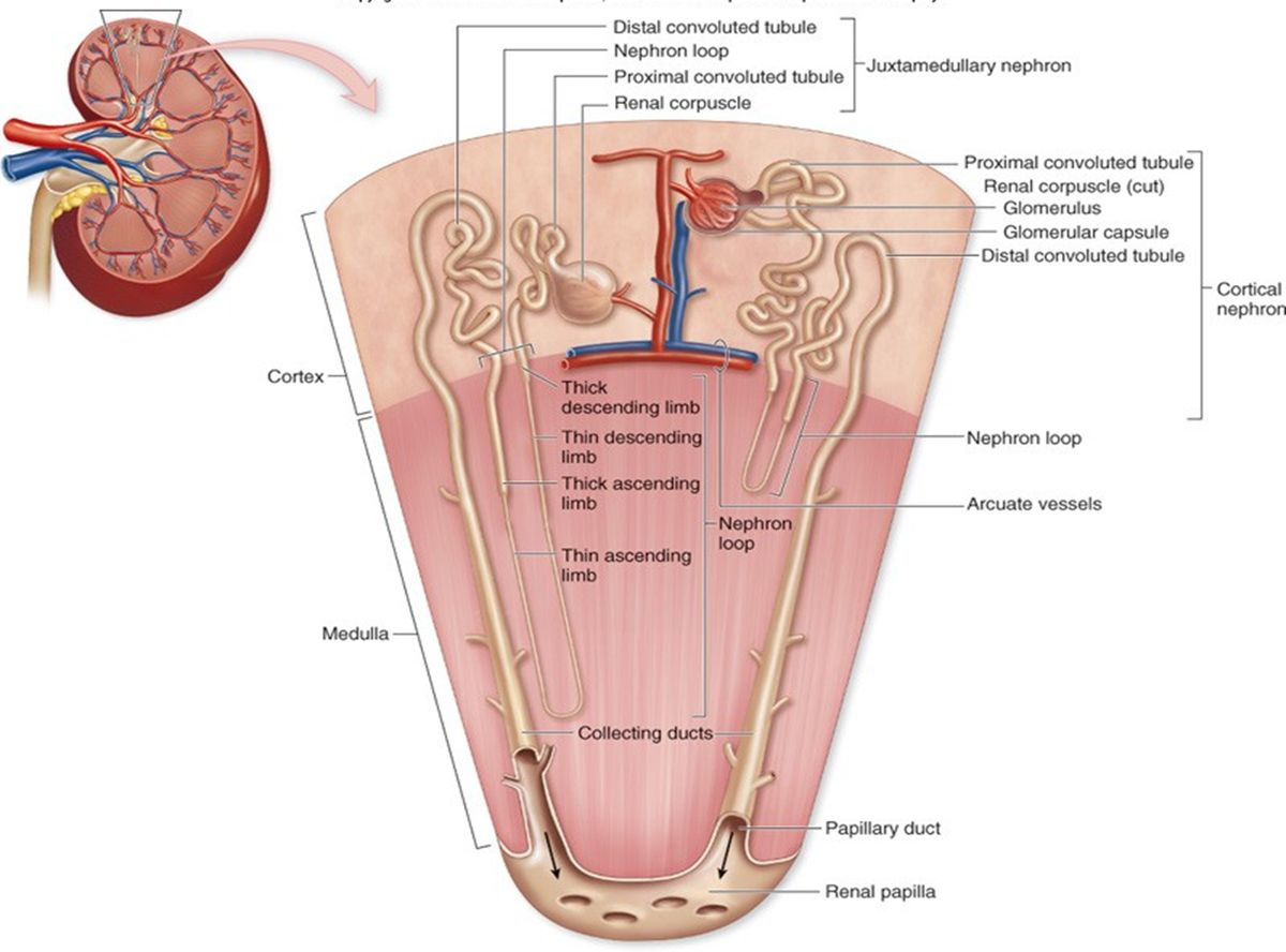
The nephron is the functional unit. Two components: the renal corpuscle (filter) and the renal tubule (processor).
Renal corpuscle: glomerulus (capillary tuft) + glomerular (Bowman's) capsule.
Renal tubule (filtrate order):
- Proximal convoluted tubule (PCT) — begins at the tubular pole; simple cuboidal with a brush border; bulk reabsorption.
- Nephron loop (loop of Henle) — descending limb (thick then thin) into the medulla; ascending limb (thin then thick) back to the cortex.
- Distal convoluted tubule (DCT) — begins at the thick ascending limb; lies next to the afferent arteriole (juxtaglomerular apparatus).
- Collecting tubule → collecting duct — receives filtrate from many nephrons; final water handling. Once in the collecting duct, no further modification; it is now officially "urine."
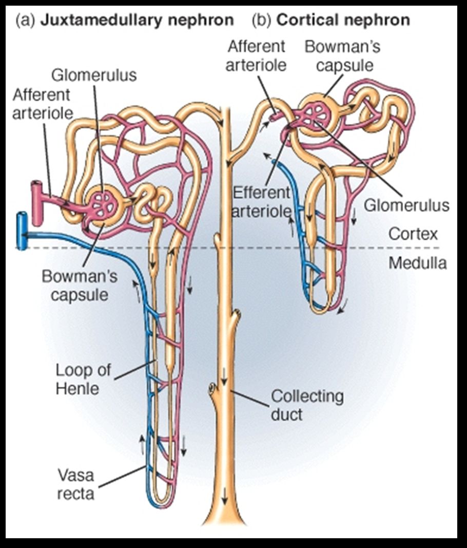
Two nephron types:
| Cortical (~85%) | Juxtamedullary (~15%) | |
|---|---|---|
| Location | Peripheral cortex | Near cortex-medulla junction |
| Loop length | Short loops | Long loops into medulla |
| Capillaries | Peritubular | Vasa recta |
| Role | "Standard" urine | Concentrate urine (recover water, Na⁺, Cl⁻) |
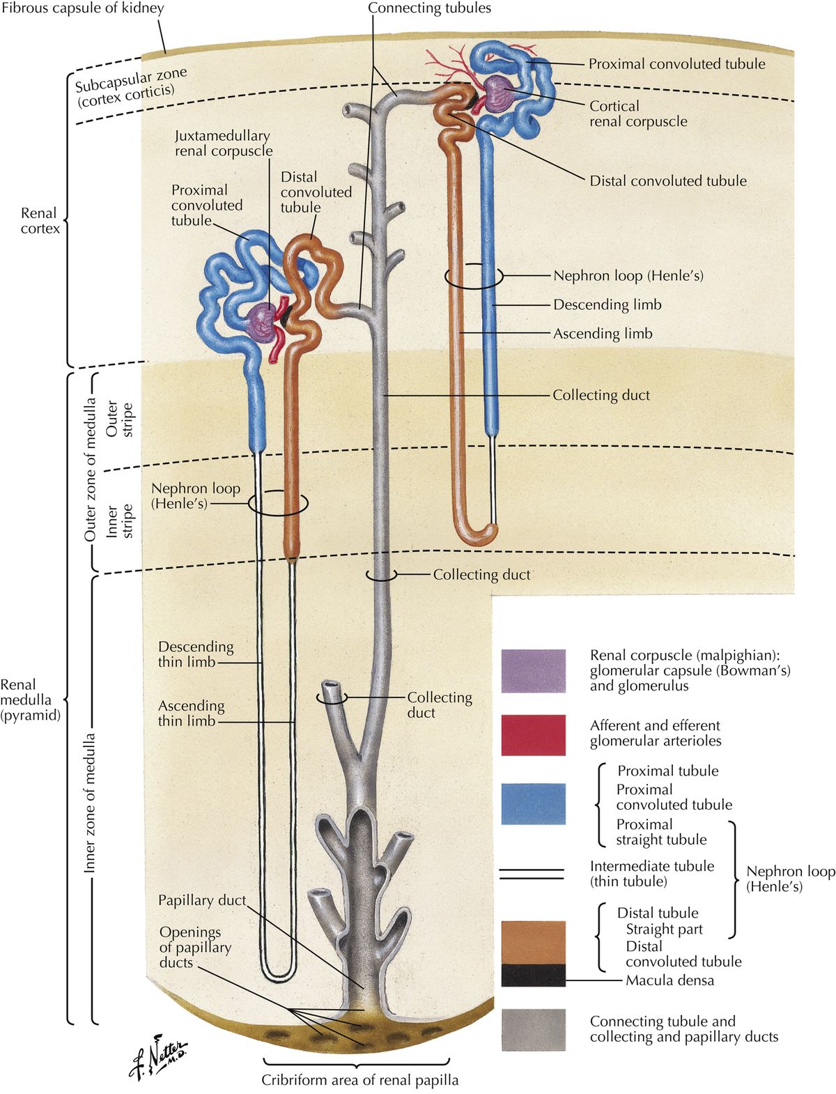
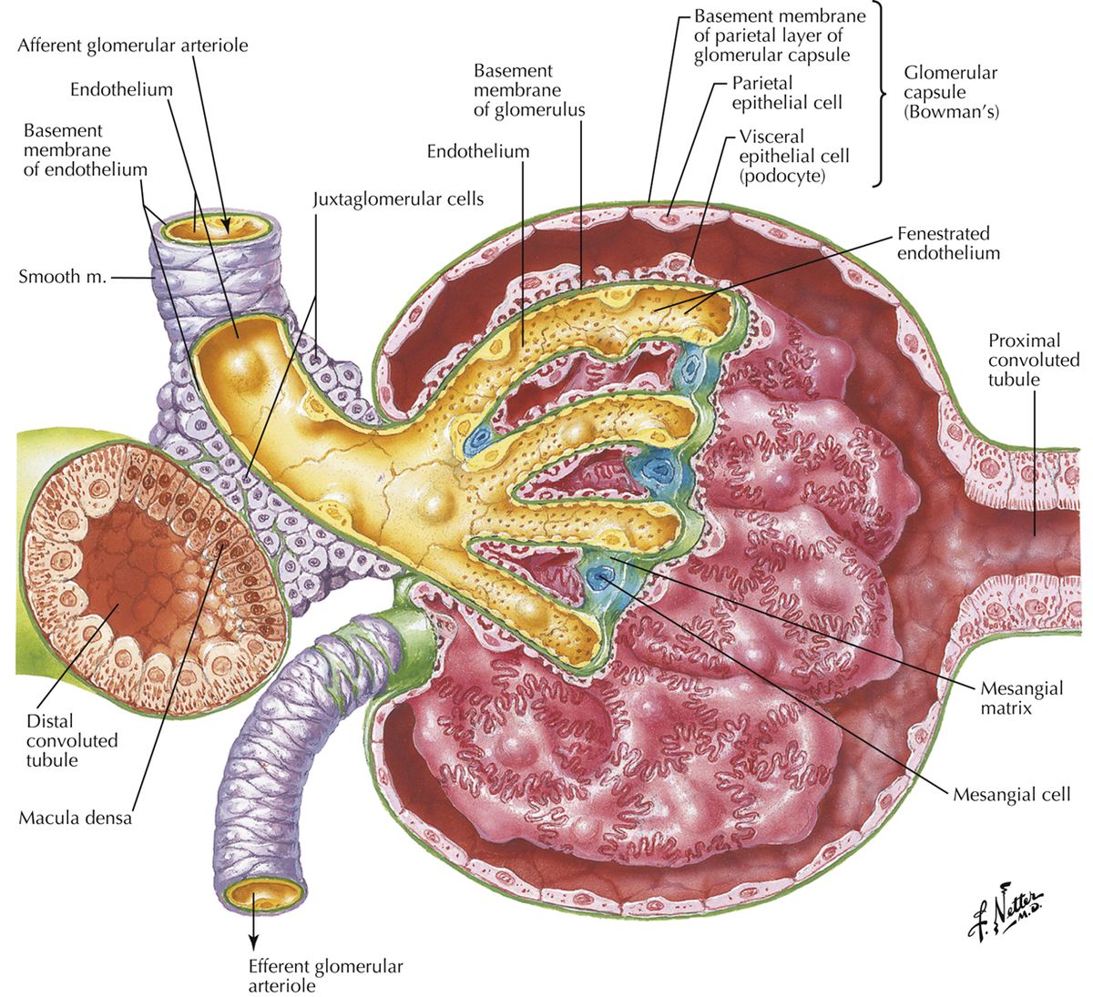
Juxtaglomerular apparatus (JGA): where the DCT contacts the afferent arteriole. Macula densa (DCT) senses NaCl/flow; juxtaglomerular (granular) cells (afferent arteriole smooth muscle) secrete renin in response to low BP or macula densa signals; EPO-producing/oxygen-sensing cells release erythropoietin.
5.5 · Objective 5 — Describe the anatomical relationships of each component of the urinary system and the surrounding anatomical structures
Kidneys — retroperitoneal against the posterior wall; the right sits below the liver, the left relates to the stomach, spleen, pancreas, and left colic flexure. Adrenal glands cap the superior poles. Renal artery off the aorta; renal vein to the IVC (the left renal vein crosses anterior to the aorta under the SMA and receives the left gonadal & suprarenal veins).
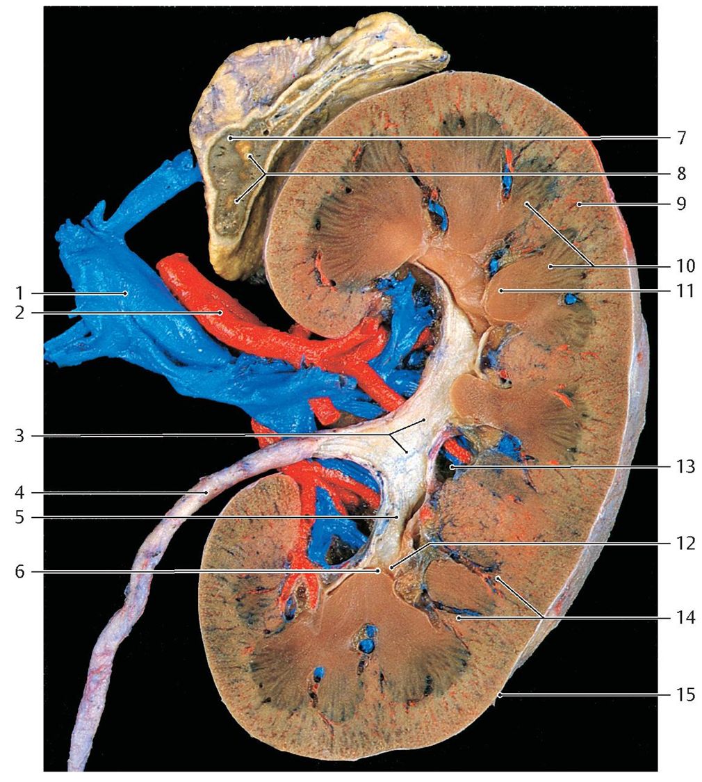
Ureters — retroperitoneal, ~25 cm; from the renal pelvis they descend on the psoas, cross the pelvic brim at the bifurcation of the common iliac vessels, and enter the posterolateral bladder. Three common stone sites: ureteropelvic junction (UPJ), crossing the common iliac vessels, and the ureterovesical junction (UVJ). Clinically the ureter passes inferior to the gonadal vessels ("water under the bridge").
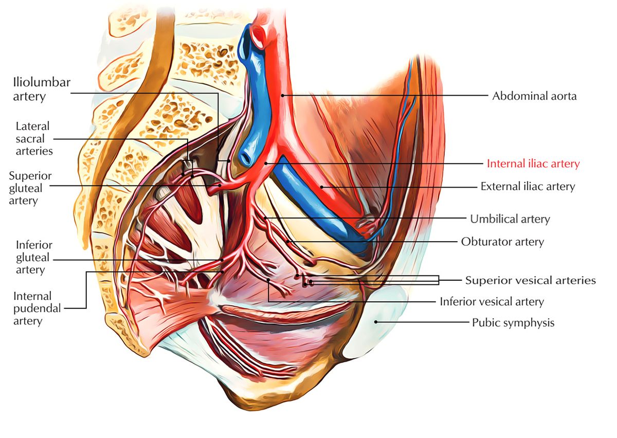
Bladder — subperitoneal, just posterior/superior to the pubic symphysis; only its superior surface is covered by peritoneum. The median umbilical ligament (urachal remnant) runs to the umbilicus.
In females: uterus is posterosuperior, vagina posteroinferior.
In males: rectum is posterosuperior, prostate lies inferiorly.
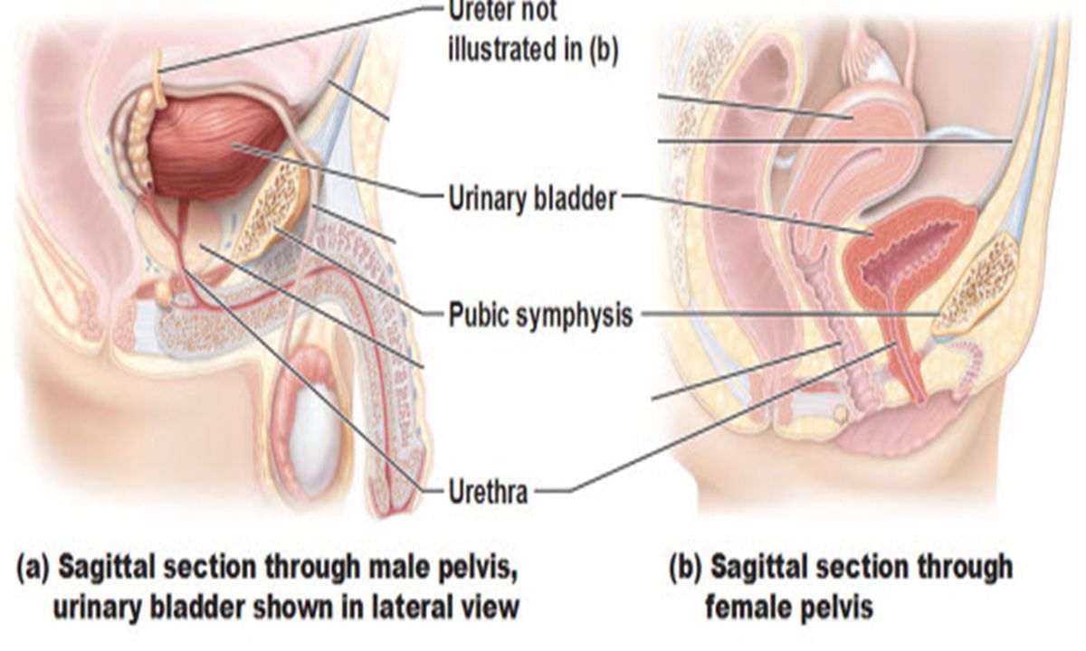
Urethra — female ~3–4 cm (opens in the vestibule, anterior to the vaginal orifice, posterior to the clitoris); male ~18–20 cm with prostatic, membranous, and spongy regions.
5.6 · Objective 6 — Compare and contrast the cell types of the urinary system
| Structure | Epithelium / cells | Why |
|---|---|---|
| Glomerular capillaries | Fenestrated endothelium | Large pores → highly permeable to plasma (not cells). |
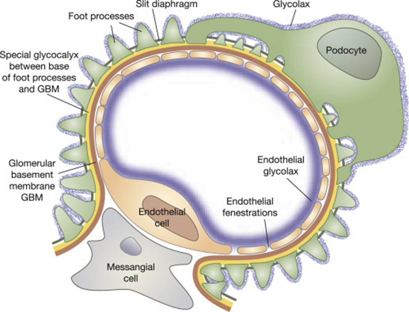
| Filtration membrane | Fenestrated endothelium + basement membrane + podocytes (with filtration slits, coated in negatively charged glycocalyx) | Size + charge barrier — repels albumin and large anions; passes water, ions, glucose, amino acids, urea. |
| Bowman's capsule — parietal layer | Simple squamous | Outer wall of the capsule. |
| Bowman's capsule — visceral layer | Podocytes (pedicles + filtration slits) | Wrap the glomerular capillaries. |
| PCT | Simple cuboidal with a brush border (microvilli) | Maximizes reabsorptive surface. |
| Thin loop of Henle | Simple squamous | Thin for passive water/ion movement. |
| DCT / collecting duct | Simple cuboidal (fewer microvilli; principal & intercalated cells in the duct) | Fine-tuned, hormone-regulated transport. |
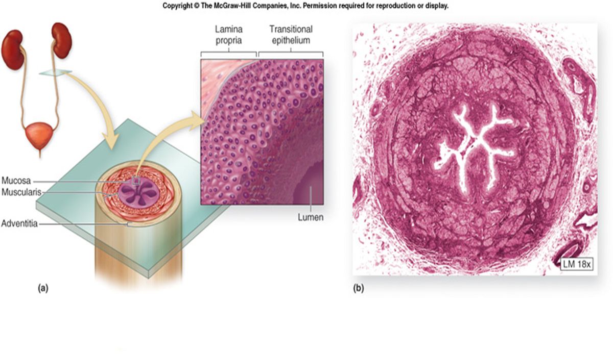
| Ureter, bladder, proximal urethra | Transitional epithelium (urothelium) | Stretches as urine volume changes; impermeable barrier. |
| Ureter/bladder muscle | Smooth muscle (bladder = detrusor; ureter = inner longitudinal + outer circular) | Peristalsis / voiding. |