1 · Common Ophthalmological Disorders
Instructional Objectives
OPHTHALMOLOGY — Common Ophthalmological Disorders
- Compare and contrast the etiologies, epidemiology, risk factors, clinical
manifestations, differential diagnosis, diagnostic testing (including ordering and
interpretation), management (acute and chronic, including applicable rehabilitative and
palliative care), appropriate referrals, patient education, and prognosis of the following
common ophthalmological disorders:
- Conjunctivitis
- Episcleritis
- Scleritis
- Keratoconjunctivitis sicca
- Ocular manifestations of herpes infections: a. Herpes simplex virus (HSV-1) · b. Varicella zoster
- Subconjunctival hemorrhage
- Pinguecula
- Pterygium
- Orbital (post-septal) and periorbital (pre-septal) cellulitis
- Blepharitis
- Chalazion
- Hordeolum
- Ectropion
- Entropion
- Dermatochalasis
- Xanthelasma
- Lacrimal disorders: a. Dacryocystitis · b. Dacryoadenitis
- Uveitis: a. Anterior uveitis (iritis/iridocyclitis) · b. Posterior uveitis
- Keratitis
- Corneal ulcer
- Identify medical care strategies for ophthalmological disorders in the lecture topic list for the following populations: 1. infant · 2. child · 3. adolescent · 4. adult · 5. elderly
Where this sits against Clinical Pathophysiology. Clin Path I Lecture 4 covers almost this exact condition list from the mechanism side — why the tissue fails. This section is the management side: what it looks like, what to order, what to give, and when to refer. If you find yourself revising the same fact twice, check which half you are actually being asked for.
A standing warning. Several of these rules are stated as absolutes but are softened in practice. Imaging is not automatic for dacryoadenitis, dacryocystitis or clearly pre-septal disease; hematology referral is not automatic for a recurrent subconjunctival hemorrhage; and perforation risk in scleritis is greatest in necrotising disease rather than uniformly. Those hedges are folded in below.
1.1 · The red eye, before you name a diagnosis
She said she will not test this section. After finishing posterior uveitis she moved on and said: “This is extra material. You can write that. I’m not going to pull test questions from this. This is not instructional objective.” It is kept here because it is the most clinically useful material in the block — but no quiz question is built on it. Read it for the ward, not for the exam.
The single most transferable thing here is a sequence rather than a fact. Complete this before naming the diagnosis:
- Visual acuity in each eye, with correction — the vital sign of the eye
- Pupils: shape, reactivity, afferent defect
- Extraocular movements, noting pain or restriction
- Corneal clarity and fluorescein staining
- Pattern of injection and discharge
- History: contact lenses, trauma, surgery, steroids
Reduced vision or an abnormal pupil is a red flag. The warning is blunt: do not let obvious redness substitute for an eye examination.
Danger signs — these stop the reflex diagnosis of conjunctivitis: moderate to severe pain or consensual photophobia (pain in the affected eye when light is shone in the other eye) · reduced acuity, a relative afferent pupillary defect, or an abnormal pupil · corneal opacity, infiltrate, ulcer or dendrite · ciliary flush (a ring of redness spreading from the corneal edge), hypopyon, or markedly raised pressure · proptosis, diplopia, or painful restricted eye movement · chemical exposure, penetrating trauma, or recent eye surgery · a contact lens wearer with pain or photophobia.
Use the pattern to localize before you name:
| Localizes to | Pattern |
|---|---|
| Conjunctiva | Itch or discharge; diffuse injection; vision preserved |
| Cornea | Pain and photophobia; fluorescein defect, infiltrate or opacity |
| Anterior chamber | Consensual photophobia; ciliary flush; irregular pupil |
| Sclera / orbit | Deep pain or painful eye movement; violaceous sclera, proptosis, restriction |
| Angle closure | Pain and headache with halos and nausea; cloudy cornea; mid-dilated pupil |
Two exceptions to the sequence. Chemical exposure — irrigate copiously first, before history or examination, then confirm the surface pH has normalized. Suspected open globe — rigid shield, no pressure, no tonometry, keep nil by mouth, emergency consultation.
Also tested
- Chemical eye injury. This is emergent: copious irrigation starts at once, before visual acuity, imaging or referral calls.
1.2 · The diagnostic modalities
| Test | What it is | What it finds |
|---|---|---|
| Slit lamp | Low-power microscope with a high-intensity slit beam | Anterior structures: lids, cornea, conjunctiva, sclera, iris |
| Ophthalmoscopy | Direct (hand-held), indirect (lens + head-worn), or slit-lamp — the last is most common because the patient is already seated there | Vitreous, retina, retinal vessels, macula, optic disc |
| Fluorescein examination | Yellow dye instilled, viewed under a Wood's lamp (ultraviolet) | Corneal abrasions, ulcers, foreign bodies |
| Fluorescein angiography | Dye injected into hand or arm, reaches the eye in 10–15 seconds, blue-flash camera. No iodine, relatively safe | Blood flow in retina and choroid: diabetic retinopathy, macular degeneration and edema, ocular melanoma, detachment, retinitis pigmentosa |
Also tested
- Demonstrating a corneal abrasion. Instill fluorescein and examine under a Wood's lamp with ultraviolet light; this surface fluorescein examination detects abrasions, ulcers and foreign bodies.
- Red flags in a red eye. Reduced vision and true pain move a red eye out of the benign category and separate a dangerous cause from a benign one.
- Fluorescein. Instilled and viewed under a blue light; it pools in epithelial defects and reveals abrasions, ulcers and foreign bodies.
1.3 · Eyelid disorders
What these look like
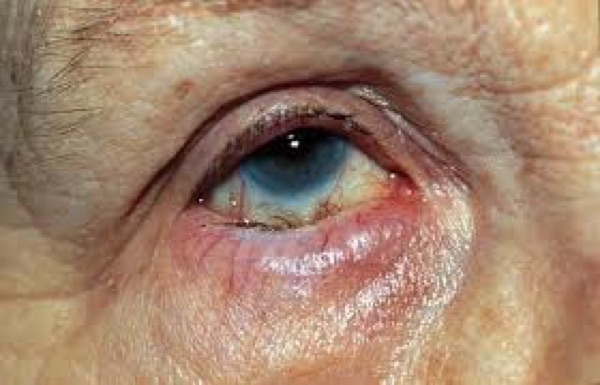
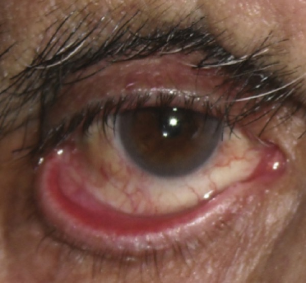
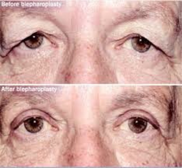
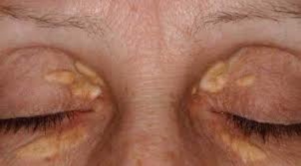
| Entropion | Ectropion | |
|---|---|---|
| Lid margin | Turns IN | Turns OUT |
| Symptom | Foreign body sensation | Tearing |
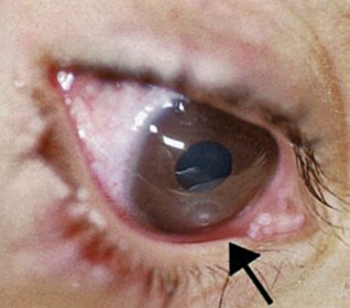
| Complication | Lashes onto the globe (trichiasis) → corneal abrasion | Exposure keratopathy |
| Causes | Aging, cicatricial (burn, surgery, trauma, chronic inflammation, scar), congenital — plus seventh nerve palsy for ectropion only | |
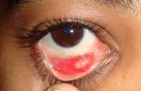
| Management | Slit lamp for corneal involvement. Preservative-free tears by day, ointment at night, tape the exposed lid. Surgery is definitive. | |
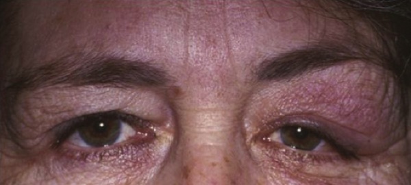
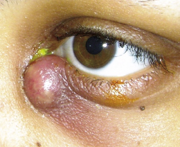
Dermatochalasis — excess loose skin with orbital fat prolapse, from aging. Patients say the lids feel heavy and they are looking through their lashes. Examine the visual fields: a demonstrated deficit is what gets blepharoplasty covered by insurance.
Xanthelasma — oval yellowish plaques, typically asymptomatic, from metabolic disorders with raised serum lipids. Work up the metabolism: lipid profile, plus fasting glucose and hemoglobin A1C for diabetes, plus liver function. Treat the underlying issue; local options are cryotherapy, laser ablation, chemical peel or excision. Recurrence is common even after effective local treatment. One caveat: many patients have entirely normal lipids, and the profile is still reasonable.
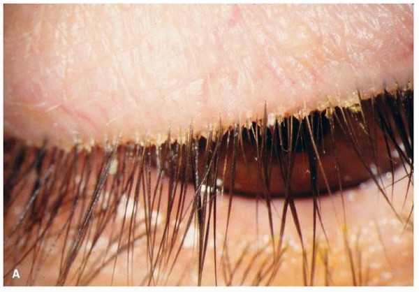
Blepharitis and meibomitis — associated with rosacea, seborrheic dermatitis, and Staphylococcus aureus colonization. Burning, dryness, grittiness, itching, foreign body sensation, tearing. Signs: crusting and scaling at the lash bases, erythematous swollen lid margins, and thick, sometimes toothpaste-like lipid secretion from the meibomian glands, with a decreased or frothy tear film. Lid hygiene first. If no improvement after two weeks, topical antibiotics, then oral. Chronic — controlled rather than cured.
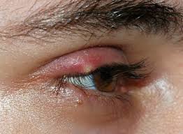
| Chalazion | Hordeolum (stye) | |
|---|---|---|
| Mechanism | Sterile obstruction of a meibomian gland | Acute infection, usually staphylococcal — meibomian (internal) or Zeis/Moll (external) |
| Onset | Days to weeks | 24 hours or overnight |
| The discriminator | NON-tender | TENDER |
| First-line | Warm compresses and gentle massage | |
| If it persists | Ophthalmology for steroid injection or curettage. Improvement may take months. | Ophthalmology for incision and drainage if no improvement in 2 weeks |
Refer a recurrent chalazion, or one persisting beyond 2–3 months, to rule out sebaceous carcinoma. If a hordeolum is accompanied by pre-septal cellulitis, treat with systemic antibiotics on that pathway.
Also tested
- Escalation for red, crusted lid margins. Lid hygiene comes first; if there is no improvement after two weeks, add a topical antibiotic, then oral antibiotics.
- Causes of ectropion. Aging, scarring and congenital causes can produce ectropion, and seventh nerve palsy causes ectropion only.
- Entropion and the cornea. The key question with entropion is always whether the lashes have damaged the cornea, assessed by slit lamp examination; an epithelial defect from lash contact means the cornea is now involved.
- Recurrent lid nodule. A nodule that keeps recurring after being treated as a chalazion raises the possibility of sebaceous carcinoma; recurrent lesions, or one persisting beyond two to three months, are referred to rule this out.
- Entropion and ectropion. Slit lamp examination is required to assess corneal involvement, i.e. whether the cornea has been damaged.
- Blepharoplasty coverage. Visual field testing is the assessment that determines coverage, because a demonstrated field deficit is what makes blepharoplasty a covered procedure.
- Seventh nerve palsy and lid position. Seventh nerve palsy causes ectropion, with the lower lid margin turned outward, watering and a red, exposed inner lid surface. Of the two malpositions, it causes ectropion only.
- Yellowish plaques near the inner canthus. Initial workup is lipid, diabetes and liver blood tests: a serum lipid profile, fasting glucose and hemoglobin A1C, and liver function tests. The lesion is a marker of the metabolic issue that is treated.
- Chalazion versus hordeolum. Tenderness separates them: the hordeolum is tender and the chalazion is not.
- Hordeolum referral. Refer to ophthalmology if persistent, for example no improvement in two weeks, for incision and drainage.
- Xanthelasma prognosis. Recurrences are common even after effective local treatment, whatever the local treatment.
- Ectropion treatment. Surgery is the definitive treatment, since only surgery restores lid position permanently; lubrication only protects the surface in the meantime.
- Yellowish plaques on the inner upper eyelids. Workup is a serum lipid profile, together with tests for diabetes mellitus and liver function; the plaques are a marker of an underlying metabolic disorder.
- Xanthelasma. Soft yellow plaques lying in the eyelid skin near the inner corner, typically on both sides.
- Entropion. It produces foreign body sensation because the inward-turned lashes rub the globe.
- Dermatochalasis. Treated with blepharoplasty, the surgical removal of the excess skin.
- Chalazion. A firm, painless, non-tender, slowly enlarging eyelid nodule is managed conservatively first, with warm compresses with gentle massage.
1.4 · Lacrimal disorders
What these look like
| Dacryoadenitis (GLAND) | Dacryocystitis (SAC) | |
|---|---|---|
| Where | Lateral one third of the UPPER lid | Nasal aspect of the LOWER lid, below the medial canthal tendon |
| Cause | Inflammatory most common; bacterial rare; viral usually bilateral | Nasolacrimal duct obstruction |
| Extra signs | Ipsilateral preauricular node, temporal conjunctival injection, fever, leukocytosis | Mucoid or purulent discharge expressible from the lower punctum |
| Treatment | Inflammatory → corticosteroids, response within 48 h. Viral → cool compresses. Cause unclear → empiric oral antibiotics 24 h then reassess | Well & reliable → oral antibiotics 10 days. Febrile, ill or unreliable → admit, IV 48–72 h then oral to complete 10–14 days |
Do not start corticosteroids for dacryoadenitis until bacterial and other infectious causes have been reasonably excluded. And a mass ABOVE the medial canthal tendon is not dacryocystitis — suspect a lacrimal sac tumor, rare as it is.
Once dacryocystitis settles, probing and irrigation are often needed to assess whether the drainage system is patent; surgery may follow. Expect improvement 24–48 hours after antibiotics start.
Also tested
- Imaging in dacryoadenitis. Contrast computed tomography of the orbits and paranasal sinuses is used when indicated, not automatically on presentation.
- Dacryocystitis needing admission. Fever, systemic illness or an unreliable patient move care to inpatient: intravenous therapy for 48 to 72 hours, then oral therapy to complete 10 to 14 days.
- Mild dacryocystitis. In an afebrile, systemically well, reliable patient it is managed as an outpatient with oral antibiotics for ten days and warm compresses.
- Corticosteroids in painful lid swelling of unclear cause. Infection must be reasonably excluded first; the alternative path is empiric oral antibiotics for 24 hours with reassessment.
- Inflammatory dacryoadenitis. Treat with oral corticosteroids, with a response expected within 48 hours.
1.5 · Conjunctiva and ocular surface
What these look like
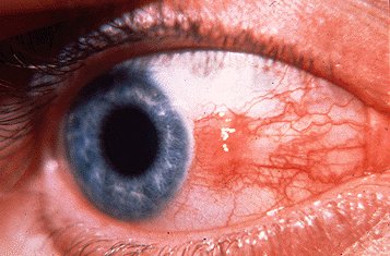
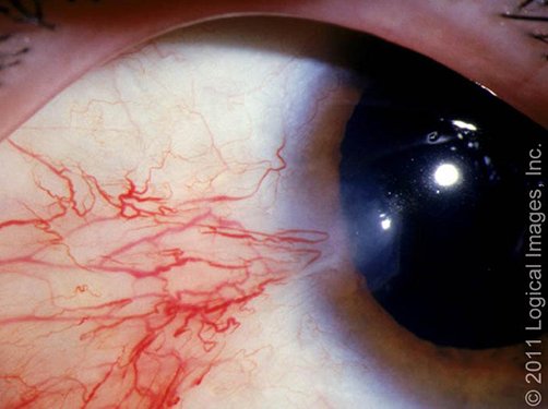
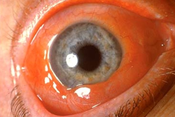
Pinguecula and pterygium both come from chronic sun and wind exposure and sit almost always at 3 o'clock or 9 o'clock. The whole distinction: the pterygium extends onto the cornea, the pinguecula does not. The mnemonic: pterodactyls fly (into the cornea), penguins can't. Protect from sun, dust and wind; lubricating drops. Conservative management will not make it resolve. Non-urgent referral if it grows or vision is affected; surgery if it distorts vision.
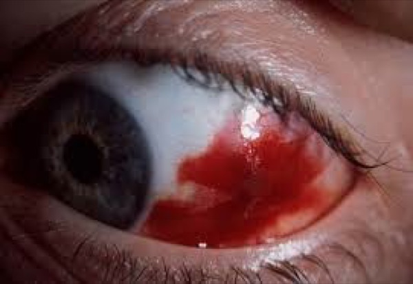
Subconjunctival hemorrhage (atraumatic) — Valsalva, bleeding disorder, antiplatelet or anticoagulant medication, hypertension. Blood under the conjunctiva, no pain, normal vision and pupil, clear cornea. History is the whole workup, and check the blood pressure if there is no explanation. Reassurance; resolves in 2–4 weeks.
The patient-education point she spent time on. The eye clears the way a bruise does — red, then purplish, then brown, then yellow. Warn them about the yellow stage: “there will be a point that they look like they’re jaundiced … so they don’t come thinking that they’re in liver failure. People do know yellow eyes and liver failure, they seem to know that association.”
And a general rule she gave alongside it: anybody who comes in with any eye condition, check their vision — with their contacts or glasses in, because what you want to know is whether it has changed.
For recurrence, what is wanted is medication review, blood pressure and targeted evaluation — not automatic hematology referral.
Chemosis is conjunctival swelling — a sign, not a diagnosis. Non-specific for irritation: allergy, infection, thyroid eye disease, angioedema, trauma, orbital cellulitis, impaired orbital venous drainage. Chemosis WITH proptosis, restricted movement, reduced vision or an afferent pupillary defect is urgent.
Also tested
- Pterygium surgery. Refer for surgical excision when a pterygium grows onto the cornea and distorts vision.
- Yellowish raised nasal conjunctival nodule not reaching the cornea. Sun, dust and wind protection with lubricating drops controls symptoms but will not resolve the nodule.
- Pinguecula versus pterygium. The pterygium extends into the cornea; the pinguecula does not involve it. Corneal involvement is the whole distinction.
- Unexplained bright red patch on the sclera. Take a careful history and check blood pressure; history is the priority, and an unexplained bleed warrants a blood pressure check.
1.6 · Conjunctivitis, all of it
What these look like
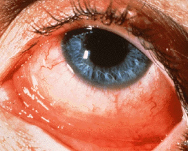
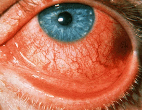
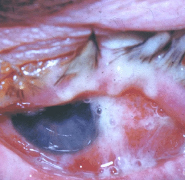
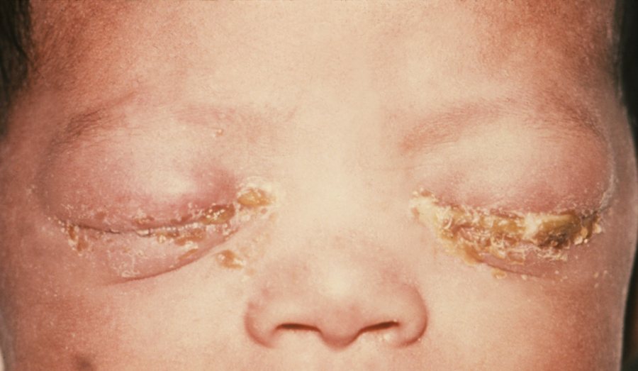
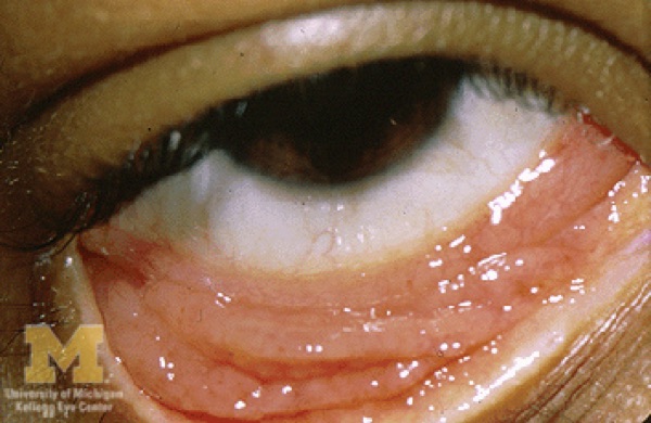
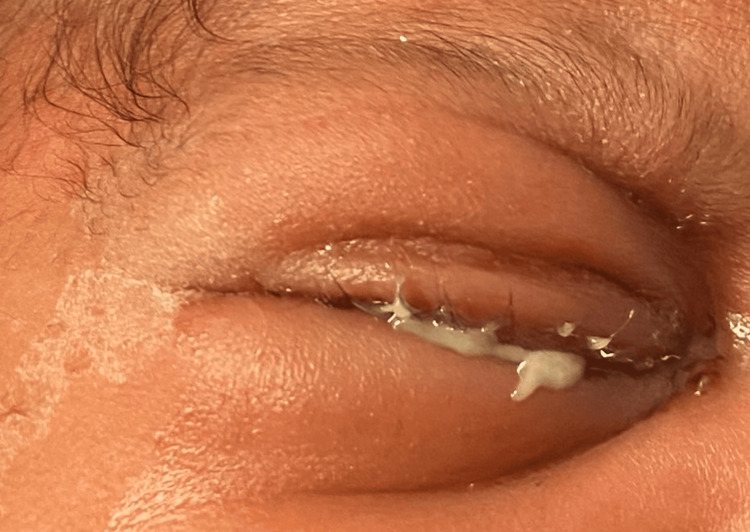
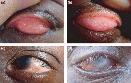
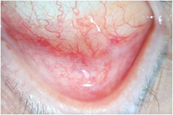
Acute is 4 weeks or less; chronic is more than 4 weeks. Two examination findings do most of the sorting:
| Finding | Appearance | Points to |
|---|---|---|
| Papillae | Red at the surface, paler at the base — her analogy: “it almost looks like a strawberry” | Bacterial (except chlamydial), allergic |
| Follicles | Pale at the surface, redder at the base | Chlamydial, viral |
| Preauricular node | — | Chlamydial, gonococcal, viral |
| Type | Giveaway | Treatment |
|---|---|---|
| Allergic | ITCH, bilateral, watery/stringy discharge, chemosis, papillae, no node | Avoid the allergen; cool compresses, artificial tears, topical histamine blocker ± mast cell stabilizer (olopatadine does both), systemic antihistamine |
| Viral | Adenovirus. Profuse watery discharge, follicles, tender preauricular node. Starts one eye, spreads to the other. Recent upper respiratory infection | Cool compresses, artificial tears, contagious precautions. Self-limiting: often worse over week one, resolving in 2–3 weeks. Refer if >3 weeks, or photophobia or vision loss after onset |
| Bacterial | Thick yellow/white discharge, often unilateral, papillae, usually no node | Immunocompetent adult: topical broad-spectrum antibiotic (e.g. fluoroquinolone) + contagious precautions |
| Gonococcal | Severe purulent discharge WITH a palpable preauricular node — the exception to the no-node rule | Newborn = emergency. Hospitalize, systemic ceftriaxone, cultures and Gram stain, test for chlamydia and dissemination. Untreated risk: corneal perforation |
| Chlamydial (adult inclusion) | Serotypes D–K. Chronic (a month or more), stringy mucoid discharge, follicles, unresponsive to topical medication. Often concurrent asymptomatic urogenital infection | Confirm with conjunctival nucleic acid amplification or direct fluorescent antibody. Doxycycline 100 mg twice daily for 7 days. Evaluate for other sexually transmitted infections; notify partners |
| Chlamydial (neonatal) | Serotypes D–K from maternal secretions; may have pneumonia | Erythromycin 50 mg/kg/day divided four times daily for 14 days. Monitor infants under 6 weeks for infantile hypertrophic pyloric stenosis — erythromycin is a motilin receptor agonist |
| Autoimmune | Recurrent/chronic hyperemia, minimal pain, NO discharge, systemic complaints. Pemphigoid, Stevens-Johnson, Sjögren, graft-versus-host | Routine ophthalmology referral |
Trachoma — serotypes A, B, C, and the leading infectious cause of blindness worldwide. Most active cases are asymptomatic. Mass drug administration with azithromycin 1 g orally as a single dose where prevalence is ≥5 percent. The chain to blindness is worth memorizing: conjunctival inflammation → eyelid scarring → entropion → trichiasis → blindness. Trichiasis needs surgery.
Urgent ophthalmology in bacterial conjunctivitis if: immunocompromised, contact lens wearer, recent eye surgery, foreign body, corneal opacity or suspected keratitis, or no improvement in 24 hours.
Also tested
- Course of viral conjunctivitis. It worsens for about a week, then clears, with resolution taking two to three weeks, and it is highly contagious throughout.
- Viral conjunctivitis education. Strict hand hygiene is the key advice, as the condition is highly contagious; contagious precautions are the central education point.
- Preauricular node in conjunctivitis. Bacterial conjunctivitis otherwise lacks a node, so severe purulent discharge with a node points to gonococcal disease.
- Neonatal purulent conjunctivitis timing. Onset on day 2 of life suggests Neisseria gonorrhoeae, since gonococcal disease presents in the first few days while chlamydial disease presents later, around days 5 to 14.
- Mucous membrane pemphigoid. Chronic red, gritty eyes with progressive scarring of the lower palpebral conjunctiva, fornix shortening and oral erosions indicate it: progressive subconjunctival fibrosis with mucosal involvement elsewhere (the ocular form).
- Trichiasis from trachoma. It requires surgical treatment, because the lashes keep abrading the cornea until they are dealt with surgically.
- Doxycycline counseling. Avoid the sun (photosensitivity) and heed food and mineral interactions: take it with a full glass of water, stay upright, and separate it from antacids and from iron, calcium or magnesium.
- Neonate with marked bilateral lid edema, copious purulent discharge and a preauricular node. Hospitalize and give systemic ceftriaxone as a single dose, with specialty consultation; the untreated risk is corneal perforation.
- Bacterial conjunctivitis risk groups. Immunocompromised, elderly, children and contact lens wearers are at particular risk. Neonates are a separate concern, where gonococcal infection is the worry.
- Allergic conjunctivitis unresponsive to topical treatment. The diagnosis may be wrong, so refer; symptoms usually settle as allergen levels fall.
- Chlamydial conjunctivitis. Beyond treating the eye, arrange evaluation for other sexually transmitted infections and notify partners for evaluation; the eye finding is a sentinel for a usually asymptomatic urogenital infection.
- Stages of trachoma. Trachoma progresses through four stages: follicles, scarring, entropion and opacity.
- Neonatal chlamydial conjunctivitis. Neonates are often treated in hospital because there is often concomitant pneumonia or pneumonitis; the lung involvement is what drives admission.
- Urgent conjunctivitis. Contact lens wear, immunocompromise, recent surgery, a foreign body, corneal opacity or no improvement in 24 hours all make it urgent.
- Trachoma. Caused by Chlamydia trachomatis; it progresses through follicles, tarsal scarring, entropion with trichiasis, and corneal opacity.
1.7 · Episcleritis and scleritis
What these look like
| Episcleritis | Scleritis | |
|---|---|---|
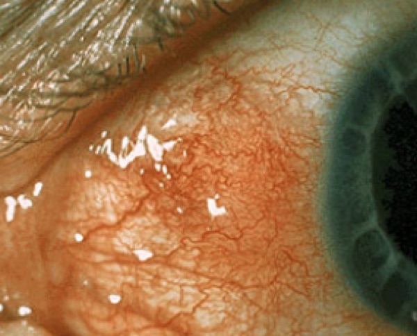
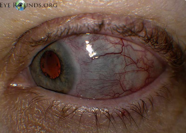
| Etiology | Often idiopathic, often no systemic association | Often systemic autoimmune |
| Pain | Mild, acute onset, focal. No discharge, no photophobia | Severe boring pain, worse at night, radiating to face and periorbital region |
| Appearance | Redness, often sectoral | Violaceous hue — choroid showing through thinned sclera. Pain with eye movement |
| Cotton-tip test | Vessels CAN be moved slightly | Vessels CANNOT be moved |
| Confirmatory test | 2.5% phenylephrine, wait 15 minutes — vessels blanch | — |
| Management | Artificial tears + oral anti-inflammatory taken WITH FOOD. Refer if no response in 2 days | URGENT referral. Slit lamp and fundoscopy, work up the systemic cause. Sclera at risk of perforation, may need a surgical patch |
| Prognosis | Usually self-limited; may recur in either eye | Decreased PAIN is the first sign of response, even if the inflammation looks unchanged |
A refinement worth carrying: non-infectious anterior scleritis commonly begins with systemic non-steroidal anti-inflammatories, with corticosteroids and immunomodulators for severe, necrotising, posterior or refractory disease — and perforation risk is greatest in necrotising disease, not uniformly.
Also tested
- Scleritis workup. Severe deep eye pain that wakes from sleep, with a bluish hue to the sclera, warrants an autoimmune workup, because scleritis is frequently associated with systemic autoimmune disease, including connective tissue disease.
- Episcleritis. The inflamed tissue is the deep subconjunctival tissue, the episcleral layer.
- Scleritis response to treatment. Decreased pain is the expected first sign of response and comes before any change in appearance, so an unchanged-looking eye is not failure.
- Scleritis referral. Refer to ophthalmology urgently, because the sclera is at risk of perforation and may need a surgical patch.
- Episcleritis. The bedside test is to instill 2.5 percent phenylephrine and look for blanching of the vessels after 15 minutes; episcleral vessels blanch, and mild painless sectoral redness fits episcleritis.
- Episcleritis. It is self-limiting and often needs no specific treatment; management is reassurance, with lubricants for comfort.
1.8 · Keratitis and corneal ulcer
What these look like
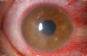
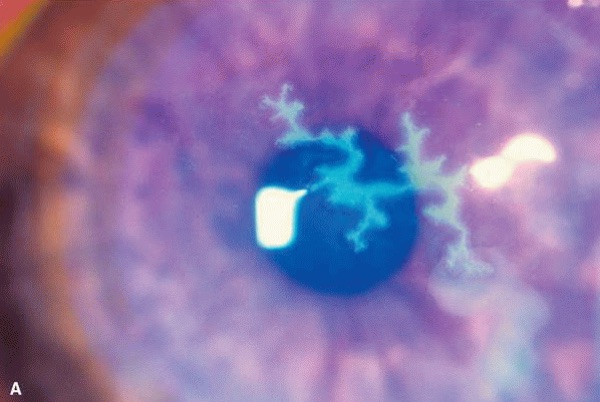
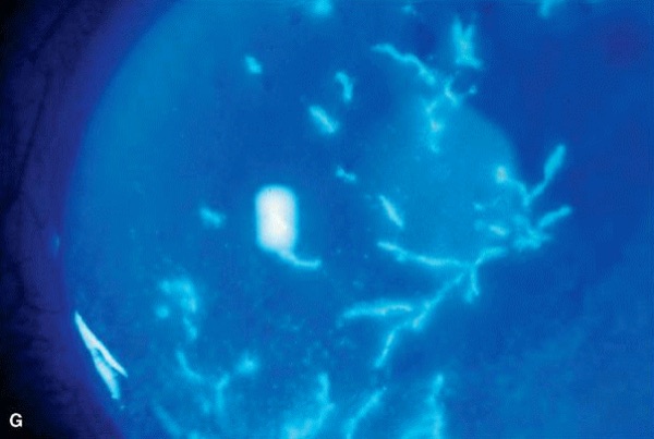
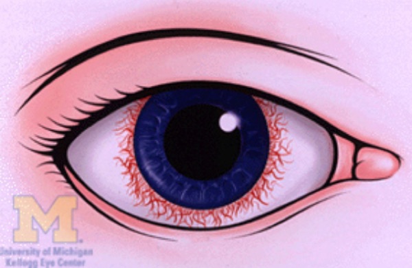
Ciliary flush — a ring of red vessels spreading from the limbus around the cornea, from the anterior ciliary arteries. It means inflammation of cornea, iris or ciliary body, and appears in corneal inflammation (ulcer, keratitis), anterior uveitis, and acute glaucoma. It is the finding that rules out simple conjunctivitis.
Keratitis. Risks: corneal trauma, dry eyes, contact lens overwear, topical ocular corticosteroids. Bacterial, viral, fungal; parasitic rare. Pain, foreign body sensation, tearing, photophobia, redness at the corneal edge, blurred vision. Signs: corneal opacification, a “broken up” corneal light reflection, ciliary flush. Classic ring infiltrate = Acanthamoeba, in lens wearers with poor hygiene such as rinsing lenses in tap water. Urgent referral within 24 hours for slit lamp with fluorescein. Undertreated → corneal scarring or perforation → endophthalmitis → possible removal of the eye.
| Herpes SIMPLEX keratitis | Herpes ZOSTER keratitis | |
|---|---|---|
| Corneal sign | TRUE DENDRITE — tree-branching, elevated edges, terminal end bulbs. Pathognomonic | PSEUDODENDRITE — lacks the branch pattern, the elevated edges and the end bulbs |
| Patient | Often younger | Often older; rare in children; also immunosuppressed |
| Skin | Not dermatomal, may not respect the midline (only ~10% of primary disease is bilateral) | Dermatomal, most often V1, respects the midline, unilateral, often spares the lower lid. Hutchinson sign (nose tip) → higher ocular risk |
| Treatment | Oral antivirals (aciclovir, valaciclovir, famciclovir) ×10 days. Ideally start within 72 hours of rash onset. IV aciclovir for severe, disseminated, orbital, retinal, central nervous system or significantly immunocompromised disease | |
| Prognosis | Good, benign, self-limited. Recurrences common under stress | Good to poor by corneal involvement. Postherpetic neuralgia can be devastating |
NO TOPICAL GLUCOCORTICOIDS BY THE PRIMARY CARE PROVIDER IN ACTIVE HERPES SIMPLEX EPITHELIAL DISEASE. That decision belongs to ophthalmology. Prevention: recombinant zoster vaccine for adults 50 and over, and immunocompromised adults 19 and over.
Corneal ulcer. Contact lens use is the major risk factor. Painful eye, the patient resists opening it, photophobia, foreign body sensation, tearing, blurred vision. Ciliary flush and a corneal defect. EMERGENT referral — a step above keratitis. Slit lamp, fluorescein, swab central or large ulcers for culture. Start a broad-spectrum topical agent (fourth-generation fluoroquinolone). Steroid drops can worsen infection if started too early — especially fungal or herpetic — so leave that to ophthalmology. Next-day follow-up; most heal in 2–3 weeks; untreated can blind; severe cases may need a transplant.
Also tested
- Contact lens corneal ulcer. Pseudomonas aeruginosa is the organism of greatest concern with contact lens wear, and it can perforate the cornea rapidly.
- Corneal ulcer with intraocular inflammation. A hypopyon, layered white cells in the anterior chamber, indicates significant intraocular inflammation.
- Vesicular rash on forehead and nose tip. The ophthalmic division (V1) of the trigeminal nerve is involved, and involvement of the nose tip signals ocular involvement.
- Hutchinson sign. Vesicles on the tip of the nose indicate a high likelihood of ocular involvement, because the nasociliary branch supplies both the nose tip and the globe.
- Herpes zoster ophthalmicus with deeper involvement. If there is retinitis, choroiditis or optic neuritis, treatment is steroids and intravenous acyclovir.
- Corneal ulcer referral. A corneal ulcer needs ophthalmology emergently, outranking keratitis's 24-hour window.
- Corneal ulcer follow-up. Review is needed the next day after an anti-infective is started, and most ulcers heal over a two to three week course.
- Herpes zoster ophthalmicus. A dermatomal vesicular rash over the forehead and upper lid seen within the ideal 72-hour window calls for an oral antiviral now, plus referral for slit lamp examination and dilated fundoscopy.
- Corneal ulcer position. Prognosis follows both size and position relative to the visual axis; ulcers inside the visual axis carry a poorer outcome.
- Herpes simplex keratitis prognosis. Good, though recurrences are common.
- Corneal ulcer on examination. The patient resists opening the affected eye, because of photophobia and foreign body sensation.
- Nasal-tip vesicle (Hutchinson sign). It indicates nasociliary branch involvement, and therefore a higher risk of ocular involvement.
- Corneal infiltrate with epithelial defect and hypopyon. Urgent referral for scraping and antibiotics is the next step. A hypopyon indicates significant intraocular inflammation and the eye is at risk; treatment is intensive topical antibiotics after corneal scraping.
- Punctate epithelial keratitis. It presents as a painful, photophobic red eye with reduced vision, with diffuse punctate fluorescein staining without an infiltrate, from exposure and surface drying.
- Herpes simplex keratitis. A true dendrite, a branching corneal lesion on fluorescein with terminal end bulbs, is pathognomonic for herpes simplex, and the treatment is oral antiviral therapy for ten days.
1.9 · Uveitis
What these look like
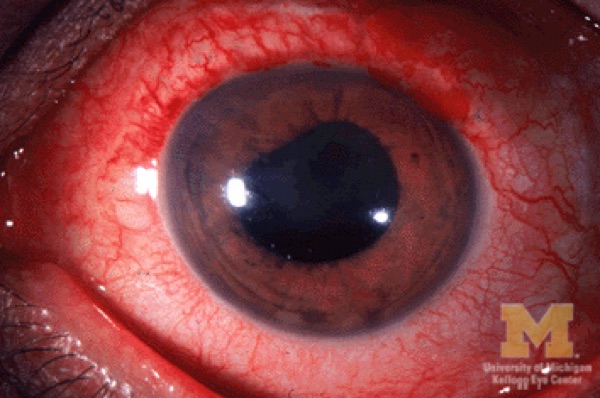
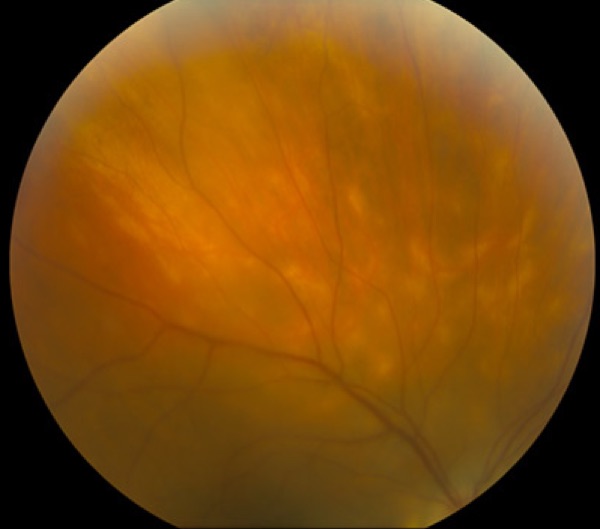
| Anterior (iritis, iridocyclitis) | Posterior (choroiditis, retinitis) | |
|---|---|---|
| Etiology | Idiopathic, autoimmune | Idiopathic, autoimmune, infectious — toxoplasmosis, cytomegalovirus |
| Pain | Yes — with photophobia and redness at the corneal edge | NO pain if isolated |
| Vision | Often preserved | Blurred, with floaters, scotomas, metamorphopsia |
| Signs | Cells in the anterior chamber, consensual photophobia, ciliary flush, variable pressure, irregular pupil stuck to lens or cornea, keratic precipitates (white cell deposits on the corneal endothelium) | Cells in the posterior vitreous, vitreous haze, retinal or choroidal inflammation |
| Referral | Urgent, within 24 h — delay may cost vision | Refer for slit lamp and dilated fundoscopy; possibly fluorescein angiography to separate active from inactive lesions |
| Treatment | Infectious → treat the organism. Non-infectious → topical corticosteroids | Does NOT respond to topical treatment — may need an intraocular corticosteroid injection |
| Course | Most acute cases respond dramatically in days to weeks | Develops far more slowly, may last years |
Recurrent uveitis, or uveitis with features suggesting systemic autoimmune disease, needs a thorough systemic evaluation. And infection must be excluded before immunosuppression.
Also tested
- Recurrent anterior uveitis with inflammatory back pain. Back pain that improves with exercise and worsens with rest points to HLA-B27 spondyloarthropathy, the ankylosing spondylitis association, which is why the systemic history matters.
- Cycloplegic in anterior uveitis. It relieves pain and prevents synechiae by resting the ciliary body and keeping the iris from adhering to the lens.
- Posterior uveitis. It involves the choroid and/or the retina, that is, choroiditis and retinitis.
- Infectious cause of posterior uveitis. Toxoplasma gondii causes chorioretinitis and is a classic infectious cause of posterior uveitis.
- Posterior uveitis treatment. Topical treatment will not work; an intraocular corticosteroid injection may be required, the key management difference from anterior uveitis.
- Posterior versus anterior uveitis course. Posterior uveitis develops a lot more slowly and may last several years, though it can also turn acute and threaten vision.
- Recurrent uveitis with systemic features. Recurrent uveitis with suggestive systemic features warrants a thorough systemic evaluation for an underlying autoimmune condition.
- Anterior uveitis. It involves the iris (iritis), or the iris and ciliary body (iridocyclitis).
1.10 · Pre-septal and post-septal cellulitis
What these look like

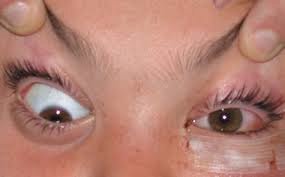
Both come from direct extension from a bacterial sinus, skin or dental infection. In diabetic, elderly or immunocompromised patients, consider fungus — aspergillosis, mucormycosis.
| PRE-septal (periorbital) | POST-septal (orbital) | |
|---|---|---|
| The giveaway | The eye itself is WHITE | The eye itself is RED and cannot move fully |
| Symptoms | Both: periocular pain, fever and chills, warmth around the eye | |
| Post-septal only | — | Pain and difficulty with eye movement, reduced vision, diplopia |
| Signs | Diffuse balloon-like lid edema, erythema, tenderness; variable conjunctival injection | Significant injection, proptosis, decreased and painful movement, possible afferent pupillary defect, decreased vision |
| Management | Mild → outpatient oral antibiotics 10–14 days against Staphylococcus (including resistant strains) and Streptococcus | ALL post-septal → hospitalize, broad-spectrum IV 48–72 h, then oral at least a week |
Also admit a pre-septal patient if: moderate-severe or toxic, poor compliance expected, a child of 5 years or younger, or no improvement after oral antibiotics started.
Workup: contrast computed tomography of orbits and paranasal sinuses, complete ocular examination with fundoscopy, Gram stain and culture of any drainage, complete blood count with differential, blood cultures. May need ear-nose-throat, oral and maxillofacial surgery, or infectious disease consults. Untreated → intracranial spread → meningitis or cavernous sinus thrombosis. Expect improvement in 24–48 hours.
Notes caveat: mild, clearly pre-septal disease with normal vision, pupils and painless full movements may be managed clinically without routine imaging.
Also tested
- Mild pre-septal cellulitis. In a systemically well child with reliable follow-up, manage with oral antibiotics and early review at 24 to 48 hours.
- Progression of pre-septal cellulitis. New pain on eye movement and mild proptosis mean the infection has progressed behind the orbital septum; painful restricted movement and proptosis indicate post-septal disease.
- Post-septal cellulitis. All post-septal disease is admitted for intravenous antibiotics and imaging; computed tomography of the orbits and sinuses defines the extent.
- Hospitalizing pre-septal cellulitis. Admit for moderate to severe or toxic disease, concern for poor compliance, a child of five years or younger, or no improvement after oral antibiotics were started.
- Orbital cellulitis. The usual source of infection is the paranasal sinuses, which is why imaging covers the sinuses as well as the orbits.
1.11 · Referral timing — explicit disposition
Same caveat as 1.1 — this table comes from the section she called extra material and said she would not pull test questions from. Worth knowing; not examinable.
| Urgency | Conditions |
|---|---|
| EMERGENT — now | Chemical injury (irrigate first), open globe, acute angle closure, orbital cellulitis, endophthalmitis |
| SAME DAY | Keratitis or corneal ulcer, anterior uveitis, scleritis, ocular herpes zoster ophthalmicus |
| URGENT — 24–48 hours | Unexplained decreased vision, persistent pain or photophobia, atypical or worsening red eye |
| ROUTINE | Uncomplicated conjunctivitis, chronic eyelid or ocular surface disease |
Routine is only appropriate when ALL of these hold: acuity preserved, pupils and movements normal, cornea clear with no fluorescein uptake or infiltrate, no significant pain or photophobia, and reliable follow-up. Every assessment ends with a documented disposition and a safety net — acuity, key negatives, suspected diagnosis, urgency, destination, and explicit return precautions.
First-line treatment
Every condition in this lecture, with what you reach for first and how fast — which in ophthalmology is half the answer. Where the deck escalates after a failed trial, only the first step is here; the full ladder and the reasoning are in the comparison chart.
| Condition | First line | How fast |
|---|---|---|
| Entropion | Preservative-free artificial tears by day, lubricating ointment at night, tape the exposed lid. | Routine |
| Ectropion | Same as entropion: tears by day, ointment at night, taping. | Routine |
| Dermatochalasis | Blepharoplasty. | Routine |
| Xanthelasma | Treat the underlying metabolic issue. Local: cryotherapy, laser ablation, chemical peel, surgical excision. | Routine |
| Blepharitis / Meibomitis | Lid hygiene first. | Routine |
| Chalazion | Warm compresses with gentle massage. | Routine |
| Hordeolum (stye) | Warm compresses with gentle massage. | Routine |
| Dacryoadenitis | Inflammatory → corticosteroids. Viral → cool compresses. Cause unclear → empiric oral antibiotics 24 h then reassess. Analgesia as needed. | Urgent |
| Dacryocystitis | Well, afebrile, reliable → oral antibiotics 10 days. Febrile, ill or unreliable → admit, intravenous 48–72 h then oral to complete 10–14 days. Warm compresses; consider drainage of an abscess. | Urgent |
| Pinguecula | Sun, dust and wind protection; lubricating drops. | Routine |
| Pterygium | Sun protection and lubricating drops. | Routine — refer non-urgently if growing or vision affected |
| Subconjunctival hemorrhage | Reassurance. Artificial tears if mildly irritated. Treat the underlying bleeding disorder or hypertension. | Routine |
| Chemosis | Treat the cause. | URGENT if with proptosis, restricted movement, reduced vision or an afferent pupillary defect |
| Allergic conjunctivitis | Avoid the allergen; cool compresses, artificial tears, topical histamine blocker ± mast cell stabilizer (olopatadine does both), systemic antihistamine. | Routine |
| Viral conjunctivitis | Cool compresses, artificial tears. Contagious precautions and hand hygiene. | Routine — refer if >3 weeks, or photophobia or vision loss after onset |
| Bacterial conjunctivitis | Immunocompetent adult → topical broad-spectrum antibiotic (e.g. fluoroquinolone). Contagious precautions. | Routine — URGENT if immunocompromised, contact lens wearer, recent surgery, foreign body, corneal opacity, or no improvement in 24 h |
| Gonococcal conjunctivitis | Newborn: hospitalize, systemic ceftriaxone once, specialty consultation. | EMERGENT |
| Chlamydial conjunctivitis — adult inclusion | Doxycycline 100 mg orally twice daily for 7 days. | Routine |
| Chlamydial conjunctivitis — neonatal | Erythromycin 50 mg/kg/day divided four times daily for 14 days. Azithromycin 20 mg/kg daily for 3 days is an alternative with more limited data. | Urgent |
| Trachoma | Mass drug administration: azithromycin 1 g orally as a single dose where prevalence is ≥5%. Trichiasis requires surgery. | Urgent |
| Autoimmune conjunctivitis | Managed by ophthalmology alongside the systemic disease. | Routine |
| Episcleritis | Artificial tears and an oral non-steroidal anti-inflammatory taken WITH FOOD. | Routine — refer if no response in 2 days |
| Scleritis | Non-infectious anterior disease commonly begins with systemic anti-inflammatories; systemic corticosteroids and immunomodulators for severe, necrotising, posterior or refractory disease. | SAME DAY — sclera at risk of perforation, may need a surgical patch |
| Pre-septal (periorbital) cellulitis | Mild → outpatient oral antibiotics 10–14 days against Staphylococcus (including resistant strains) and Streptococcus. | Urgent — admit if moderate-severe or toxic, poor compliance, child ≤5 years, or no improvement on orals |
| Post-septal (orbital) cellulitis | Hospitalize — broad-spectrum intravenous antibiotics 48–72 h, then oral for at least a week. May need ear-nose-throat, oral and maxillofacial surgery, or infectious disease consults. | EMERGENT |
| Keratitis | Treat the underlying cause to prevent persistent inflammation and scarring, guided by ophthalmology. | SAME DAY — urgent referral within 24 h |
| Herpes simplex keratitis | Oral antivirals (aciclovir, valaciclovir, famciclovir) for 10 days. | SAME DAY |
| Herpes zoster keratitis | Oral antivirals for 10 days, ideally within 72 hours of rash onset. Intravenous aciclovir for severe, disseminated, orbital, retinal, central nervous system or significantly immunocompromised disease. | SAME DAY |
| Corneal ulcer | Broad-spectrum topical agent (fourth-generation fluoroquinolone) to start; then agent-specific therapy per ophthalmology. | EMERGENT |
| Anterior uveitis (iritis, iridocyclitis) | Infectious → treat the organism. Non-infectious → topical corticosteroids. | SAME DAY — urgent within 24 h, because delay may cost vision |
| Posterior uveitis (choroiditis, retinitis) | Does NOT respond to topical treatment — may require an intraocular corticosteroid injection. | Urgent |
1.12 · The eye in three layers
Every disorder in this block sits in one of three tunics, and naming the layer usually names the kind of problem.
| Layer | Structure | What it does |
|---|---|---|
| Fibrous outer |
Cornea | Transparent front surface; provides most of the eye's focusing power |
| Sclera | Tough white coat; maintains shape and gives the muscles their attachment | |
| Vascular (uvea) middle |
Iris | The colored part; controls pupil size and so regulates light entry |
| Ciliary body | Produces aqueous humor and controls lens accommodation | |
| Choroid | Highly vascular; nourishes the retina | |
| Neural inner |
Retina | Rods and cones convert light into neural signals |
| Macula | Central vision | |
| Fovea | Highest visual acuity | |
| Optic disc | Where the optic nerve exits — the blind spot |
Useful shorthand: uveitis is inflammation of the middle layer, which is why it takes iris, ciliary body and choroid together; scleritis and episcleritis sit in the outer; and the vascular occlusions and detachments are all inner-layer disease.
2 · Neuro-Ophthalmology
Instructional Objectives
OPHTHALMOLOGY — Neuro-ophthalmology
- Describe nystagmus
- Describe the afferent pupillary light pathway.
- Describe the parasympathetic (efferent pupillary) pathways.
- Describe the sympathetic pathways (oculo-sympathetic).
- Describe the accommodation reflex.
- Compare and contrast the etiologies, epidemiology, risk factors, clinical
manifestations, differential diagnosis, diagnostic testing (including ordering and
interpretation), management (acute and chronic, including applicable rehabilitative and
palliative care), appropriate referrals, patient education, and prognosis of the following
common neuro-ophthalmological disorders:
- Relative afferent pupillary defect (Marcus Gunn pupil)
- Horner syndrome
- Argyll Robertson pupil
- Adie tonic pupil
- Cranial nerve III, IV, and VI palsies
- Ptosis
- Anisocoria
- Differentiate the clinical presentation of pre-and post-chiasm lesions.
- Identify medical care strategies for ophthalmological disorders in the lecture topic list for the following populations: a. adult · b. elderly
How to read this section. Almost every diagnosis here is decided by which pupil is the abnormal one and what it does in the dark. Get the four pathways straight first — afferent light, parasympathetic efferent, sympathetic, and near/accommodation — and the named pupils fall out of them rather than needing to be memorized as a list.
2.1 · Nystagmus Objective a
What these look like
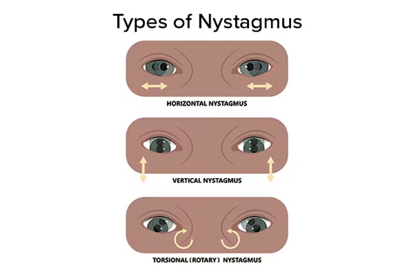
Definition: an involuntary, biphasic, rhythmic, tremor-like oscillating movement of the eyes. It is either congenital (infantile or latent) or acquired.
It is usually symptomatic — unless it was acquired before the age of 8. That single exception is the reason a child with long-standing nystagmus may report nothing at all. Symptoms, when present:
| Symptom | What the patient means |
|---|---|
| Vertigo | The patient or the room is spinning. Often the primary symptom, and commonly points to a vestibular problem. |
| Oscillopsia | The environment or an object appears to move back and forth. |
| Blurred vision | The retinal image is being dragged by the oscillation. |
| Abnormal head position | A compensation for the blurring, not a separate problem. |
Jerk nystagmus is classified by trajectory and named for the direction of the FAST beat — vertical, horizontal or torsional. It increases with gaze in the direction of the fast phase. Horizontal jerk nystagmus is the most common form: the eyes drift slowly to one side and snap quickly back.
Who gets referred and worked up. Infants and young children with nystagmus; nystagmus acquired during adolescence or adulthood; and concerning or non-physiologic nystagmus in adults. The pathway is ophthalmology for a complete ophthalmic exam → initial imaging → labs if relevant, and in every case the underlying etiology must be addressed. Slide 5
Also tested
- Acquired nystagmus in an adult. It suggests an underlying neurological cause; it is not benign and requires investigation.
- Nystagmus present since birth. It warrants evaluation for a cause of poor vision, since congenital nystagmus often reflects an underlying visual deficit.
- Early-onset nystagmus. It is usually asymptomatic if acquired before age eight, which is why onset age matters.
- Monocular or asymmetric nystagmus. Refer for work-up; it is a concerning, non-physiologic pattern.
- Nystagmus in infants and young children. It warrants referral for work-up.
- Upbeat nystagmus. New upbeat nystagmus is classified as concerning and non-physiologic, and it warrants work-up in adults.
2.2 · The three pupillary pathways Objectives b, c, d
Start with pupil size itself. Pupillary size is set by the AVERAGE of the illumination detected by each eye. Cover one eye of a normal person and watch the other: the pupil enlarges, because the average illumination has effectively been halved. That one fact explains why the swinging flashlight test works at all.
| Pathway | Route | Effect |
|---|---|---|
| Afferent light (objective b) | Retinal ganglion cells → CN II → optic tracts → synapse in the pretectal nuclei. Each pretectal nucleus sends axons to BOTH Edinger-Westphal nuclei. | This bilateral crossing is why light in one eye constricts both pupils. |
| Parasympathetic efferent (objective c) | Preganglionic fibers from the Edinger-Westphal nucleus travel along CN III to the ciliary ganglion, then short ciliary nerves to the iris sphincter. | MIOSIS |
| Sympathetic (oculo-sympathetic) (objective d) | First order hypothalamus → brainstem and cervical cord, synapsing at the ciliospinal center of Budge, C8–T2. Second order exits the cord, loops up through the brachial plexus over the lung apex to the superior cervical ganglion. Third order follows the carotid to the radial muscle of the iris. | MYDRIASIS |
Why the three-neuron sympathetic chain is worth the effort. The whole of Horner syndrome localization below is just which of those three neurons is damaged. The second-order neuron's detour over the lung apex is why a Pancoast tumor causes it, and the third-order neuron's ride along the carotid is why a dissection does.
Opioids are parasympathomimetics. Overdose gives reduced mental status, respiratory depression under 12 per minute, reduced tidal volume, and pupillary constriction. It is the mental status and the respiration that matter clinically, not the pupil. Reversal is naloxone, preferably intravenous, now available over the counter as an intranasal preparation. Slide 12
Also tested
- Light reflex, afferent limb. Afferent impulses travel along the second cranial nerve: retinal ganglion cells to the optic nerve and tracts.
- Opioid toxicity. The features that matter clinically are reduced consciousness and respiratory depression; the miosis is a clue rather than the danger.
- Pupillary muscles. The iris sphincter constricts and the radial muscle dilates, producing miosis and mydriasis respectively.
- Naloxone reversal. Naloxone may need repeating, because it lasts about two hours and respiratory depression can return.
2.3 · The accommodation reflex, and light-near dissociation Objective e
On shifting gaze from far to near, three things happen — to the lens, the pupil and the globes. All three share one afferent limb: retinal input via CN II and the optic tract to the lateral geniculate nucleus and visual cortex.
| Component | What happens | Result |
|---|---|---|
| Lens | Ciliary smooth muscle contracts | Lens becomes more convex → increased refractive power |
| Pupil | Constriction | Improves up-close focus |
| Globes | Both eyes adduct | Convergence |
The key asymmetry. The pupillary near response travels from visual cortex to iris sphincter skipping a portion of the route the pupillary light pathway takes. That difference is the entire basis of “light-near dissociation” — a pupil that will not react to light but still constricts to near. Argyll Robertson and Adie pupils both live in that gap.
Also tested
- Pupillary light-near dissociation. It is defined by no reaction to light with a preserved reaction to near.
- Near response. On shifting gaze from distance to near, the lens thickens, the pupils constrict and the eyes converge; it acts on lens, pupil and globes together.
2.4 · Anisocoria — deciding which pupil is abnormal Objective f7
The governing principle. The efferent limb of the pupillary reflex is bilateral, so both pupils receive the same command and should always be the same size. They should only be unequal when the efferent pathways are not working properly.

| Ask | If yes | Then the abnormal pupil is |
|---|---|---|
| Is the difference greater in the DARK? | The smaller pupil is failing to dilate | The SMALL one — think Horner, opioids, Argyll Robertson |
| Is the difference greater in the LIGHT? | The larger pupil is failing to constrict | The LARGE one — think CN III palsy, Adie, pharmacologic mydriasis |
| Is it equal in light and dark? | Both pupils are working | Physiologic anisocoria — the most common cause, usually under 0.4 mm difference |
When the large pupil is abnormal, the causes are an ocular condition preventing constriction, or pharmacologic mydriasis. Anticholinergics give a pupil that is usually 8 mm or more and does not constrict to light at all — atropine and other anticholinergic mydriatic or cycloplegic drops are the classic culprits.
Also tested
- Pharmacologically dilated pupil. Anticholinergics such as atropine, ipratropium, scopolamine and glycopyrrolate can cause it, giving a large pupil that is unreactive to light.
- Physiologic anisocoria. The size difference between the pupils is typically under about half a millimeter and is equal in light and dark.
- Sympathomimetic versus anticholinergic pupil. A sympathomimetic-dilated pupil is only mildly enlarged, unlike the very large anticholinergic pupil.
- Scopolamine patch mydriasis. A unilateral fixed dilated pupil after handling a scopolamine patch will resolve as the drug wears off; pharmacologic mydriasis is transient and needs no imaging.
- Physiologic anisocoria. Mild anisocoria, equal in light and dark, with normal lids and movements, needs only reassurance and no work-up.
2.5 · The four named pupils Objectives f1–f4
What these look like
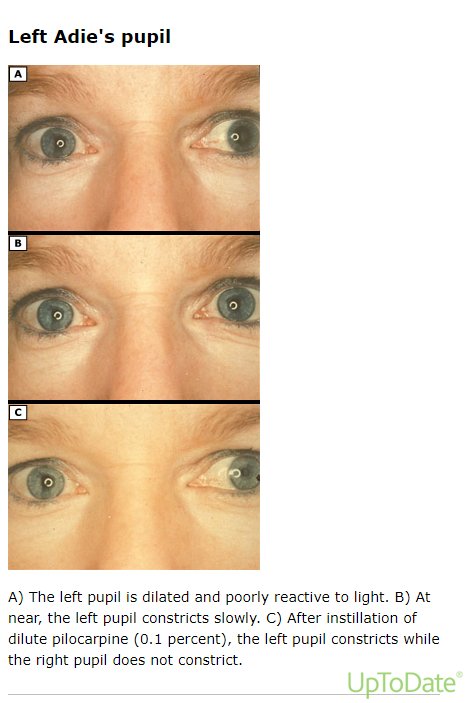
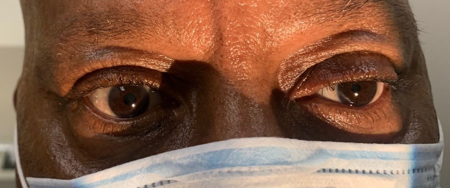
| Marcus Gunn (RAPD) | Horner syndrome | Argyll Robertson | Adie tonic | |
|---|---|---|---|---|
| Limb at fault | Afferent | Sympathetic | Pretectal / dorsal midbrain | Parasympathetic (ciliary ganglion) |
| Pupil size | Equal at rest | Miosis, unilateral | Miosis, BILATERAL | Mydriasis, often unilateral |
| Light reaction | Both pupils DILATE when the light swings to the affected eye | Normal | Absent | Poor |
| Near reaction | Normal | Normal | Brisk — light-near dissociation | Present but slow and tonic |
| Hallmark | Positive swinging flashlight test | DILATION LAG | Bilateral small pupils sparing near | Sector paralysis of the iris on slit lamp |
| Classic cause | Retina or optic nerve lesion | Damage anywhere along the sympathetic chain; often idiopathic | Tertiary syphilis | Inflammation of the ciliary ganglion, then aberrant reinnervation |
Marcus Gunn pupil. An afferent defect, usually at the level of the retina or optic nerve. Moving a bright light from the unaffected eye to the affected eye makes both eyes dilate, because the ability to perceive that light is diminished — the average illumination the brain is working from has just dropped.
Horner syndrome — dilation lag is the hallmark. The anisocoria is most evident in the first 4–5 seconds after dimming the lights, which is exactly when a normal sympathetic pathway would be actively dilating. After 10–15 seconds the pupil does dilate a little — but that is passive relaxation of the iris sphincter, not sympathetic function returning. Slide 29
Classic triad: ptosis, miosis, and anhidrosis — with the caveat that anhidrosis may be absent depending on where along the pathway the lesion sits. Diagnostic test: dilute apraclonidine drops, which are ineffective in a normal pupil but cause mydriasis in the Horner pupil.
| Horner — neuron order | Causes |
|---|---|
| First order | Brainstem strokes and tumors; spinal cord lesions above T1 |
| Second order | Pancoast tumor (superior pulmonary sulcus), thyroid cancer |
| Third order | Carotid dissection, cavernous sinus pathology |
| Often idiopathic — emphatically so, and a negative workup is a real outcome rather than a failure. | |
The one that must not be missed. Carotid artery dissection, traumatic or spontaneous, can present with focal neurologic complaints and Horner syndrome — a third-order Horner. Cases have also been reported after COVID-19 with intubation and mechanical ventilation. Slide 15
Argyll Robertson pupil. Bilateral miosis; the pupils do NOT constrict to light but DO constrict quickly to near accommodation — light-near dissociation in its purest form. Classically tertiary syphilis, and those patients have other findings: tabes dorsalis from posterior column involvement, with sensory loss. The lesion is suspected to sit in the pretectal area of the superior colliculus (dorsal midbrain).
Adie tonic pupil (Holmes-Adie syndrome). Inflammation — infectious or autoimmune — damages the ciliary ganglion or the short ciliary nerves, followed by aberrant reinnervation. The affected pupil shows mydriasis with a poor light response. Typically women in their 30s, often unilateral; there may be photophobia and blurred vision but it is often asymptomatic. Look for sector paralysis on slit lamp, decreased regional corneal sensation, and absent Achilles or patellar reflexes.
Also tested
- Horner syndrome. A droopy lid, small pupil that reacts to light and dry face (ptosis, miosis, anhidrosis) in a smoker with a months-long cough means an apical lung tumor, a Pancoast tumor, must be excluded.
- First-order Horner syndrome. Brainstem stroke fits this level, along with spinal cord lesions above T1.
- Holmes-Adie syndrome. It is accompanied by absent deep tendon reflexes, often the Achilles or patellar reflex.
- Argyll Robertson pupils. Syphilis remains the classic association, but diabetes, alcohol, sarcoidosis and multiple sclerosis can also cause them.
- Horner syndrome anisocoria. The difference between the pupils is most evident within the first few seconds after the lights are dimmed, when a normal pupil would dilate rapidly and actively.
- Apraclonidine in Horner syndrome. It dilates a Horner pupil through alpha-two greater than alpha-one activity, acting on a denervated, supersensitive pupil.
- Adie pupil versus third nerve palsy. An Adie pupil has no ptosis and no extraocular weakness; a third nerve palsy also weakens the levator and extraocular muscles, giving ptosis and a down-and-out eye.
- Adie tonic pupil. It is often benign and asymptomatic, though photophobia and blurred near vision can occur.
- Absent anhidrosis with ptosis and miosis. It does not exclude the diagnosis, since it depends on lesion level: the triad may be incomplete depending where along the pathway the lesion sits.
- Adie pupil. Dilute pilocarpine drops support the diagnosis: the denervated pupil is supersensitive and constricts.
- Relative afferent pupillary defect. Optic neuritis can explain it, because the defect lies in the afferent pathway, which is the optic nerve.
- Acute Horner syndrome with neck pain after chiropractic manipulation. The next step is urgent vascular imaging, because carotid dissection must be identified before it embolizes.
- Argyll Robertson pupil. Small, irregular pupils in both eyes that do not react to light but constrict briskly on near effort: light-near dissociation, "accommodates but does not react".
- Marcus Gunn pupil. The lesion is pre-chiasmal, in the retina or optic nerve.
- Relative afferent pupillary defect. Detected with the swinging flashlight test: moving the light to the affected eye makes both pupils dilate.
- Argyll Robertson pupils. Look for the associated finding of sensory ataxia, from tabes dorsalis with posterior column involvement.
2.6 · Cranial nerve III, IV and VI palsies Objective f5
What these look like
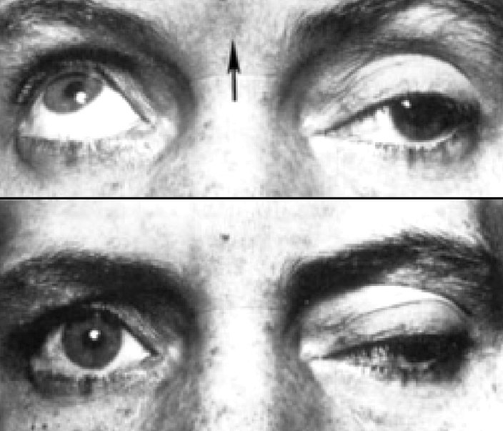
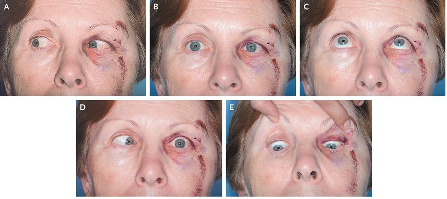
| CN III (oculomotor) | CN IV (trochlear) | CN VI (abducens) | |
|---|---|---|---|
| Muscle | Superior division: levator palpebrae, superior rectus. Inferior division: inferior rectus, medial rectus, inferior oblique, and parasympathetics to the ciliary ganglion | Superior oblique — intorts and depresses | Lateral rectus — abducts |
| Diplopia | With ptosis and mydriasis | VERTICAL, binocular, worse with postural head change | HORIZONTAL, binocular |
| Most common cause | Microvascular — diabetes, hypertension | Congenital when isolated, even in adults. Acquired → trauma, even mild, or microvascular | Children: intracranial tumors, especially brainstem and posterior fossa. Adults: microvascular; also major trauma or skull base fracture |
| The dreaded cause | Compression by an enlarging intracranial aneurysm, most commonly of the posterior communicating artery — threat of rupture within hours to days | — | — |
A third nerve palsy is classified two ways at once: complete versus incomplete, and pupil-INVOLVED versus pupil-SPARED.
| Pupil involved | Pupil spared |
|---|---|
| STAT CTA head or MRA brain — this is the vascular pathway, and the aneurysm is what you are ruling out | Reassurance and imaging, but it does not need to be STAT |
The anatomical reason: the parasympathetic fibers run on the outside of the nerve, so an external compression reaches them first, while microvascular disease infarcts the core and spares them. Slide 41
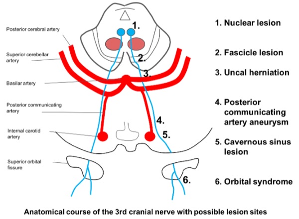
CN IV is the odd one out anatomically: it is the only nerve arising from the dorsal surface of the brainstem, and it crosses — the left trochlear nucleus sends fibers to the RIGHT eye. Clinically the patient tilts the head to the opposite side of the problem eye to compensate.
| Situation | Management |
|---|---|
| Isolated, atraumatic CN IV or CN VI palsy | MRI brain with and without contrast. Check hemoglobin A1C if there are risk factors and no known diabetes. |
| Traumatic | Observe roughly 6 months before considering corrective treatment. In the interim, patch one eye to relieve binocular diplopia. |
| Congenital | Patching. |
Also tested
- Sixth nerve palsy. Binocular horizontal diplopia that worsens on gaze to one side, with under-abduction of that eye, means a sixth nerve palsy on that side: the lateral rectus abducts, and the deficit is on the side of gaze.
- Patching in congenital palsy. Patching of a young child with a congenital palsy should be supervised by ophthalmology, especially under about five years of age.
- Head tilt in fourth nerve palsy. Tilting the head compensates for the weak muscle: the patient tilts away from the affected side to reduce the vertical deviation.
- Third nerve palsy with a normal, reactive pupil. A normal pupil in ptosis with the eye deviated down and out suggests a microvascular ischemic cause. Pupillary fibers run peripherally and are spared by ischemia but compressed by an aneurysm.
- Sixth nerve palsy. It is a false localizing sign because of its long intracranial course; the palsy does not indicate where the lesion is, since raised pressure anywhere can affect it.
- Third nerve palsy causes. Severe head trauma is a cause, alongside microvascular disease and aneurysm.
- Superior division oculomotor lesion. It causes ptosis and impaired elevation, because the superior division supplies levator palpebrae and superior rectus.
- Third nerve palsy. The eye is "down and out" because of unopposed lateral rectus and superior oblique: the muscles the third nerve does not supply are the ones left working.
- Fourth cranial nerve. It supplies the superior oblique, which depresses the adducted eye, hence vertical diplopia worse on downgaze.
- Peripheral fourth nerve lesion. A peripheral lesion after the nerve exits gives an ipsilateral deficit, in the same side eye.
- Acquired fourth nerve palsy. The likely causes are trauma and microvascular disease, with even mild trauma sufficient.
2.7 · Ptosis Objective f6
Definition: drooping of the upper eyelid from a congenital or acquired abnormality of the muscles that elevate it. Three structures hold the lid up, and knowing which is which is how you localize the cause:
| Structure | Innervation | Role |
|---|---|---|
| Levator palpebrae superioris | CN III | Elevates the upper lid |
| Müller's muscle | Sympathetic | An additional 1–2 mm of elevation |
| Orbicularis oculi | CN VII | Closes the lids — the antagonist |
| Levator function | Laterality | The giveaway | |
|---|---|---|---|
| CN III palsy | Reduced | Usually unilateral | Impaired extraocular movement and mydriasis in the same eye |
| Horner syndrome | NORMAL — only Müller's is out, hence the mild droop | Usually unilateral | Miosis in the same eye |
| Myasthenia gravis | Reduced | Unilateral or bilateral | Variable throughout the day — fatigability is the whole diagnosis |
Ptosis plus a SMALL pupil is Horner. Ptosis plus a LARGE pupil is a third nerve palsy. That one line resolves most of the exam questions in this objective, and the third possibility — ptosis that comes and goes through the day with a normal pupil — is myasthenia.
Also tested
- Eyelid closure. The muscle that closes the eyelids, orbicularis oculi, is supplied by the seventh (facial) cranial nerve.
- Fatigable bilateral ptosis with variable diplopia. This pattern points away from a structural palsy and toward myasthenia, so the next step is referral for neurologic evaluation.
- Ptosis causes. Horner syndrome and third nerve palsy both drop the lid, and each also disturbs the pupil, so ptosis appears alongside anisocoria.
- Ptosis of third nerve palsy versus Horner syndrome. Levator function distinguishes them: it is reduced in third nerve palsy and normal in Horner syndrome.
2.8 · Pre- and post-chiasm lesions Objective g
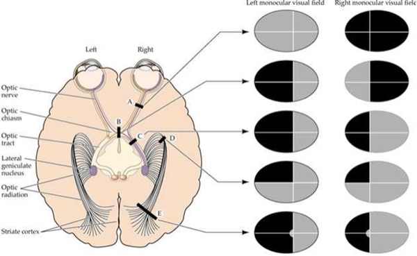
| Lesion site | Field defect | Pre- or post-chiasm |
|---|---|---|
| Optic nerve | Total blindness of that eye | PRE — monocular, because the fibers have not crossed yet |
| Optic chiasm | Bitemporal (heteronymous) hemianopsia | At the chiasm — the crossing nasal fibers are hit |
| Optic tract | Left homonymous hemianopsia (for a right tract lesion) | POST |
| Optic radiation | Left superior quadrantanopia | POST |
| Striate cortex | Left homonymous hemianopsia WITH MACULAR SPARING | POST |
The rule that generates all five rows. A monocular defect is pre-chiasmal — one eye's fibers, before any crossing. A bitemporal defect is at the chiasm. Anything homonymous — the same side of the field in both eyes — is post-chiasmal, and the further back the lesion, the more congruent and the more likely to spare the macula.
Also tested
- Homonymous hemianopsia with macular sparing. This pattern suggests a lesion of the occipital cortex; macular sparing is the cortical signature.
- Monocular field defect. The lesion lies anterior to the chiasm; a monocular defect localizes pre-chiasmal.
2.9 · Care strategies, adult and elderly Objective h
The summary, in order: begin with a thorough history and physical examination; labs and imaging (head CT or brain MRI) if indicated; ophthalmology referral; and appropriate specialist referral — neurology, vascular surgery, or neurosurgery. Slide 54
| Presentation | Urgency |
|---|---|
| CN III palsy with pupil involvement | EMERGENT — STAT CTA or MRA for aneurysm |
| Horner syndrome with neck pain, trauma or focal neurology | EMERGENT — carotid dissection |
| New nystagmus acquired in adolescence or adulthood | Work up — ophthalmology, imaging, labs |
| Isolated atraumatic CN IV or VI palsy | MRI with and without contrast; A1C if at risk |
| Pupil-sparing CN III palsy | Imaging, but not STAT |
First-line treatment
Every condition in this lecture, with what you reach for first and how fast — which in ophthalmology is half the answer. Where the deck escalates after a failed trial, only the first step is here; the full ladder and the reasoning are in the comparison chart.
| Condition | First line | How fast |
|---|---|---|
| Relative afferent pupillary defect (Marcus Gunn) | Treat the underlying retinal or optic nerve disease. | Urgent |
| Horner syndrome | Treat the cause. Levator function is normal, so the ptosis is mild. | Emergent |
| Argyll Robertson pupil | Treat the underlying syphilis. | Urgent |
| Adie tonic pupil | Reassurance; treat symptoms. Dilute pilocarpine constricts it through denervation supersensitivity. | Routine |
| Cranial nerve III palsy | Depends on etiology. Traumatic: observe about 6 months, patching in the interim. | Emergent |
| Cranial nerve IV palsy | Traumatic: observe about 6 months before corrective treatment; patch one eye meanwhile. | Urgent |
| Cranial nerve VI palsy | Traumatic: observe about 6 months; patch one eye in the interim. | Urgent |
| Nystagmus | Ophthalmology for a complete ophthalmic exam → imaging → labs if relevant. The underlying etiology must be addressed. | Urgent |
Also tested
- Tonic pupil in an otherwise well person. Management is reassurance once causes are excluded; it is benign, though the anisocoria may persist.
- Neuro-ophthalmological work-up. After history and examination, head computed tomography or MRI is used if indicated.
2.10 · Nystagmus — the two classification groups
Nystagmus splits into two groups, and the split is the first thing to establish.
| Jerk | Pendular | |
|---|---|---|
| Phases | A slow phase and a fast phase | Both phases equal in velocity and amplitude — no fast phase at all |
| Named for | The direction of the FAST beat — vertical, horizontal or torsional | Not named by beat, since there is no fast beat |
| Behavior | Increases with gaze toward the fast phase | Most often horizontal |
| Context | Most common form is horizontal jerk — eyes drift slowly one way, snap back | Usually congenital, or follows prolonged bilateral blindness beginning in childhood |
The horizontal jerk subtypes: normal physiologic gaze-evoked, infantile, spasmus nutans, and latent nystagmus.
Upbeat nystagmus is ALWAYS abnormal. It means a cerebellar or medullary lesion, and less commonly drug intoxication. It is present only on upward gaze. This is the one direction that carries its own alarm.
3 · Acute Vision Loss
Instructional Objectives
OPHTHALMOLOGY — Acute Vision Loss
- Compare and contrast the etiologies, epidemiology, risk factors, clinical
manifestations, differential diagnosis, diagnostic testing (including ordering and
interpretation), management (acute and chronic, including applicable rehabilitative and
palliative care), appropriate referrals, patient education, and prognosis of the following
acute vision loss disorders:
- Amaurosis fugax
- Acute angle closure glaucoma
- Optic neuritis
- Retinal detachment
- Retinal vascular occlusion: a. Central retinal artery occlusion (CRAO) · b. Branch retinal artery occlusion (BRAO) · c. Central retinal vein occlusion (CRVO) · d. Branch retinal vein occlusion (BRVO)
- Anterior ischemic optic neuropathy (AION): a. Arteritic AION, associated with Giant cell (temporal) arteritis · b. Non-arteritic AION
- Identify medical care strategies for acute vision loss in the lecture topic list for the following populations: 1. adult · 2. elderly
Every single person who has sudden acute vision loss is having a stroke until proven otherwise. That is why everyone gets an MRA, even with no weakness, no numbness and no speech difficulty — because you can have a stroke with no other symptom than acute vision loss. She repeated the sentence and added that Professor Alaya will ask this question when you reach the emergency department rotation.
3.1 · The four questions that separate these diagnoses
Jaquith opened by telling the class which details to highlight, because these conditions overlap heavily and the discriminating information is in the history rather than the fundus:
| Ask | Answer | Points toward |
|---|---|---|
| One eye or both? | One | Nearly all of them — amaurosis fugax, occlusions, detachment, optic neuritis, AION |
| Both | Papilledema, or a lesion at or behind the chiasm | |
| Sudden or gradual? | Seconds, then recovers | Amaurosis fugax |
| Seconds, and stays | CRAO | |
| Hours to days | Optic neuritis (and detachment advances over days) | |
| Central or peripheral? | Peripheral first, central preserved | Chronic open-angle glaucoma — “tunnel vision” |
| Central, with color desaturation | Optic neuritis | |
| Painful or painless? | PAINFUL | Acute angle-closure glaucoma (severe, at rest), or optic neuritis (on eye movement) |
| PAINLESS | Everything else — amaurosis fugax, all four occlusions, detachment, AION, papilledema |
The word “curtain” belongs to TWO diagnoses. Jaquith flagged this explicitly: “there's a couple of conditions where they use the word curtain, so don't confuse it.” Amaurosis fugax — the curtain comes down and then lifts within seconds to minutes. Retinal detachment — the curtain comes down and stays, advancing over days. Duration is the discriminator, not the word.
Also tested
- Assessing acute vision loss. The initial assessment turns on four questions: one or both eyes, duration, pain, and a description of the symptoms.
3.2 · Amaurosis fugax Objective a1
Transient monocular vision loss, also called “fleeting blindness”. Lasts a few seconds to minutes. It can involve both eyes but is usually one.
If it lasted hours, it was not a transient ischemic attack. Jaquith was explicit. Duration is the first filter you apply.
| Facet | Content |
|---|---|
| Etiology | Retinal emboli of carotid (TIA) or cardiac origin; also retinal vascular spasm. Most commonly a TIA. |
| Risk factors | Older age, diabetes, hypertension, atherosclerosis, cardiac valve disease, intravenous drug use, sickle cell, coagulation disorders, Raynaud's |
| Manifestations | A “curtain” coming down, transient. Anything from mild blurring or fogging to complete blackness. May involve part or all of the field — upper or lower half, temporal or nasal, small central or paracentral areas, or the whole field. Painless. |
| Diagnosis | Carotid Doppler if a carotid source is suspected. Echocardiogram if cardiac. MRA to evaluate all the arteries for emboli — and per the flag above, everyone gets it. |
| Treatment | Treat the underlying cause — she called this the essential point. To reduce stroke risk: aspirin and clopidogrel. Carotid emboli → carotid endarterectomy. Raynaud's or vascular spasm → calcium channel blockers. |
| Prognosis | About 85% recover fully; the rest progress to a central retinal artery occlusion. Early evaluation reduces the risk of permanent visual loss. |
| Education | A TIA is a major warning sign that a stroke is coming. Transient does not mean benign. |
Also tested
- Amaurosis fugax source. Painless monocular blackouts lasting under a minute most likely arise from a carotid or cardiac embolus, most commonly a transient ischemic attack of retinal origin.
- Treatment of amaurosis fugax from carotid emboli. Antiplatelet therapy is given for stroke risk, together with endarterectomy for the carotid source.
- Amaurosis fugax. A transient curtain-like loss of vision in one eye with full recovery is a transient ischemic attack of the eye, carries the same stroke risk, and calls for carotid imaging and urgent stroke workup.
- Amaurosis fugax. Loss that descends over the field of one eye like a shade from above is attributed to retinal ischemia.
- Transient monocular vision loss lasting minutes. The mechanism is transient retinal ischemia, usually embolic, most often from carotid atheroma.
- Curtain coming down over vision. This description is a warning sign because more than one condition uses it; it fits amaurosis fugax and retinal detachment.
- Amaurosis fugax. The term for fleeting blindness, meaning monocular episodic visual loss.
- Amaurosis fugax outlook. About 85% recover fully, and the remainder progress to a central retinal artery occlusion.
3.3 · Glaucoma — acute closed-angle and chronic open-angle Objective a2
What these look like
| ACUTE — closed angle | CHRONIC — open angle | |
|---|---|---|
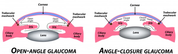
| Mechanism | The iris blocks the drainage circuit → intraocular pressure rises dramatically | Trabecular meshwork abnormality next to the canal of Schlemm, secondary to aging → optic nerve damage, with or without raised pressure |
| Frequency | Less common | MUCH more common |
| Pain | Severe and sudden eye pain | Painless |
| Symptoms | Decreased vision, colored halos around lights, headache, nausea and vomiting | Asymptomatic in most patients. Peripheral field goes first — patients say “tunnel vision” — then complete blindness |
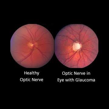
| Signs | Pupillary dilation, hazy cornea, bilateral narrow or occluded angle | Optic nerve cupping, rim pitting, bayoneting (vessels with narrow angulations), splinter hemorrhages, rim thinning, field defects |
| Pressure | 40–80 mmHg on tonometry or gonioscopy | May be normal OR elevated |
| Other risks | Systemic anticholinergics (atropine), nebulized bronchodilators, prior anterior uveitis, lens dislocation, African American race, obstruction from tumor or scarring | African American race, Hispanic ethnicity, adults over 40, diabetes, age, family history, hypertension, myopia |
| First-line | Topical pilocarpine (alpha-blocker) or timolol (beta-blocker); IV acetazolamide then mannitol or isosorbide | Latanoprost, tafluprost, timolol drops |
| Definitive | Laser peripheral iridotomy, 1–2 days after onset — lets fluid pass from the posterior to the anterior chamber | Laser trabeculoplasty if refractory or advanced. Surgery is definitive for both forms. |
A patient went to the emergency department with an intractable headache — everything had been tried, nothing worked — and was admitted. After multiple doctors, a PA assessed him properly, found a red eye, and diagnosed glaucoma. The chief complaint was a headache, and it was localized behind the eye. Glaucoma is also a leading cause of blindness worldwide, and untreated it ends in blindness.
Patient education, and she meant it personally: everyone should have annual eye examinations — that screening is what finds asymptomatic chronic open-angle disease before the field is gone. Some patients stay on drops for life and never have surgery, when the surgical risk outweighs the benefit over their life expectancy.
Also tested
- Secondary angle-closure glaucoma. Drainage can be obstructed by tumor, scarring or other mechanical causes.
- Anticholinergics in angle closure. Systemic anticholinergic medications such as atropine are a risk factor for acute angle closure, in which the iris blocks the drainage circuit and pressure rises, so they can worsen angle-closure glaucoma.
- Intraocular pressure in glaucoma. Pressure may be either normal or elevated in this disease.
- Why open-angle glaucoma is missed. Loss is gradual and the other eye compensates, which is why it is found on screening rather than by symptoms.
- Chronic open-angle glaucoma cause. Idiopathic optic nerve damage, from trabecular meshwork change with aging.
- Evaluating glaucoma. Tonometry and gonioscopy are the two tests: tonometry measures pressure, and gonioscopy views the angle.
- Painful red eye with a hazy cornea. Intraocular pressure is measured first, by tonometry or gonioscopy; expect 40 to 80 mmHg.
- Acute severe eye pain with halos, hazy cornea and fixed dilated pupil. With a shallow anterior chamber and ciliary injection, tonometry is the immediate next step: measuring the intraocular pressure confirms the diagnosis.
- Acute angle-closure glaucoma. A shallow anterior chamber predisposes to it: a crowded angle is what allows closure when the pupil dilates.
3.4 · Optic neuritis Objective a3
What these look like
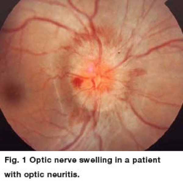
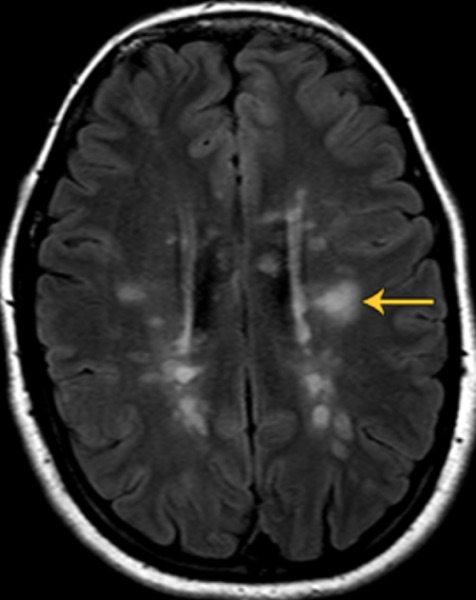
| Facet | Content |
|---|---|
| Epidemiology | 18 to 45 years old, 75% female — a much younger group than everything else in this block |
| Etiology | Inflammation due to multiple sclerosis, autoimmune disorders, postviral, or idiopathic |
| Symptoms | Unilateral vision loss over hours to several days, with PAINFUL EYE MOVEMENT. Often central vision loss and loss of color vision |
| Signs | Often a normal-appearing disc. Relative afferent pupillary defect (Marcus Gunn) |
| Diagnosis | Refer to ophthalmology. Complete ophthalmic exam — slit lamp, dilated fundoscopy, color vision assessment — plus a neurological exam. MRI brain AND orbits, with and without contrast |
| Escalation | If MRI shows at least 2 characteristic demyelinating lesions, ophthalmology treats and refers to neurology or neuro-ophthalmology |
| Treatment | Corticosteroids if a demyelinating cause is found |
| Prognosis | Spontaneous recovery is the rule. Without treatment vision begins improving within a few weeks, may continue for months, and is usually normal within a year |
| Education | Recurrence carries a greater risk of multiple sclerosis. Do not treat the episode and stop — find out why it happened, or you miss the bigger problem |
Also tested
- Optic neuritis epidemiology. About 75 percent of cases occur in women, in patients aged 18 to 45.
- Optic nerve pallor without cupping. This favors ocular ischemic syndrome, which is underlain by carotid stenosis.
- Optic neuritis course. Vision usually recovers substantially over weeks, though color desaturation may persist.
- Imaging in suspected optic neuritis. Magnetic resonance of the brain and orbits is ordered, with and without contrast.
- Optic neuritis course. Improvement within weeks, normal vision within a year, often without treatment.
- Optic neuritis patient profile. Typically aged 18 to 45 and mostly female; about 75% are women.
- Optic neuritis with demyelinating lesions on imaging. Neurology referral with corticosteroids follows; steroids are given if a demyelinating cause is found. Neuro-ophthalmology is equally appropriate.
3.5 · Retinal detachment Objective a4
What these look like
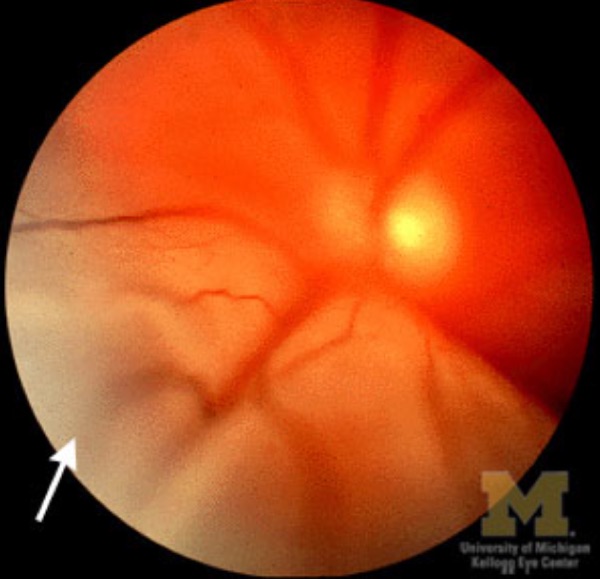
| Facet | Content |
|---|---|
| Pathophysiology | Traction detachment that commonly follows a retinal tear or hole. Three types: rhegmatogenous, traction, serous/exudative |
| Epidemiology | Most common after age 50 — the vitreous humor shrinks with age |
| Risk factors | Myopia (contracted ciliary muscle), trauma, cataract extraction, diabetes, tumor, connective tissue disease, family history |
| Early symptoms | New flashing lights and floaters — these represent the retinal TEAR, not the detachment itself |
| Then | Gray or black shadows in the peripheral field, which may cover the whole eye within days. A “curtain or dark cloud” across the field |
| The escalation | If the macula is involved → sudden loss of vision in that eye |
| Odd but useful | The visual loss can change with head position, because the detached retina is floating loose |
| Diagnosis | Direct and dilated ophthalmoscopy: retina elevated, gray cloud with folds, a pigmented well-demarcated area, and tears that are orange and crescent shaped. Ultrasound is MORE SENSITIVE than the fundoscopic exam and determines the type |
| Treatment | EMERGENCY — refer immediately. Surgical repair urgently or within a week depending on type. Options: laser photocoagulation, cryotherapy, pneumatic retinopexy, vitrectomy, scleral buckle |
Her disposition, verbatim in substance: this one needs to be seen right now. If no ophthalmologist can take them, they go to a hospital that has one. Not tomorrow.
Also tested
- Posterior vitreous detachment without a retinal break. Advise return if a curtain or shadow appears; that warning converts this from observation to emergency.
- Holding text further away with unchanged distance vision. The lens stiffens, so accommodation fails.
- Myopia risk factors. Family history and near work are risk factors, and trauma displacing the lens forward can cause myopia.
- Retinal detachment symptoms. Vision changes with head position, which is unusual among causes of acute vision loss, because the detached retina moves.
- Suspected retinal detachment with a poor fundus view. Ocular ultrasound is more sensitive than fundoscopy, and it also types the detachment.
- Retinal detachment with the macula still attached. Central vision is salvageable before the macula goes, so macula-on detachment is the more urgent repair.
- Types of retinal detachment. Rhegmatogenous, traction and serous; the serous form is also called exudative.
3.6 · The four retinal vascular occlusions Objective a5
| CRAO — artery | CRVO — vein | |
|---|---|---|
| Mechanism | Embolus — arteriosclerosis, atherosclerosis, carotid or cardiac emboli | Thrombus occluding the central retinal vein |
| Frequency | Less common | MORE common than CRAO |
| Onset | Painless profound loss over a few SECONDS | Sudden and painless; sometimes gradual over days to weeks |
| Acuity | Counting fingers to light perception; an “island” of vision in the temporal field | Blurring to loss |
| Pupil | Slow to direct light, but BRISK when the other eye is illuminated — she called this a huge clinical clue | — |
| Fundus | Pale swelling of the posterior segment with a CHERRY-RED SPOT at the fovea; emboli visible in the central artery | “BLOOD AND THUNDER” — disc swelling, venous dilation, cotton wool spots, retinal hemorrhages |
| Risk factors | Shared: hypertension, diabetes, hyperlipidemia, Raynaud's, age over 50, hypercoagulable disorders, giant cell arteritis, endocarditis, atrial myxoma, obesity. CRAO adds atrial fibrillation | |
| Later | Neovascularization occurs weeks to months after the occlusion in both | |
| Confirmatory | Color fundus photography and fluorescein angiography | |
| Treatment | Prompt. High-concentration inhaled oxygen and digital massage over the eyelid; IV acetazolamide to lower pressure; anterior chamber paracentesis; thrombolytic infusion into the ophthalmic artery within 8 hours | Urgent ophthalmology referral to restore blood flow; evaluate and treat the underlying disorders |
CRAO is a stroke in the eye. Irreversible retinal damage may occur after 90 minutes. And the systemic implication matters as much as the eye: the retinal arteries are tiny, so if plaque broke off the carotid and reached one, there is a great deal more plaque still in that carotid — stroke risk rises at the onset of a retinal artery occlusion.
The branch forms. BRAO and BRVO are the same disease as their central counterparts, differing only in where the blockage sits. A branch vessel is occluded rather than the main trunk, so only part of the retina is affected and the field loss is partial and localized rather than widespread. Everything else — risk factors, workup, referral — is the same. Jaquith was explicit that this is all there is to it.
Also tested
- Central retinal artery occlusion and general health. Stroke risk is markedly increased, since plaque reaching the retinal artery implies more in the carotids.
- Branch versus central retinal vein occlusion. In a branch occlusion a smaller branch vein is blocked, so only part of the retina is affected, unlike central retinal vein occlusion.
- Sudden painless monocular vision loss. Immediate ophthalmology referral is the next step; it is a stroke of the eye, and the window is measured in minutes to hours.
- Branch versus central retinal vein occlusion. In the branch form the clot is in a branch vein, so hemorrhage is confined to that territory and only part of the retina is affected.
- Central retinal vein occlusion complication. Neovascular glaucoma is monitored for, because ischemia drives new vessel growth at the iris and angle.
- Cherry-red spot. It is the intact choroid showing through the fovea; the surrounding retina is pale from infarction, and the fovea has no inner retinal layers to become edematous.
- Central retinal artery occlusion. Anterior chamber paracentesis is performed to lower the intraocular pressure; fluid is withdrawn from the eye with a needle.
- Central retinal vein occlusion. Look for hypertension, diabetes and hyperviscosity; the venous side reflects systemic vascular and rheological disease.
- Frequency of retinal vascular occlusions. Vein occlusion is more common than central retinal artery occlusion.
- Central retinal artery occlusion. It causes sudden profound painless loss of vision, with a pale, swollen retina and a cherry-red spot at the fovea.
- Arterial versus venous retinal occlusion on fundoscopy. Arterial occlusion shows pallor with a cherry-red spot at the fovea rather than hemorrhages.
- Branch retinal artery occlusion. It causes sudden painless loss of part of the visual field, with a pale wedge of retina in one artery's territory producing loss of the opposite field.
- Central retinal artery occlusion. Profound painless loss of vision in one eye over seconds that does not recover, with a pale retina.
- Central retinal vein occlusion. Sudden painless loss of vision with the "blood and thunder" fundus: disc swelling, dilated, engorged veins, cotton wool spots and hemorrhages in every quadrant.
3.7 · Papilledema
What these look like
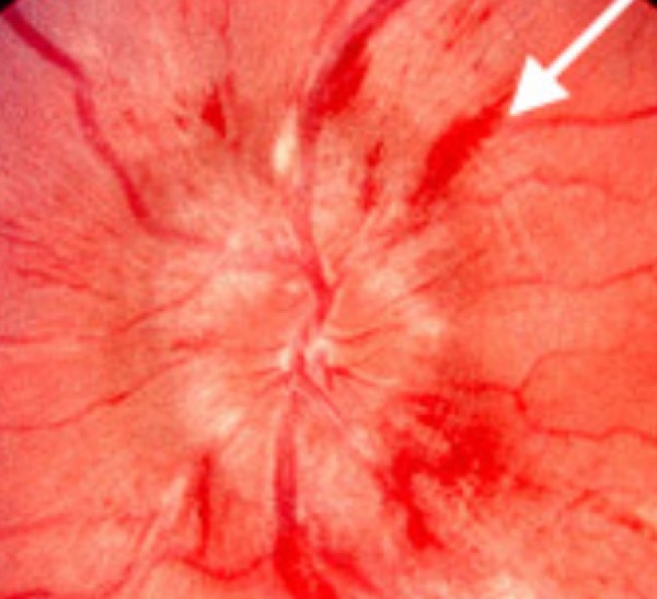
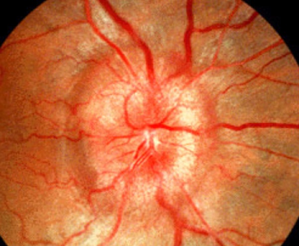
This is the one condition here that is NOT about intraocular pressure. Papilledema is swelling of the optic disc caused by raised INTRACRANIAL pressure — pressure inside the skull pushing on the optic disc. Every other pressure in this block is inside the globe. She drew the line explicitly.
| Facet | Content |
|---|---|
| Causes | Tumor, trauma, intracranial infection (meningitis), hemorrhage, vitamin A toxicity |
| Visual symptoms | Non-specific: flickering vision, blurry vision, double vision |
| Systemic symptoms | Non-specific signs of raised intracranial pressure: nausea, vomiting, headache |
| Fundus | Engorged retinal veins, swollen optic disc, with or without retinal hemorrhages |
| Diagnosis | Lumbar puncture — an increased opening pressure confirms raised intracranial pressure. MRI and/or CT head to rule out a mass lesion |
| Treatment | Treat the underlying disorder |
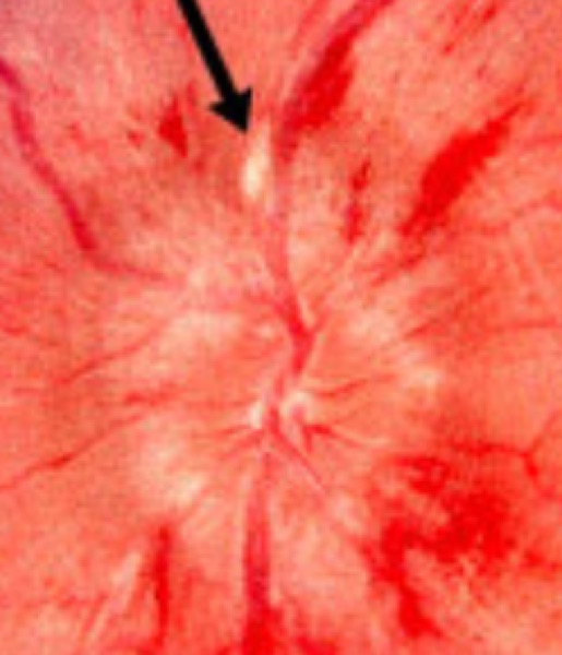
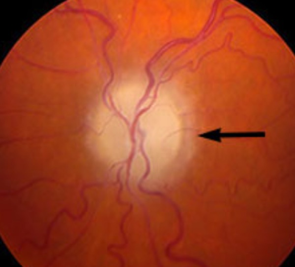
Papilledema pushes the disc OUT. Glaucoma cups it IN. She stressed how distinctly different the two look, and it is the single most reliable way to tell them apart at the fundus.
Also tested
- Papilledema versus optic neuritis. Acuity is preserved in papilledema and reduced in optic neuritis; papilledema is bilateral, while optic neuritis is usually unilateral with an afferent pupillary defect.
- Lumbar puncture in idiopathic intracranial hypertension. With papilledema, the expected finding is a raised opening pressure with normal constituents.
3.8 · Anterior ischemic optic neuropathy Objective a6
Definition: sudden loss of blood flow to the front part of the optic nerve — the optic disc, causing rapid, painless vision loss. Common to both forms: sudden painless loss of side or central vision, swelling and PALENESS of the optic nerve head, and involvement of one eye first, though the second eye is at risk.
| ARTERITIC (AAION) | NON-ARTERITIC (NAION) | |
|---|---|---|
| Share of cases | The minority | 90–95% |
| Age | 55 and older | 40 to 60 |
| Cause | Giant cell (temporal) arteritis — inflammation of the blood vessels | Linked to a small structural optic disc — a “disc at risk” |
| Associations | Systemic: malaise, weight loss, fever, headache in the temporal or occipital region, scalp tenderness (classically on combing the hair), jaw claudication on eating or chewing | Hypertension, diabetes, high cholesterol, sleep apnea |
| Urgency | MEDICAL EMERGENCY. Refer emergently any patient over 50 with sudden visual loss | Not emergent once arteritis is excluded |
| Workup | ESR and CRP — the tests used to rule giant cell arteritis in or out. Temporal artery biopsy is the gold standard | A diagnosis of EXCLUSION. The workup is identical — you are making sure there is no giant cell arteritis — then medical evaluation for hypertension, diabetes and anemia, with neuroimaging if unclear |
| Treatment | IV methylprednisolone for 3 days, then a slow oral taper to the lowest dose that suppresses the disease, guided by symptoms and labs — typically 6 to 12 months. Add famotidine for gastrointestinal ulcer prophylaxis | Observation and cardiovascular risk factor modification. Consider avoiding antihypertensives at bedtime, since nocturnal hypotension can worsen it |
| Prognosis | Depends on how long it has been present and when corticosteroids started. Early therapy is critical — untreated it leads to blindness. Follow up with ophthalmology | Follow up with ophthalmology |
Her boundary on giant cell arteritis. “I'm not going to test you on GCA directly, I promise you — but it's kind of hard when I know GCA is what's causing this not to explain GCA to you.” So learn it as the cause of arteritic AION and the reason that presentation is an emergency, not as a rheumatology topic in its own right.
The picture she painted: an elderly woman, usually Caucasian, over 55, with no history of headaches, who suddenly has a temporal headache. That change from no headaches to a new one is the warning sign.
Also tested
- Inflammatory markers in arteritic optic neuropathy. They help rule the diagnosis in or out; erythrocyte sedimentation rate and C-reactive protein are markedly raised.
- Arteritic optic neuropathy outlook. The outlook depends on the duration of symptoms and when steroids began, which is why early therapy is critical.
- Antihypertensive timing in non-arteritic anterior ischemic optic neuropathy. The antihypertensive dose may be moved away from bedtime to avoid nocturnal hypotension, which can worsen the condition.
- Optic disc in anterior ischemic optic neuropathy. Sudden painless vision loss with a swollen, pale disc and loss of side or central vision is the expected picture.
- Arteritic anterior ischemic optic neuropathy. With markedly raised inflammatory markers, treatment starts with intravenous methylprednisolone for three days, then a slow oral taper; early therapy is critical.
- Non-arteritic share of anterior ischemic optic neuropathy. The non-arteritic form accounts for ninety to ninety-five percent of cases and is by far the commoner form.
- Suspected arteritic vision loss. Sudden painless vision loss with scalp tenderness, jaw aching on chewing and a markedly raised erythrocyte sedimentation rate calls for high-dose corticosteroids immediately; treatment precedes biopsy because the other eye is at risk within days.
- Arteritic anterior ischemic optic neuropathy. It affects people aged 55 years and older.
- Non-arteritic anterior ischemic optic neuropathy. It typically affects people aged 40 to 60 years, accounting for 90 to 95 percent of cases.
- Giant cell arteritis with vision loss. Start treatment today rather than waiting for the biopsy, because the fellow eye can be lost within days.
- Non-arteritic anterior ischemic optic neuropathy. Altitudinal field loss on waking, with normal inflammatory markers and no arteritic symptoms such as scalp tenderness or jaw claudication.
3.9 · Care strategies — adult and elderly Objective b
| Presentation | Disposition |
|---|---|
| Sudden painless monocular loss, any cause | Stroke until proven otherwise — MRA, and urgent ophthalmology |
| CRAO | Emergency department NOW — the retina is lost after 90 minutes |
| Retinal detachment | Same day, to a hospital with an ophthalmologist if clinic cannot take them |
| Acute angle-closure glaucoma | Emergent — lower the pressure medically, iridotomy in 1–2 days |
| Any patient over 50 with sudden vision loss | Emergent ophthalmology, plus ESR and CRP for arteritic AION |
| CRVO / BRVO / BRAO | Urgent ophthalmology to restore flow; treat the underlying disorder |
| Optic neuritis | Refer to ophthalmology; MRI brain and orbits; neurology if 2 or more lesions |
| Papilledema | Imaging to exclude a mass, then lumbar puncture; treat the cause |
| Chronic open-angle glaucoma | Routine — but annual eye exams are how it gets found at all |
First-line treatment
Every condition in this lecture, with what you reach for first and how fast — which in ophthalmology is half the answer. Where the deck escalates after a failed trial, only the first step is here; the full ladder and the reasoning are in the comparison chart.
| Condition | First line | How fast |
|---|---|---|
| Amaurosis fugax | Treat the underlying cause. Aspirin and clopidogrel for stroke risk; carotid endarterectomy for carotid emboli; calcium channel blockers for vasospasm. | Urgent |
| Acute angle-closure glaucoma | Topical pilocarpine or timolol; IV acetazolamide then mannitol or isosorbide. Definitive: laser peripheral iridotomy, 1–2 days after onset. | Emergent |
| Chronic open-angle glaucoma | Latanoprost, tafluprost or timolol drops. | Routine |
| Optic neuritis | Corticosteroids if a demyelinating cause is found. At least 2 demyelinating lesions → neurology or neuro-ophthalmology. | Urgent |
| Retinal detachment | Refer immediately. Surgery urgently or within a week by type: laser photocoagulation, cryotherapy, pneumatic retinopexy, vitrectomy, scleral buckle. | Emergent |
| Central retinal artery occlusion (CRAO) | High-concentration inhaled oxygen and digital massage, IV acetazolamide, anterior chamber paracentesis, and thrombolytic into the ophthalmic artery within 8 hours. | Emergent |
| Branch retinal artery occlusion (BRAO) | As for CRAO — the management does not change. | Emergent |
| Central retinal vein occlusion (CRVO) | Urgent ophthalmology referral to restore blood flow. Evaluate and treat the underlying disorders. | Urgent |
| Branch retinal vein occlusion (BRVO) | As for CRVO — urgent referral, and treat the underlying disorder. | Urgent |
| Arteritic AION (giant cell arteritis) | IV methylprednisolone ×3 days, then a slow oral taper to the lowest suppressive dose, typically 6–12 months. Add famotidine for ulcer prophylaxis. Do not wait for the biopsy. | Emergent |
| Non-arteritic AION | Observation and cardiovascular risk factor modification. Consider avoiding antihypertensives at bedtime. | Urgent |
| Papilledema | Treat the underlying disorder. | Emergent |
Also tested
- Sudden painless monocular vision loss. Urgent ophthalmology involvement is the non-negotiable step, because each of these diagnoses is sight-threatening and time-dependent.
4 · Chronic Vision Loss & Tumors of the Eye
Instructional Objectives
OPHTHALMOLOGY — Chronic Vision Loss and Tumors
- Compare and contrast the etiologies, epidemiology, risk factors, clinical
manifestations, differential diagnosis, diagnostic testing (including ordering and
interpretation), management (including applicable rehabilitative and palliative care),
appropriate referrals, patient education, and prognosis for the following chronic
vision loss disorders and tumors of the eye:
- Cataract — congenital, acquired
- Age related macular degeneration — wet, dry
- Primary open angle glaucoma
- Astigmatism
- Hyperopia
- Myopia
- Strabismus
- Amblyopia
- Idiopathic intracranial hypertension
- Neoplasms, malignant: retinoblastoma, melanoma, squamous cell carcinoma
- Neoplasms, benign: nevus
- Identify medical care strategies for common ophthalmological disorders in infant, adolescent, adult, and elderly populations.
4.1 · Idiopathic intracranial hypertension
Also called pseudotumor cerebri, and both terms are used. It is almost exclusively a disease of overweight women of childbearing age, and it comes on so slowly that the symptoms sound like everyday complaints.
| Symptoms | Intractable headache of variable character · transient visual loss that comes and goes · intracranial noises — pulsatile tinnitus · pain behind the eyes · mild pain on eye movement or globe compression |
|---|---|
| Signs | Papilledema · visual field loss · abducens (sixth nerve) palsy. Not all are present at once — it depends how long it has been running. |
| Order first | MRI brain with MR venography. A new or changing headache always prompts imaging, and venography is there to exclude a cerebral venous sinus thrombosis, which raises pressure the same way. |
| Then confirm | Lumbar puncture — elevated opening pressure. That is the diagnosis. Note this is intracranial, not systemic, hypertension: the blood pressure may be perfectly normal. |
| Management | Acetazolamide promptly, plus a weight reduction program with a dietitian. Surgical CSF diversion — optic nerve sheath fenestration or a shunt — only for those who fail or cannot tolerate medical therapy. |
| Prognosis | Followed jointly by ophthalmology and neurology. Weight loss is the only thing that permanently fixes it; medication covers the patient while that happens. |
Beck's framing: if an overweight woman of childbearing age comes in with vague headache, keep this in the back of your mind. The symptoms are ordinary; the demographic is the clue.
Also tested
- Stakes of untreated idiopathic intracranial hypertension. Permanent vision loss from optic nerve damage is the risk; the vision, not the headache, drives treatment.
- Idiopathic intracranial hypertension workup. Imaging with venography comes first; when it is normal, the next step is lumbar puncture to measure opening pressure, which confirms the diagnosis.
- Idiopathic intracranial hypertension. Classic picture: a woman with a raised body mass index, daily headaches, pulsatile tinnitus, transient visual obscurations, papilledema with bilateral disc swelling, and normal neuroimaging.
4.2 · Primary open-angle glaucoma
The most common form of glaucoma, and the mirror image of the acute angle-closure form in section 3: painless, slow, and silent until damage is done.
| The angle | Open-angle means the angle between iris and cornea is wide. Closed-angle is the narrow, acute one. That geometry is the whole distinction. |
|---|---|
| What is lost | Peripheral field first, with central acuity initially spared — vision narrows like a tunnel. Macular degeneration does the opposite. |
| Signs | Cupping (excavation) of the optic nerve, characteristic field loss on perimetry, and often a raised intraocular pressure. |
| Risk factors | Raised intraocular pressure · advanced age · race · family history. |
| Treatment | Drops that either make less aqueous (beta blockers such as timolol, carbonic anhydrase inhibitors, alpha-2 agonists) or drain more (prostaglandin analogs), then laser or surgery. |
| Prognosis | Damage already done cannot be reversed. Treatment preserves what is left. That is the entire argument for screening someone who feels fine. |
Beck on referrals: an ophthalmologist is a physician and surgeon; an optometrist handles refraction and lens fitting. Medical eye disease — and every diabetic, annually — goes to ophthalmology. Make that explicit when you write the referral.
Also tested
- Why screen for open-angle glaucoma. It is asymptomatic at onset, and the damage it does cannot be reversed.
- Eye care in diabetes. Annual ophthalmology review should be arranged, because a medical eye risk needs the medical specialist.
- Why open-angle glaucoma goes unnoticed. It is painless and spares central vision early, which is why screening exists.
- Glaucoma drops. Drops protect the vision that remains, because damage already done cannot be reversed.
- Fellow eye in acute angle closure. The other eye receives prophylactic iridotomy, because it shares the crowded anatomy and is anatomically at risk.
- Glaucomatous optic disc damage. Defined by cupping, an excavated disc, with a raised cup-to-disc ratio.
- Who needs a yearly ophthalmologist. A patient with diabetes needs a yearly ophthalmologist rather than an optometrist, because a medical eye problem needs the medical specialist.
- Cupped discs on routine examination. Refer to ophthalmology for assessment; good central vision does not exclude damage.
- Most common form of glaucoma. Primary open-angle glaucoma is the most common form, and it is characteristically painless and slow.
- First-line treatment of open-angle glaucoma. A prostaglandin analog; prostaglandins or beta blockers lead, and cholinergic agents are third or fourth line.
4.3 · Age-related macular degeneration
What these look like
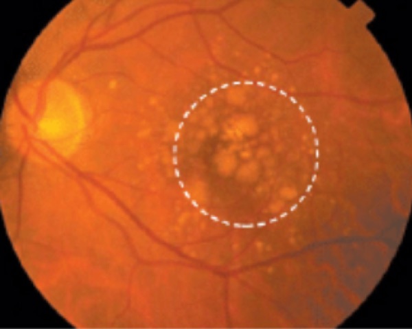
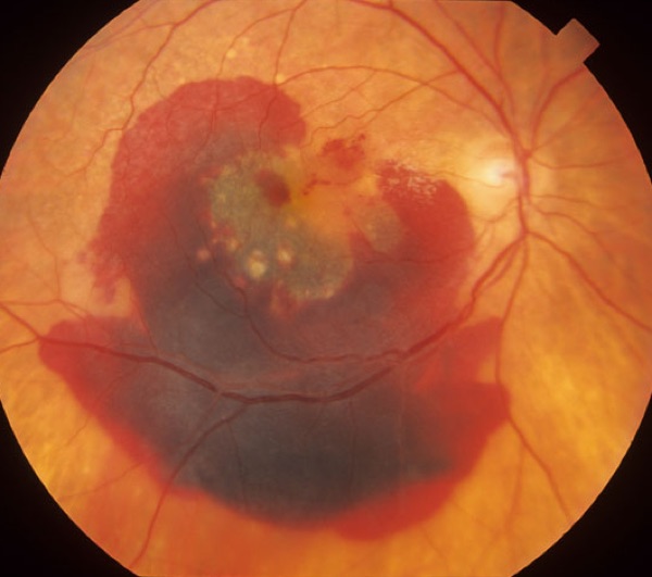
80% dry, 20% wet. Risk factors are age, cigarette smoking and probably genetics; the exact cause is unknown.
| Dry (non-exudative) | Drusen, pigmentary change and atrophy. Blurred central vision with a central scotoma. |
|---|---|
| Wet (exudative) | Choroidal neovascularization bleeds into the retina, and the blood organizes into a disciform scar. That scar is why the central vision goes. Once it has bled it stays classified as wet. |
| How dry turns wet | Waste products accumulate under the macula and disrupt the retina; new vessels grow behind it, causing swelling, bleeding and scarring — hence the rapid change on a slow background. |
| Monitoring | Amsler grid at home and serial slit lamp review. For wet, add fluorescein angiography and optical coherence tomography. |
| Management | STOP SMOKING. Dry: AREDS2 — vitamins C and E, zinc, copper, lutein, zeaxanthin. Wet: intravitreal anti-VEGF, thermal laser, photodynamic therapy. |
| Prognosis | Treatment slows progression but does not reverse vision already lost. |
Also tested
- Dry versus wet macular degeneration. About 80 percent of macular degeneration is the dry form, leaving roughly 20 percent wet.
- Sharp worsening of central blur in dry macular degeneration. A rapid change over a week raises concern for conversion to the wet form, in which new vessels bleed.
- Amsler grid. It is a self-monitoring tool to detect distortion or a missing area, which suggests conversion of dry macular degeneration to the wet form.
- Dry form of macular degeneration. About eighty percent of age-related macular degeneration is dry; the wet form accounts for the remaining twenty.
- Gradual painless vision loss in older patients. The three commonest causes, and so the top of the differential, are cataract, macular degeneration and open-angle glaucoma.
- Dry macular degeneration. Defined by drusen, pigment change and atrophy. Neovascular bleeding defines the wet form instead.
- Dense macular scar from previous bleeding. This is wet macular degeneration, because it began with neovascular bleeding. The scar is the end stage of the wet process.
- Wet macular degeneration treatment. Intravitreal anti-VEGF injection, with photodynamic therapy and laser as alternatives.
- Wet macular degeneration with acute distortion and central gray patch. Straight lines distort, with subretinal fluid and hemorrhage at the macula. Give urgent referral for intravitreal anti-VEGF therapy; the wet form is treatable and the window matters.
- Dry to wet macular degeneration. In the wet form, new vessels from the choroid bleed into retina, and the blood organizes into a scar.
4.4 · Refractive errors
Emmetropia is normal refraction, and is not an error. The three errors differ only in where the image lands.
| Myopia — nearsighted | “Long eyeball”, generally an over-protruding cornea. Too much refractive power, so distant objects focus in front of the retina. Corrected with a concave, negative diopter lens that scatters light and moves focus back. |
|---|---|
| Hyperopia — farsighted | “Short eyeball”, generally a flat cornea. Too little power, so objects focus behind the retina. Corrected with a convex, positive diopter lens that converges light forward. Risk factors are anything shortening axial length — trauma pushing the lens back, or a mass behind the globe. |
| Astigmatism | Uneven curvature of cornea or lens, so no single point focus forms — multiple focal points, blur at every distance. Corrected with a toric lens. |
| Presbyopia | Listed as a non-refractive error — the age-related loss of accommodation. |
Refer isolated refractive error to optometry. And the discriminator that matters clinically: refractive blur corrects fully with lenses. Beck's version — “if you have schmutz, putting glasses in front of the schmutz isn't going to help.”
Also tested
- Long eyeball. A long eyeball predicts blurred distance vision with clear near vision, because myopia focuses distant objects in front of the retina.
- Lens correcting myopia. Myopia is corrected by a concave lens of negative diopter, which moves the focus back.
- Astigmatism. Uneven curvature gives several focal points, so no single point focus forms, causing blur at every distance.
- Farsightedness. A convex lens corrects it because it converges light onto the retina; the farsighted eye focuses behind the retina.
- Hyperopia. The eye is too short, and accommodative effort at near produces headaches and eye strain when reading, with normal distance vision.
- Hyperopia and ocular emergencies. Hyperopic patients are warned about acute angle-closure glaucoma, because the short eye has a crowded anterior segment.
4.5 · Strabismus
Misalignment, from any level: brain, cranial nerves, neuromuscular junction, or the extraocular muscles themselves.
| The adult symptom | Diplopia that disappears on covering either eye — that is what makes it binocular, and therefore mechanical. |
|---|---|
| Signs | One eye fails to track or fixate, or does not move fully in some direction. Cover test elicits fixation of the misaligned eye. In moderate to large deviations the corneal light reflex is displaced. |
| Cranial nerve causes | Three — ptosis, pupil involvement, exotropia with vertical deviation. Four — vertical diplopia, worse on downgaze. Six — horizontal diplopia, esotropia. |
| Other differentials | Myasthenia gravis — variable ptosis and diplopia, mimics any pattern. Thyroid eye disease restricts movement. Down syndrome carries a higher prevalence of esotropia; cerebral palsy associates with strabismus and nystagmus. |
Also tested
- Double vision that resolves when either eye is covered. Binocular diplopia means misalignment until proven otherwise, so the next step is to assess ocular alignment and cranial nerves.
- Third nerve palsy with pupil involvement. Sudden ptosis, a dilated pupil and an eye deviated down and out require urgent exclusion of an aneurysm compressing the third nerve; pupil involvement makes this an emergency.
- Levels causing strabismus. The cause can lie in the brain, nerve, junction or muscle; causes are found at all four levels.
4.6 · Amblyopia
What these look like
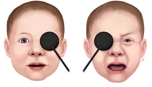
The most common cause of vision loss in children, affecting 3–5%. One eye fails to reach normal acuity even with correction, because the brain has stopped listening to it.
| Three routes in | Strabismus — misalignment, so the brain suppresses one image. Anisometropia — a large difference in prescription between eyes. Deprivation — anything blocking the view: cataract, ptosis, corneal opacity, an eyelid hemangioma. |
|---|---|
| Occlusion objection test | The child is unbothered when the amblyopic eye is covered and becomes fussy when the good eye is covered. Losing the eye they actually use is what upsets them. |
| Defining it | Unilateral amblyopia is a two-line or greater difference in best corrected acuity between the eyes. |
| Screening | The US Preventive Services Task Force and the American Academy of Pediatrics recommend screening all children under five. Pre-verbal children need optotype tests rather than a Snellen chart. |
| Treatment | Patch the good eye, or blur it with atropine drops, forcing the weaker eye to work. Beck's analogy: you cannot build the weak arm by bench pressing with both — the strong one takes over. |
| Prognosis | Good if treated before seven, better before five. Many do not reach fully normal acuity. |
Also tested
- Amblyopia outcome with patching. Outcome is good, though vision is not always fully normal, and starting treatment before age five is better still.
- Objection to covering one eye. A toddler who cries only when the right eye is covered sees poorly with the left eye, because he objects when his good eye is taken away.
- Occlusion objection test. A child who objects when one eye is covered has amblyopia in the other eye, because the child objects when the good eye is covered.
- Amblyopia treated late. An adult may have good acuity but struggle with 3D films because stereo vision never developed; the brain still uses one eye at a time.
- Amblyopia mechanism. The brain favors one eye and the other fails to develop. Strabismus, refractive difference or deprivation all do this.
- Patching in amblyopia. A patch over the good eye makes the weaker eye work; the stronger eye otherwise does everything.
- Unilateral amblyopia. It is defined by two or more lines of difference between the eyes on best corrected acuity.
- Eyelid hemangioma covering the visual axis. The priority is prompt referral to prevent amblyopia, because the obstruction has to be cleared while vision can still develop.
4.7 · Cataract
What these look like
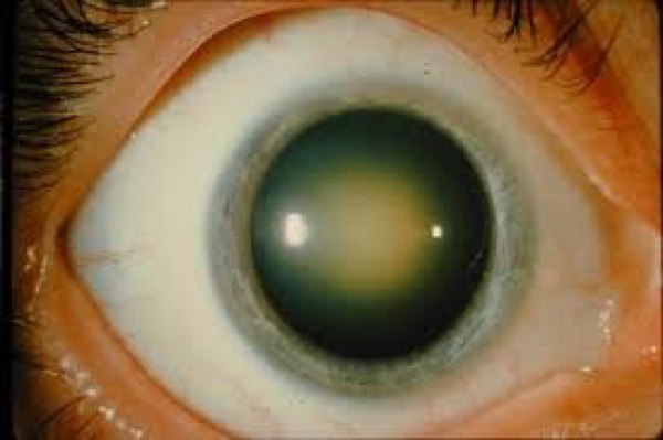
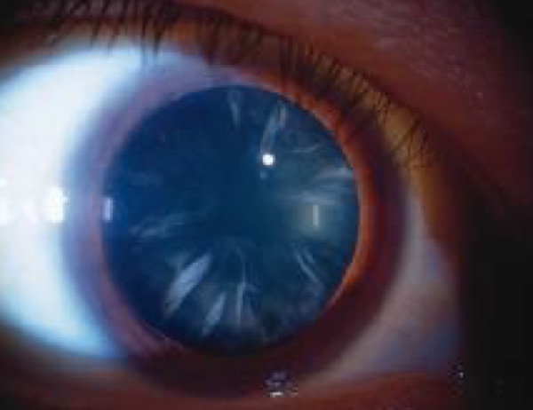
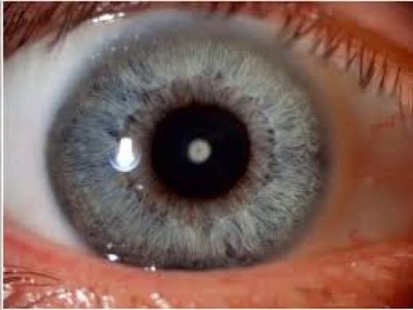
Lens proteins break down and clump, clouding the lens. Beck's image: like looking through a foggy window. Aging is the commonest cause overall; the commonest pediatric cause is idiopathic. Add diabetes, corticosteroid use, smoking and ultraviolet exposure.
| Acquired — nuclear | Brown discoloration of the central lens. Blurs distance more than near — a myopic shift, which is why some patients find they can read without glasses again. |
|---|---|
| Acquired — posterior subcapsular | Plate-like opacity at the back of the lens; slit lamp shows a dark shadow against the red reflex. Glare and trouble reading, and characteristically better after dilation. More rapid onset, classically under 50, and typically on corticosteroids or diabetic. |
| Acquired — cortical | Radial, spoke-like opacities in the periphery. Asymptomatic until it reaches the center, at which point glare is the most common complaint. |
| Pediatric | Zonular is the commonest — white opacity surrounding the nucleus. Polar is less common but central, so it is caught more easily. Also nuclear, and posterior lenticonus. |
| Signs | Early: slit lamp. Advanced: loss of the red reflex, leukocoria, a pupil that looks gray or white. A cataract alone does NOT cause a relative afferent pupillary defect — if there is one, look further. |
| Management | Non-urgent referral unless secondary to uveitis or glaucoma. Surgery is the only treatment and is deferred until it interferes with daily activities — except in a neonate, where it is done early to prevent amblyopia, or where the cataract blocks diagnosis or treatment of another eye disease. |
| Prognosis | Recovery is prompt and near-complete in 99% of eyes with isolated cataract. Complications are infrequent: retinal detachment in a myopic eye, and posterior capsule opacification — the “cataract came back” — treated with a YAG laser. |
Beck's test of whether to operate when glaucoma coexists: yes — because you have to see past the lens to assess the nerve behind it.
Also tested
- Timing of cataract surgery. Surgery can be deferred until the cataract affects daily life, unless another eye disease forces the issue.
- Cataract referral urgency. A cataract with uveitis in the same eye needs urgent referral, whereas an isolated cataract is a routine referral.
- Recovery after cataract surgery. Recovery is prompt and near-complete in almost all isolated cases, with complications infrequent overall.
- Improved near vision with nuclear cataract. The lens shifts toward myopia, so suddenly reading without glasses is explained, because nuclear change blurs distance more than near.
- Absent red reflex in an infant. It demands urgent ophthalmology referral, because cataract and retinoblastoma both present this way.
- Age and amblyopia treatment. Treatment works less well as maturity approaches; the earlier amblyopia is treated, the better the outcome.
- Manifest misalignment in a child. An eye that turns inward, with the other eye moving to take up fixation on the cover test, needs referral to ophthalmology to prevent amblyopia.
- Deciding on cataract surgery. The decision is driven by the effect on daily functioning, that is, symptoms and function, rather than by an acuity threshold alone.
- Improved near vision in cataract. A myopic shift from nuclear sclerosis ('second sight') explains it: the hardening lens increases its refractive power.
- Cataract that blocks the retinal exam. Surgery is needed to see behind it, because the cataract blocks diagnosis of other eye disease.
- Posterior vitreous detachment. Cataract surgery, myopia and ocular trauma all make it more likely.
4.8 · The blurry-vision differential
A single framework for everything that clouds vision slowly, and what separates each.
| Corneal opacity | Scarring from trauma or infection. On the cornea, not the lens. |
|---|---|
| Refractive error | Corrects fully with lenses. |
| Posterior capsule opacification | Post-operative, treated with YAG laser. |
| Macular degeneration | Central loss with distortion; the lens stays clear. |
| Glaucoma | Peripheral loss; may coexist with cataract. |
| Diabetic retinopathy | Retinal hemorrhages and exudates — retinal, not lenticular. |
| Uveitis | Pain, redness, photophobia; can cause a secondary cataract. |
| Optic neuropathy | Reduced acuity and color vision, with a normal lens. The only one here that takes color — think of it first when a patient reports colors washing out. |
| Functional visual loss | Normal examination, normal pressure, clear lens, and symptoms that do not fit anatomy. |
Also tested
- Treatment of cataract. Surgery to replace the lens, deferred until the cataract interferes with daily activities.
- Posterior capsule opacification. Vision clouds again after cataract surgery because residual lens epithelial cells cloud the capsule.
4.9 · Retinoblastoma
What these look like
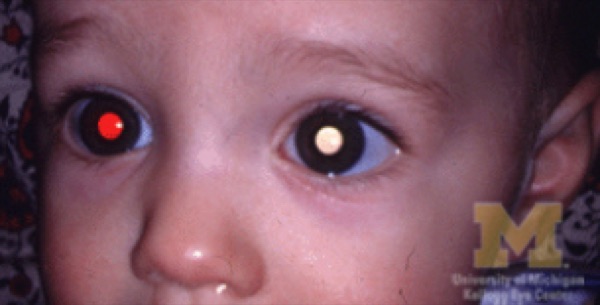
Rare, and almost exclusively in young children. A genetic mutation that is often recessive, so a clean family history does not reassure.
| Presentation | Poor vision or a turned eye. The sign is leukocoria — and the classic story is a parent noticing one white pupil and one red one in a photograph. |
|---|---|
| Diagnosis | Dilated examination plus imaging. No biopsy — pathology is not needed and sampling risks seeding the tumor through the local tissue. |
| Primary care role | Check a red reflex in every infant and young child, and screen for subnormal vision. Refer suspicious cases within weeks — and chase it. |
| Family history | Referred to an ophthalmologist experienced in retinoblastoma, or an ocular oncologist, within the first eight weeks of life, even with a normal red reflex. |
| Prognosis | Untreated, close to 100% fatal. Treated, over 95% five-year survival. Metastasis usually appears within a year if untreated, and no recurrence at five years counts as cured — unusual language for a cancer. |
Also tested
- Delayed appointment for suspected retinoblastoma. Call the specialist and get the appointment brought forward, and document the request if they will not.
- Untreated retinoblastoma. It is almost always fatal, which is why referral cannot wait.
- Biopsy in suspected retinoblastoma. Biopsy is avoided because it risks seeding tumor cells, so diagnosis rests on examination and imaging.
- Presentation of retinoblastoma. The two most common presenting features are leukocoria (a white pupil) and strabismus (a squint).
- Bilateral retinoblastoma. It implies a heritable germline mutation, with implications for siblings and for second malignancies.
4.10 · Uveal melanoma, nevus and conjunctival melanoma
What these look like
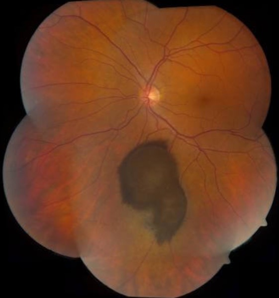
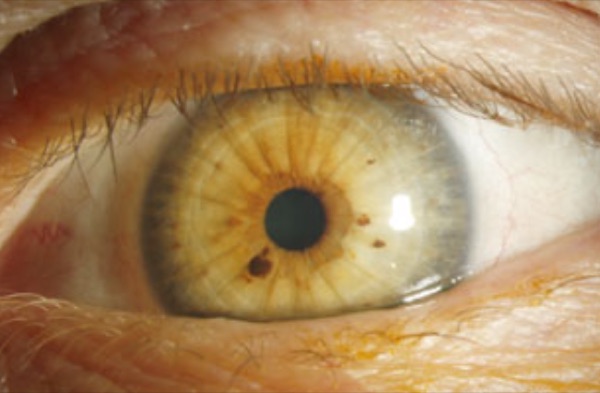
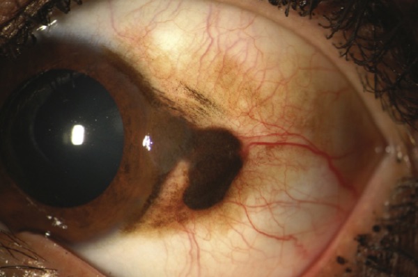
Uveal melanoma is the most common eye cancer in adults, arising from melanocytes of the choroid, ciliary body or iris. It is a distinct entity from cutaneous melanoma and from conjunctival melanoma.
| Symptoms | Typically none — most are found incidentally on routine surveillance. |
|---|---|
| Iris melanoma signs | Slow-growing dark brown or translucent mass on the anterior iris, usually the inferior half, usually unilateral, over 3 mm at the base and over 1 mm deep, with a prominent feeder vessel. May pull the pupil out of shape (corectopia). |
| Nevus, by contrast | Apparent around puberty, asymptomatic, inferior iris, typically does not grow, flat or minimally elevated, uncommonly over 3 mm, and not vascular. |
| The discriminator | A cancer builds its own blood supply; a freckle does not. Growth, size and vascularity are what separate them. |
| Diagnosis | History, ophthalmic examination and imaging. Fine needle aspiration is for molecular prognostic testing and risk stratification, not to make the diagnosis. |
| Treatment | Radiation therapy is now the commonest, with enucleation less often needed — the eye is saved where possible. |
| Prognosis | Local control is usually effective, but many have already metastasised at diagnosis. The liver is the most common site. Ten-year mortality around 32% overall, but iris melanoma only 4–10% — because it is visible, and therefore found earlier. |
| Conjunctival melanoma | Raised and vascular, against a conjunctival nevus that is flat with clear cysts. Differentials include primary acquired melanosis (flat, patchy, can be premalignant) and racial melanosis (bilateral and symmetric). |
| Lisch nodules | Tan, bilateral, multifocal, non-progressive — and a pointer to neurofibromatosis type 1, alongside café-au-lait spots. |
How Beck says to raise it with a patient: ask whether the freckle has always been there, then — without alarming them — send them to ophthalmology so its size can be documented and watched. Most are exactly what they look like.
Also tested
- Iris lesion size concerning for melanoma. A lesion over 3 mm across and over 1 mm deep raises concern for melanoma; a nevus is usually smaller and flat.
- Uveal melanoma outlook by site. Iris melanoma carries a better outlook than ciliary body melanoma, with roughly 4 to 10 percent mortality for the iris against far worse for the ciliary body.
- Growing vascular iris lesion. A raised pigmented iris lesion with a feeder vessel that has grown raises real concern and warrants referral to ophthalmology, likely ocular oncology.
- Ciliary body versus iris melanoma. Ciliary body melanoma does worse because it is hidden and found later, whereas an iris lesion is visible and caught earlier.
- Stable iris nevus. The risk of transformation to cancer is low, but surveillance continues because the risk is not zero.
- Enlarging vascular conjunctival lesion. A raised pigmented conjunctival lesion that has enlarged and has its own feeding vessels needs urgent referral for biopsy, since growth and intrinsic vascularity are the concerning features.
- Stable flat pigmented iris lesion. A lesion unchanged on photographs over five years, with no pupil distortion, is managed with photographic monitoring, as documented stability supports a benign nevus.
- Treated choroidal melanoma follow-up. Surveillance for liver metastasis, because the liver is where it goes.
- Raised pigmented choroidal lesion. With orange pigment and associated subretinal fluid, the next step is urgent ophthalmology referral; orange pigment, thickness and subretinal fluid separate melanoma from a benign nevus.
- Small flat uniformly pigmented choroidal lesion. With drusen on its surface and no subretinal fluid, management is photographic surveillance; flatness, drusen and absent fluid all favor a benign nevus.
- Iris freckles. Flat, multifocal, bilateral surface pigment on the irides, with no stromal involvement.
4.11 · The differentials — what else looks like this
Each tumor and each macular disease comes with a list of mimics, and a vignette is far more likely to ask you to separate them than to name the obvious one.
Also tested
- Small flat pigmented iris spot. A flat, 2 mm, avascular pigmented iris spot is likely benign but should be referred to ophthalmology to document and monitor, as a baseline is needed.
- Enlarging pigmented conjunctival lesion. Refer to ophthalmology for assessment, because growth in a pigmented lesion demands specialist review.
- Iris nevus follow-up. Surveillance with dilated review is more frequent at first, then annual; the early checks confirm the nevus is not growing.
- Ocular trauma red flag. A relative afferent pupillary defect converts a routine review into an emergency referral, because it indicates optic nerve compromise.
Macular dystrophies that mimic age-related macular degeneration
| Condition | What gives it away |
|---|---|
| Stargardt disease (late-onset) | Inherited macular dystrophy with yellow-white flecks and central vision loss |
| Sorsby fundus dystrophy | Autosomal dominant; choroidal neovascularization that looks like wet degeneration |
| North Carolina macular dystrophy | Congenital and non-progressive macular changes resembling degeneration |
| Best disease (vitelliform) | Lipofuscin accumulation mimics drusen, but presents earlier in life |
The common thread: all four are inherited and most present younger than age-related degeneration. A macular picture in a patient too young for it is the cue.
Leukocoria — what else besides retinoblastoma
| Condition | What gives it away |
|---|---|
| Coats' disease | Idiopathic retinal telangiectasia with exudation, often unilateral |
| Persistent fetal vasculature | Congenital anomaly with remnants of fetal vasculature, typically unilateral |
| Toxocariasis | Parasitic infection causing granulomatous retinal inflammation |
| Retinal astrocytoma | Benign glial tumor, often with tuberous sclerosis |
| Medulloepithelioma | Rare tumor of the nonpigmented ciliary epithelium |
| Congenital cataract | Produces a white pupillary reflex without any tumor |
| Ocular toxoplasmosis | Retinal scarring and inflammation |
Uveal melanoma by site
Uveal melanoma is named for where in the uvea it sits — iris, ciliary body or choroid — and an iris melanoma may be melanotic or partly amelanotic, so pigment is not required. Two signs point to a ciliary body lesion in particular: a sentinel vessel (a dilated episcleral vessel overlying the tumor) and extrascleral extension.
Iris nevus, and the pigmented iris differential
An iris nevus is a small pigmented, benign spot, usually apparent around puberty and asymptomatic. It sits in the inferior half of the iris, does not typically grow, is flat or minimally elevated (under 1 mm) and uncommonly over 3 mm across, and is usually not vascular. It may pull the pupil out of shape — corectopia.
| Lesion | What separates it |
|---|---|
| Iris freckle | Flat, superficial pigment; no stromal involvement or distortion; usually bilateral and multifocal |
| Lisch nodules | Tan nodules of neurofibromatosis type 1; bilateral, multifocal, non-progressive |
| Melanocytoma | Dark brown, granular; usually benign but can cause secondary glaucoma through pigment dispersion |
| Melanocytosis | Congenital uveal pigmentation, sectoral or diffuse; raises the risk of uveal melanoma |
| Cogan-Reese (ICE) syndrome | Pigmented iris nodules with corneal endothelial abnormality; secondary glaucoma |
| Iris melanoma | Nodular or diffuse growth; may show vascularity, ectropion uveae, sectoral cataract or seeding |
| Metastatic carcinoma | Secondary deposit rather than a primary iris lesion |
Risk factors that push an iris lesion toward melanoma: inferior location, diffuse configuration, and blood in the anterior chamber.
Non-pigmented conjunctival lesions
Conjunctival melanoma is pigmented, so the useful differential is the lesions that are not.
| Lesion | What gives it away |
|---|---|
| Squamous cell carcinoma | Gelatinous or leukoplakic; can be mistaken for an amelanotic melanoma |
| Conjunctival lymphoma | Salmon-pink, painless mass, typically in the fornix or bulbar conjunctiva |
| Kaposi sarcoma | Reddish-purple vascular lesion, associated with HIV/AIDS |
| Pyogenic granuloma | Rapidly growing red mass, often after trauma or surgery |
First-line treatment
Every condition in this lecture, with what you reach for first and how fast — which in ophthalmology is half the answer. Where the deck escalates after a failed trial, only the first step is here; the full ladder and the reasoning are in the comparison chart.
| Condition | First line | How fast |
|---|---|---|
| Idiopathic intracranial hypertension | Acetazolamide promptly, plus a supervised weight reduction program. Surgical CSF diversion (optic nerve sheath fenestration or shunt) only if medical therapy fails. | Urgent |
| Age-related macular degeneration — dry | STOP SMOKING. AREDS2 supplement: vitamins C and E, zinc, copper, lutein, zeaxanthin. | Routine |
| Age-related macular degeneration — wet | Intravitreal anti-VEGF injection, thermal laser photocoagulation, photodynamic therapy. | Same day |
| Cataract — nuclear | Surgery is the only treatment — lens extraction with implant, deferred until it interferes with daily activities. | Routine |
| Cataract — cortical | Lens extraction when it interferes with function. | Routine |
| Cataract — posterior subcapsular | Lens extraction. Review the steroid burden where that is the cause. | Routine |
| Cataract — pediatric | Surgery is NOT deferred in a neonate — it is done early to prevent amblyopia. | Emergent |
| Myopia | Concave (negative diopter) lens — scatters light and moves focus back onto the retina. | Routine |
| Hyperopia | Convex (positive diopter) lens — converges light forward onto the retina. | Routine |
| Astigmatism | Toric lens. | Routine |
| Strabismus | Treat the cause; ophthalmology referral. Untreated in a child it causes amblyopia. | Urgent |
| Amblyopia | Patch or atropinise the GOOD eye to force the weaker one to work. Treat the underlying cause. | Urgent |
| Retinoblastoma | Multimodal therapy under ocular oncology, with genetic counseling. Enucleation for large tumors. | Emergent |
| Uveal melanoma | Radiation therapy is now the commonest treatment; enucleation is less often needed. | Urgent |
| Iris nevus | Surveillance. More frequent initially to establish it is not growing, then annual dilated review. | Routine |
| Conjunctival melanoma | Specialist management under ocular oncology. | Urgent |
5 · Ocular Trauma
Instructional Objectives
OPHTHALMOLOGY — Ocular Trauma
- Compare and contrast the etiologies, epidemiology, risk factors, clinical
manifestations, differential diagnosis, diagnostic testing (including ordering and
interpretation), management (acute and chronic, including applicable rehabilitative and
palliative care), appropriate referrals, patient education, and prognosis for ocular
trauma:
- Foreign body
- Corneal abrasions
- Globe rupture
- Hyphema
- Lid lacerations
- Orbital and lid contusion
- Periorbital hematoma
- Retinal and vitreous detachments
- Blowout fractures
- Orbital fracture
- Basilar skull fracture
- Identify medical care strategies for common ophthalmological disorders in the lecture topic list for the following populations: 1. infant · 2. child · 3. adolescent · 4. adult · 5. elderly
Before the eye, the patient. ABCs first — airway patent, breathing adequate, circulation present — then vital signs, neurological evaluation and the rest. An injured eye usually arrives attached to major trauma involving the brain, and the eyes may be assessed second. Always reason from the anatomy to what else could be damaged.
One point she added aloud that is not on a slide: the Glasgow Coma Scale runs from 3 to 15, not from 0. Three is the floor because each of its three components scores a minimum of one.
5.1 · The four rules that apply before any diagnosis
These come first because breaking one of them makes the injury worse.
| Rule | Why |
|---|---|
| DO NOT REMOVE a penetrating object | It may be tamponading a wound. Removal in the emergency department can extrude intraocular contents. |
| CT orbit — never MRI | If the object is metallic, a magnetic field will move it through the eye. |
| NEVER dilate the eye when ocular trauma is suspected | It removes the pupil examination, which is one of the few windows onto what is happening inside. |
| Check tetanus status | Any penetration with metal or organic material — wood, leaf, dirt — carries the risk. |
When is imaging automatic? Any loss of consciousness from trauma or alcohol, confusion, tachypnea, apneic breathing, anticoagulant use, or eye penetration → CT without contrast.
Epidemiology worth carrying: ocular trauma is the leading cause of monocular blindness in young adult men in the United States.
Also tested
- Embedded object in the eye. Leave it in place and arrange surgical repair, because it may be tamponading the wound and removal risks extruding intraocular contents.
- Ocular trauma. Dilating the pupil must be avoided in any suspected ocular trauma, because it removes the pupil examination, one of the few windows onto the inside of the eye.
- Ocular trauma epidemiology. Ocular trauma is the leading cause of monocular blindness in young adult men in the United States, so eye protection matters most for them.
- Possible intraocular metal fragment. Computed tomography of the orbit is ordered, since it answers the question without moving the fragment.
- Lid laceration with globe risk. The globe beneath is assessed before any lid repair, since two thirds of full-thickness lid lacerations have an accompanying globe injury.
- Automatic imaging after facial trauma. Anticoagulation (such as warfarin) and confusion both make imaging automatic; computed tomography without contrast is the choice.
- Periorbital penetrating injury. Penetration with metal or organic material carries tetanus risk, so confirm that tetanus immunization is up to date.
5.2 · Assessing the patient
Establish the circumstance: who, what, where, when, and if you can, why. Blunt or sharp? High or low velocity? Prior ocular trauma? Nil-by-mouth status — what and when they last ate and drank — because surgery may follow. If the patient is intoxicated or has an altered level of consciousness, the history comes from family, friends or bystanders.
The ophthalmic examination is done without causing further damage and depends on how cooperative the patient is: inspect the eyes and periorbital tissues for lacerations, ecchymosis, proptosis, corneal clouding and hyphema; assess pupil size, shape and response to direct and consensual light; and if the patient is conscious and cooperative, test visual acuity and confrontation fields. Consult ophthalmology for anything needing further evaluation.
A practical aside: if no lid retractor is to hand, an unfolded paper clip bent with a hemostat will retract a lid.
Also tested
- Mechanism in ocular trauma. Whether the injury was blunt or sharp, and high or low velocity, most changes the level of concern, because it predicts the injury pattern.
5.3 · Open globe injury
A full-thickness defect in cornea and/or sclera, so the intraocular compartments are open to the outside. It splits into full-thickness eye wall laceration and globe rupture.
The signs to recognize: pupillary distortion, usually toward the wound; a flat anterior chamber; extraocular protrusion of uveal tissue; massive hemorrhagic chemosis; a soft eye; deep eyelid laceration; and intraocular blood as hyphema or vitreous hemorrhage.
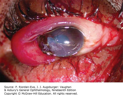
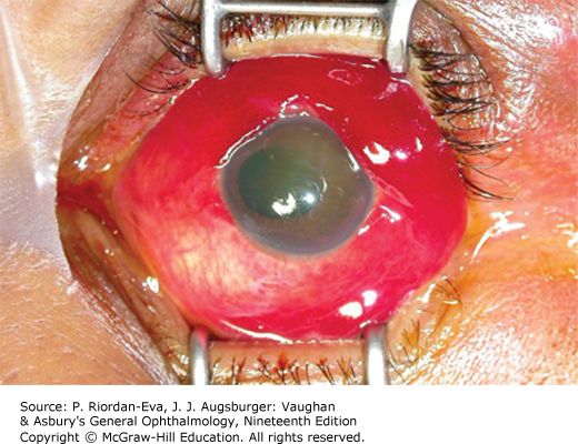
| Full-thickness eye wall laceration | Globe rupture | |
|---|---|---|
| Mechanism | Sharp object or high-velocity projectile — fishing hook, knife | Severe blunt force — airbag, fist, baseball |
| What happens | Cuts clean through cornea, sclera or both. The object may have been withdrawn, may be retained, or may have passed through with entry and exit wounds | Splits or tears at a weak point: posterior to the extraocular muscle insertions (especially superonasal), old surgical incisions, the lamina cribrosa |
| Extra | A cut lens capsule leaves the lens hydrated, edematous and opaque; a fragment can extrude forward and inflame the anterior chamber. Lensectomy is required but often deferred. Posterior foreign bodies are left alone initially | Suspect it whenever blunt trauma gives massive hemorrhagic chemosis or a soft eye |
The moment an open globe is suspected: a rigid protective shield taped over the eye, ophthalmology called immediately, antiemetics and analgesia so the patient does not strain or vomit, tetanus, and CT to exclude a foreign body. Then surgical repair.
Also tested
- Suspected globe rupture. Immediate management is a rigid shield, nil by mouth, antiemetics and urgent referral; avoid anything that raises intraocular pressure and apply no pressure to the eye.
- Deferring lensectomy after full-thickness eye wall laceration. It is often deferred to let the hyphema and inflammation settle and to measure accurately for an implant.
- Antiemetics in suspected open globe. They are given alongside analgesia because vomiting raises intraocular pressure; keeping the patient from straining protects the eye.
- Suspected open globe. Alongside the shield, give antiemetics, analgesia and tetanus, and obtain computed tomography; keeping the patient from straining protects the eye.
- Lensectomy timing in open globe injury. Lensectomy for a cut lens capsule is required but usually deferred from the globe repair, being performed later once hyphema and inflammation settle.
- Globe rupture. A soft eye with massive hemorrhagic chemosis after blunt force is a globe rupture until proven otherwise; shield the eye and call ophthalmology.
- Globe rupture sites. Ruptures commonly occur behind the muscle insertions, especially superonasally, and also along incisions from earlier intraocular surgery and at the lamina cribrosa.
- Corneal laceration with iris prolapse. Do not attempt repositing the prolapsed iris; manipulation risks further extrusion and infection. Shield the eye and refer in the emergency department.
- Globe rupture risk. Previous intraocular surgery makes rupture more likely at lower force, because old incisions leave a permanent weak point.
- Lens capsule laceration. The lens becomes hydrated, edematous and opaque, as aqueous entering the capsule clouds it.
- Globe rupture. A peaked, teardrop-shaped pupil points toward the rupture site, and the anterior chamber deepens with a posterior rupture.
5.4 · Corneal abrasion and foreign body
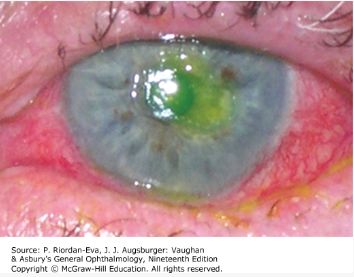
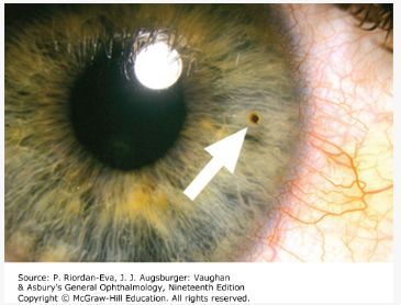
Corneal abrasion — scraped corneal epithelium, one of the commonest ocular injuries, typically from a fingernail or handling a contact lens. Severe foreign body sensation, tearing, photophobia, blurred vision. Slit lamp with fluorescein stains the exposed basement membrane and shows the extent. Treat with a topical broad-spectrum antibacterial; no patch (slide 18 says patching may ease pain, but it neither speeds healing nor relieves pain); re-examine to confirm healing.
Never give a patient topical anesthetic drops to take home. They delay healing, mask worsening symptoms, and can cause a corneal ulcer. This is the single most testable instruction in the section.
Foreign body — an object with too little momentum to pass through the eye wall lodges in cornea or conjunctiva. The history is grinding or striking metal. Linear vertical corneal epithelial defects mean the object is in the tarsal conjunctiva of the UPPER lid, so evert the lid and look. Removal: topical anesthetic, slit lamp, sterile 27-gauge needle; a rust ring from iron or copper comes out with a battery-operated burr. Then broad-spectrum antibiotic and abrasion care. Refer if the object may have passed through the cornea — that is an open globe.
Also tested
- Corneal abrasion follow-up. Periodic re-examination is required, alongside a topical broad-spectrum antibacterial, to confirm healing and exclude infection.
- Uncomplicated corneal abrasion. A well-defined epithelial defect with no infiltrate is treated with topical antibiotic ointment and analgesia, with follow-up to confirm healing.
- Numbing drops for corneal abrasion. They should not be given to take home, because repeated use is toxic and delays healing, and it masks worsening infection.
- Rust ring. A rust ring remaining after removal of a metallic foreign body is removed with a burr tip drill.
- Conjunctival foreign body. One visible on diffuse light examination is removed with a cotton-tipped applicator, since a conjunctival object lifts off with a swab.
- Corneal abrasion at home. Give analgesia and a topical antibiotic; take-home anesthetic delays healing, masks worsening symptoms and can cause a corneal ulcer.
- Metallic corneal foreign body with rust ring. Remove it and the rust ring, then give antibiotics. The rust ring is removed too, as it perpetuates inflammation.
- After removing a superficial corneal foreign body. Check that there is no deeper injury, because a superficial fragment can accompany a penetrating one.
- Vertical linear corneal scratches. Evert the upper lid to find the object; vertical linear defects mean it is in the tarsal conjunctiva of the upper lid, scraping with each blink.
- Corneal rust ring. A rust ring from iron or copper is removed with a battery-operated burr.
- Corneal abrasion that worsens. Worsening pain and a white spot on the cornea mean a corneal ulcer has developed. This is why abrasions are re-examined, and why anesthetic drops are never sent home.
- Corneal foreign body. Photophobia, tearing and foreign body sensation with nothing on diffuse light call for slit lamp examination as the next step: larger objects show on diffuse light, smaller ones need the slit lamp.
5.5 · Hyphema
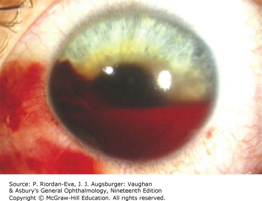
Blood in the anterior chamber from injured vessels, after blunt or penetrating trauma. It can itself be a sign of open globe. Blurred vision, eye pain, photophobia. Diagnosed on diffuse light for a gross hyphema, then slit lamp and full ophthalmic examination.
Measure the intraocular pressure — unless a penetrating globe injury is suspected — and treat it if high, with beta blockers, pilocarpine, acetazolamide and osmotic agents if needed.
The whole of management aims at preventing a rebleed: bed rest with the head of the bed slightly elevated, antiemetics, ocular hypotensives, topical or oral corticosteroids, cycloplegic drops (atropine, homatropine, scopolamine) to rest the ciliary body, and oral aminocaproic acid, an antifibrinolytic that slows clot breakdown.
Most rebleeding happens in the first 72 hours, and a secondary hemorrhage is what costs the vision permanently. Avoid aspirin and antiplatelet drugs. Risk is increased in sickle cell disease.
Also tested
- Hyphema instructions. Strenuous activity and bending must be avoided, as both raise the risk of rebleeding.
- Hyphema management. Use upright positioning, a rigid shield and urgent ophthalmology review; keeping the head up lets the blood settle inferiorly and clear the visual axis.
- Hyphema with sickle cell disease. Sickle cell anemia raises the risk of complications in hyphema, where a secondary hemorrhage can cause permanent visual loss.
- Hyphema screening. Sickle cell testing is particularly important in a patient of African ancestry; sickling in the anterior chamber obstructs outflow and raises pressure at lower blood volumes.
- Hyphema. A layer of blood with a fluid level in the anterior chamber is hyphema, and it can itself indicate an open globe; it is not always an isolated injury.
- Hyphema discharge instructions. Avoid aspirin, antiplatelets and straining, since both raise the risk of a secondary hemorrhage.
- Complication of traumatic hyphema. Patients are monitored for rebleeding with a rise in intraocular pressure; the rebleed risk peaks in the first three to five days.
5.6 · Lid lacerations, contusion and periorbital hematoma
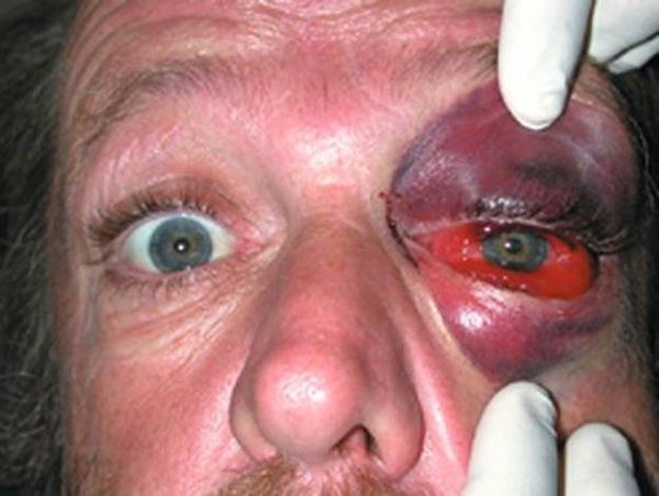
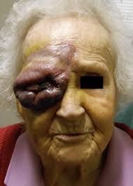
Lid lacerations. Consult ophthalmology for any laceration that involves the lid margin, sits within 6 to 8 mm of the medial canthus, involves the lacrimal duct or sac, involves the inner lid surface, is associated with ptosis, or involves the tarsal plate or levator palpebrae.
- Full-thickness — comes with a corneal laceration or globe rupture in about two thirds of cases. Always look underneath.
- Partial-thickness — anything not meeting the criteria above. Can be repaired in the emergency department, with ophthalmology follow-up in 2 to 3 days.
A deep laceration to the medial third may transect the canalicular system, and if that is not repaired properly the patient has chronic tearing for good. Facial lacerations may be left open for 24 hours before closure, because the face is so well vascularized.
Orbital contusion is soft tissue swelling without hemorrhage. The tarsal plate and septal margin act as a wall holding blood in the anterior tissues, so it presents as preseptal ecchymosis or hematoma. Management is supportive through to surgery depending on the patient — and rule out brain trauma.
Periorbital hematoma is bleeding within the bony orbit. It is not always traumatic: orbit and eyelid surgery, peribulbar injections, orbital varices, lymphangiomas and arteriovenous malformations, anticoagulants, sickle cell disease, orbital pseudotumour and idiopathic causes all produce it. Management is canthotomy with cantholysis — exposing the lateral canthal tendon and cutting its inferior branch — to let the blood out.
Also tested
- Lid margin laceration. A laceration through the lid margin needs specialist repair with meticulous approximation, because a poor repair leaves a permanent notch and chronic irritation.
- Lid laceration near the medial canthus. The canalicular drainage may be cut, and a missed canalicular injury leaves the person tearing for life.
- Simple orbital contusion. When vision, pupils, eye movements and the globe are all normal, management is supportive: ice, analgesia and review if symptoms change.
- Delayed facial laceration closure. A facial laceration may be left open for 24 hours before closure because the face is highly vascular, which is what permits the delay.
- Partial-thickness lid laceration. One meeting none of the referral criteria is repaired at the initial site, with ophthalmology follow-up in 2 to 3 days; it does not need the specialist to close it.
- Periorbital swelling and ecchymosis after trauma. Beyond supportive care, brain trauma should be excluded, because the force that bruised the orbit reaches the brain.
- Full-thickness lid laceration. Beyond repairing the lid, the globe underneath must be examined, because about two thirds of these lacerations come with a globe injury.
- Lid lacerations repairable in the emergency department. A superficial skin cut sparing the margin can be repaired there; partial-thickness lacerations meeting none of the referral criteria are repaired in the emergency department.
- Black eye after a punch. A relative afferent pupillary defect makes it more than a simple contusion; it indicates optic nerve involvement and changes the urgency entirely.
- Orbital contusion versus periorbital hematoma. Contusion has no hemorrhage; the hematoma bleeds in the orbit. The septum is what keeps the contusion in front of the eye.
- Lid laceration referral. A laceration through the lid margin goes to ophthalmology, because margin involvement is one of the explicit referral criteria.
- Orbital and lid contusion. Lid bruising and swelling with normal vision, equal reactive pupils and full eye movements indicate soft tissue injury with normal ocular function.
- Orbital contusion. Blunt injury fills the surface with hemorrhage and swells the lids, with no open wound.
5.7 · Retinal and vitreous detachment
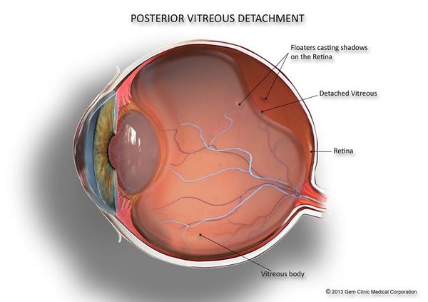
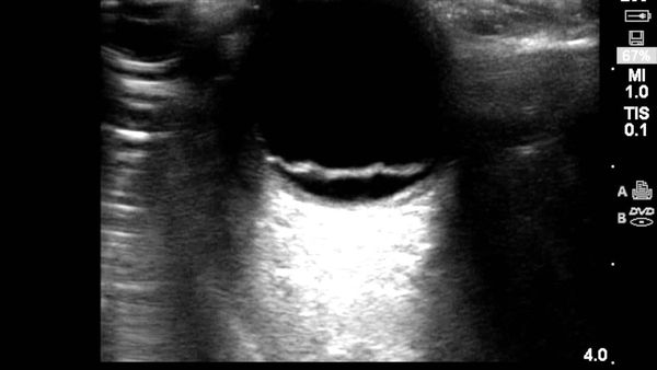
The presentation is the same one that appears in section 3: a shadow or curtain descending over the eye, cloudy or smoky vision, floaters and momentary flashes of light, a monocular field defect, and central acuity dropping once the macula is involved. Diagnosis is history plus a dilated eye examination. The patient must be seen by ophthalmology within 24 hours.
What Lecture 14 adds is the three types, which do not share a treatment.
| Type | Mechanism | Management |
|---|---|---|
| Rhegmatogenous most common |
One or more full-thickness breaks in the sensory retina, vitreous traction, and liquefied vitreous passing into the subretinal space. Usually preceded by posterior vitreous detachment. Mostly a spontaneous age-related event, brought forward by myopia, cataract surgery or trauma | Surgical. Ophthalmology STAT, pain control, antiemetics, head of bed at 30–40 degrees |
| Traction | Most commonly proliferative diabetic retinopathy. More localized and concave; begins along the vasculature then spreads to retina and macula | Surgical |
| Exudative (serous) | Neither a retinal break nor traction. Associated with systemic vascular or inflammatory disease, or an intraocular tumor | Manage the underlying condition |
Also tested
- Macular involvement in retinal detachment. Field defects come first; a drop in central visual acuity indicates that the macula has become involved.
- Retinal detachment urgency. A fresh detachment must be seen by an ophthalmologist within 24 hours; the referral is immediate.
- Retinal detachment presentation. Flashes of light, a shower of floaters and a curtain moving across the vision of one eye, with myopia as the risk factor, suggest rhegmatogenous retinal detachment.
- Rhegmatogenous retinal detachment. The most common type of retinal detachment: full-thickness retinal breaks let liquefied vitreous behind the retina.
5.8 · Orbital floor (blowout) fracture
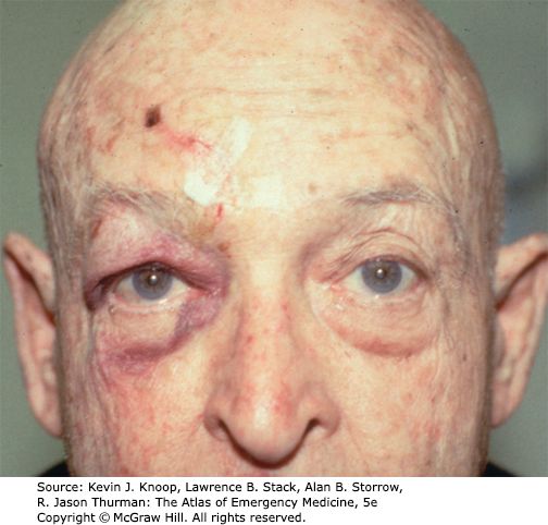
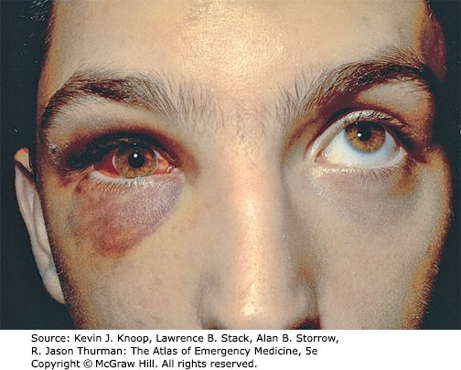
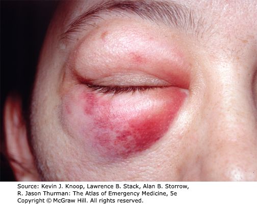
Two mechanisms. A true blowout, where a blunt object — a fist, a ball — transmits energy to the globe, raising orbital pressure until the floor (most often) or medial wall gives way. Or force transmitted to the infraorbital rim, buckling the floor.
Findings: periorbital ecchymosis and lid edema; chemosis, subconjunctival hemorrhage, and infraorbital numbness from injury to the infraorbital nerve; corneal abrasion, hyphema, enophthalmos, proptosis, iridoplegia, dislocated lens, retinal tear or detachment, ruptured globe; and periorbital subcutaneous emphysema.
The gaze rule. Diplopia on UPWARD gaze means entrapment of the inferior rectus or its supporting structures. Diplopia on LATERAL gaze means entrapment of the medial rectus. Entrapment also brings severe pain and autonomic disturbance — bradycardia and vomiting on attempted eye movement.
In children, an entrapped muscle may come with no orbital soft tissue signs at all — the “white-eyed blowout”. A quiet-looking eye does not exclude it.
Diagnosis: CT of the orbits and midface. She gave the reason the midface is included: “to make sure there aren’t any additional structures that are also fractured … if their orbital floor is fractured, sure, okay, but we want to make sure we’re not missing any other fractures.” Management runs by severity:
- No eye injury or entrapment — ice and analgesia, follow up in 2 to 3 days.
- Blood in the maxillary sinus — antibiotics, and note that they are PROPHYLACTIC. Her reasoning: the eye is sterile, so blood in the sinus means the sinus has been disrupted and now has a portal of entry. “It’s not that we’re treating anything … it’s to make sure that it doesn’t become infected.”
- True blowout fracture — ophthalmology, because 30% sustain a significant globe injury.
- Muscle entrapment — facial trauma surgeon STAT, because a compromised blood supply means muscle necrosis.
If the optic nerve is already damaged, that damage is unlikely to improve and surgery may worsen it. Where surgery is planned, it is often delayed 1 to 2 weeks to let orbital swelling settle, so intraorbital pressure during the operation is lower.
Also tested
- Blowout fracture with entrapment. Urgent surgical referral is required because entrapped muscle becomes ischemic and fibroses, and delay risks permanent restriction of eye movement.
- Orbital floor fracture without entrapment or ocular injury. Management is ice, analgesia and review in two to three days.
- Orbital floor fracture. Patients are advised not to blow the nose because it forces sinus air into the tissues; periorbital subcutaneous emphysema is a recognized feature of this fracture.
- Suspected orbital fracture. Computed tomography of the orbits and midface is the imaging study for this fracture.
- Orbital floor fracture with inferior rectus entrapment. A facial trauma surgeon is contacted urgently, because an entrapped muscle can necrose if its blood supply is compromised.
5.9 · Basilar skull fracture
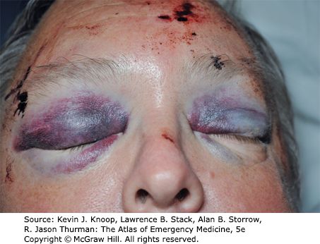
A linear fracture of the skull base — cribriform plate of the ethmoid, orbital plate of the frontal, petrous or squamous temporal, sphenoid, or occipital bone. Trauma there often produces no symptoms of its own, so it is found on indirect signs:
- Visible bleeding from the fracture into the soft tissue at the base of the head
- Raccoon eyes (periorbital ecchymosis) and Battle sign (retroauricular ecchymosis)
- Bleeding into the middle ear or sphenoid sinus; hemotympanum
- Cerebrospinal fluid leak — clear or pink rhinorrhea
Two bedside tests for cerebrospinal fluid. A dextrose stick may be positive. And fluid placed on filter paper — or found on the bedsheet — shows a halo or double ring sign: an inner ring of blood with an outer ring of cerebrospinal fluid.
How to actually do it. Hold a bedsheet, paper or tissue under the nostril, let the drip fall onto it, and watch as it dries — two rings appear, one inside the other. She was emphatic about this one: “that’s a very classic and that’s a very important sign for us to not miss.”
Diagnosis: CT orbits, though the fracture is not always evident on it. A cerebrospinal fluid leak means a neurosurgery consult and admission. Otherwise admission turns on the clinical condition, the associated injuries, and any brain injury seen on CT. Antibiotics for a cerebrospinal fluid leak are controversial, because of the risk of selecting resistant organisms.
First-line treatment
Every condition in this lecture, with what you reach for first and how fast — which in ophthalmology is half the answer. Where the deck escalates after a failed trial, only the first step is here; the full ladder and the reasoning are in the comparison chart.
| Condition | First line | How fast |
|---|---|---|
| Open globe injury | Rigid protective shield taped over the eye and ophthalmology called immediately. Antiemetics and analgesia so the patient does not strain, plus tetanus. Surgical repair. | Emergent |
| Full-thickness eye wall laceration | Surgical repair. Lensectomy is required but is often deferred, to let hyphema and inflammation settle and to measure accurately for an intraocular lens. Posterior-segment foreign bodies are left alone at first assessment. | Emergent |
| Globe rupture | Shield, ophthalmology immediately, antiemetics, analgesia, tetanus. Immediate surgical repair with wound exploration. | Emergent |
| Corneal abrasion | Topical broad-spectrum antibacterial. No patch (the slide suggests one; patching neither speeds healing nor eases pain). Re-examine periodically to confirm healing and exclude infection. | Urgent |
| Corneal or conjunctival foreign body | Topical anesthetic, then removal with a sterile 27-gauge needle. A rust ring (iron or copper) comes out with a battery-operated burr. Broad-spectrum antibiotic plus abrasion care. | Urgent |
| Hyphema | Goal is preventing a rebleed: bed rest with the head elevated, antiemetics, ocular hypotensives, topical or oral corticosteroids, cycloplegic drops, and oral aminocaproic acid to stop clot breakdown. Treat a raised pressure. | Emergent |
| Lid laceration | Ophthalmology for any of: lid margin involvement, within 6–8 mm of the medial canthus, lacrimal duct or sac, inner lid surface, associated ptosis, tarsal plate or levator. Partial-thickness can be repaired in the emergency department with ophthalmology follow-up in 2–3 days. | Emergent |
| Orbital contusion | Supportive, through to surgery depending on the patient's condition. | Urgent |
| Periorbital hematoma | Canthotomy with cantholysis — releasing the lateral canthal tendon and cutting its inferior branch — to let the blood drain. | Emergent |
| Retinal detachment — rhegmatogenous | Surgical. Ophthalmology STAT, pain control, antiemetics, head of bed at 30–40 degrees. | Emergent |
| Retinal detachment — traction | Surgical. | Emergent |
| Retinal detachment — exudative | Treat the underlying condition — not primarily a surgical problem. | Urgent |
| Orbital floor (blowout) fracture | No injury or entrapment: ice, analgesia, review in 2–3 days. Blood in the maxillary sinus: antibiotics. True blowout: ophthalmology, because 30% have a significant globe injury. Entrapment: facial trauma surgeon STAT — the muscle can necrose. | Emergent |
| Basilar skull fracture | Cerebrospinal fluid leak: neurosurgery consult and admission. Admission otherwise turns on the clinical picture, associated injuries and any brain injury on CT. | Emergent |
Also tested
- Basilar skull fracture. After head trauma, raccoon eyes, Battle sign and hemotympanum together should raise suspicion of a basilar skull fracture; they are the indirect signs.
- Basilar skull fracture. Suggested by raccoon eyes (bruising around both eyes), Battle sign (bruising behind the ear) and hemotympanum (blood behind the eardrum) together.