1 · Hypertension
Instructional Objectives
HYPERTENSION — Monique Jaquith, DMSc, PA-C
- Classify hypertension.
- Define subtypes of hypertension.
- Identify effects of hypertension on target organs (i.e. end-organ damage)
- Compare and contrast the etiologies, epidemiology, risk factors, clinical manifestations,
differential diagnosis, diagnostic testing (including ordering and interpretation), management
(acute and chronic, including applicable rehabilitative and palliative care), appropriate
referrals, patient education, and prognosis of the following hypertensive conditions:
- Primary (Essential) hypertension
- Secondary hypertension
- Hypertensive urgency
- Hypertensive emergency
- Identify medical care strategies for hypertension in the lecture topic list for the
following populations.
- infant
- child
- adolescent
- adult
- elderly
This is the first lecture of the Exam 4 block. Hypotension (section 2), atherosclerosis and lipid disorders (section 3), valvular heart disease (section 4), coronary artery disease (section 5) and heart failure (section 6) follow — every lecture of the block (Lectures 20–25) is now in this guide.
Built from the slides. The lecture was recorded in two parts on 17 September, and both parts were transcribed on 20 September. The lecturer's own emphasis has not been folded into this guide yet; until it is, everything here comes from the slides and their speaker notes.
Where each objective is answered
| Objective | Answered in |
|---|---|
| a. Classify hypertension | 1.1 · the four adult blood pressure categories |
| b. Define subtypes of hypertension | 1.1 · subtype table; resistant hypertension in 1.8 |
| c. Effects on target organs | 1.5 |
| d. a. Primary (essential) hypertension | 1.2 and 1.3, then assessment (1.6) and treatment (1.7) |
| d. b. Secondary hypertension | 1.4 |
| d. c. Hypertensive urgency | 1.9 · the deck says the term is no longer used |
| d. d. Hypertensive emergency | 1.10 |
| e. Populations | 1.11 · the deck has no strategy for infants, children or adolescents |
1.1 · Definitions and classification
The goal and the diagnostic threshold are the same number, which is what makes the national control figure directly interpretable: treat to below 130/80, with values closer to 120/80 encouraged where appropriate.
Also tested
- Hypertension prevalence. Close to half of adults (47.7 percent) have hypertension, using the threshold of 130/80 or current antihypertensive treatment.
- Hypertension control. Among adults with hypertension only about one in five (20.7 percent) achieves below 130/80; awareness and treatment figures are far higher, so the gap is between being treated and being controlled.
- Blood pressure goal. Intensify therapy when average blood pressure is above the goal of below 130/80, since values closer to 120/80 are encouraged where appropriate; the same threshold defines the condition and measures control.
Adult blood pressure categories
Four categories, applied to properly obtained average readings rather than one borderline reading. When the systolic and diastolic values fall into different categories, use the higher category: 128/78 is elevated, and 138/92 is stage 2.
| Category | Systolic (mm Hg) | Diastolic (mm Hg) | Action |
|---|---|---|---|
| Normal | <120 | <80 | Reassess in 1 year |
| Elevated | 120–129 | <80 | Lifestyle intervention |
| Stage 1 | 130–139 | 80–89 | Risk-based decision |
| Stage 2 | ≥140 | ≥90 | Lifestyle plus medication |
For stage 1, medication is started in adults with cardiovascular disease, diabetes, chronic kidney disease, or an estimated 10-year cardiovascular risk of 7.5% or more, with the PREVENT equation used to stratify that risk.
Subtypes
| Subtype | Definition |
|---|---|
| Primary (essential) | No single identifiable underlying cause. The vast majority of adult cases. |
| Secondary | An identifiable underlying condition drives the pressure. |
| Resistant | Above goal despite three complementary drugs at maximally tolerated doses. |
| Hypertensive emergency | Severe elevation with acute hypertension-mediated organ injury. |
| White coat | Raised office readings, normal out-of-office readings. |
| Masked | Normal office readings, raised out-of-office readings — and real cardiovascular risk. |
1.2 · Epidemiology
Hypertension is common, under-recognized and under-controlled: roughly one in five of those affected nationally have it controlled to goal. Prevalence rises sharply with age: 23.4% at 18–39 years, 52.5% at 40–59 years and 71.6% at 60 years and over.
1.3 · Primary (essential) hypertension
Risk factors: advancing age · family history · excess adiposity · high dietary sodium · physical inactivity · excess alcohol · dyslipidemia · insulin resistance. Low potassium intake and poor sleep belong on the modifiable list too.
1.4 · Secondary hypertension
| Cause | Mechanism | Clues | Test |
|---|---|---|---|
| Chronic kidney disease | Impaired sodium excretion expands extracellular volume; renin-angiotensin-aldosterone and sympathetic activation add to it | Requires an abnormality persisting ≥ 3 months. Relationship is bidirectional — do not assume which came first, and evaluate diabetes as an alternative cause | Creatinine with filtration rate, urine albumin-to-creatinine ratio |
| Renovascular disease | Atherosclerotic stenosis in older vascular patients, often bilateral and progressive; fibromuscular dysplasia in the young | Resistant or abrupt elevation · abdominal bruit · asymmetric kidneys · recurrent flash pulmonary edema · marked creatinine rise after starting an ACE inhibitor | Renal duplex ultrasound. Refer early for flash pulmonary edema, progressive renal decline despite optimal therapy, or truly refractory control |
| Primary aldosteronism | Autonomous aldosterone production independent of renin | Resistant pressure · unexplained hypokalemia · adrenal incidentaloma · early or familial hypertension | Morning aldosterone to renin ratio; positive above roughly 20:1 with an adequate aldosterone concentration |
| Obstructive sleep apnea | Recurrent airway obstruction → intermittent hypoxemia and arousal → sustained sympathetic activation | Loud snoring, witnessed apneas, daytime sleepiness, obesity | Polysomnography or validated home sleep apnea testing |
| Drugs and substances | Varied | Ask beyond the prescription list: over-the-counter drugs, recreational substances, licorice, herbal supplements. Excess alcohol raises pressure and withdrawal can raise it acutely | Medication review |
| Thyroid disease | Hyperthyroidism raises cardiac output; hypothyroidism raises vascular resistance | Hyper → systolic elevation · hypo → diastolic elevation | Thyroid-stimulating hormone (part of the baseline panel) |
| Cushing syndrome | Glucocorticoid excess promoting sodium retention | Proximal muscle weakness · easy bruising · thin skin · broad purple striae · central adiposity | Low-dose dexamethasone suppression test |
| Pheochromocytoma | Catecholamine excess from an adrenal medullary tumor | The five Ps: pressure, perspiration, palpitations, pallor, tremor — paroxysmal, with well intervals between | Plasma free metanephrines, or 24-hour urinary fractionated metanephrines |
| Coarctation of the aorta | Mechanical obstruction raising upper limb pressure | Radial-femoral delay; leg systolic lower than arm (normally it is higher) | Echocardiography; refer to a vascular surgeon or structural cardiologist |
Also tested
- Renal artery stenosis referral. Recurrent flash pulmonary edema, progressive renal decline despite optimal therapy, and truly refractory control trigger early referral, rather than the mere presence of narrowing on imaging.
- Coarctation of the aorta. Suspect it with upper-extremity hypertension and weak or delayed femoral pulses; comparing arm with leg pressures confirms it, since normally the leg systolic is higher and a reversal is the telling finding.
- Pheochromocytoma. Catecholamine-induced vasoconstriction produces volume contraction, so the patient can be hypertensive and orthostatic at once.
- Clues to secondary hypertension. Early or abrupt onset, resistance, or lost control should prompt a search for a secondary cause, since each departs from the gradual course expected of primary hypertension.
- Obstructive sleep apnea and blood pressure. Recurrent airway obstruction causes repeated hypoxemia and arousal; intermittent hypoxemia drives sympathetic activation, which persists into waking hours.
- Common contributors to secondary hypertension. They are kidney disease, aldosteronism, sleep apnea and drugs, with renovascular disease also on the list; each has a first test attached, so history clues point straight to which test to send.
- Elevated aldosterone to renin ratio. The ratio is a screen rather than a diagnosis, so refer to endocrinology for confirmatory testing; confirmation and subtype work belong to the specialist, which determines whether the patient is a surgical candidate.
- Creatinine rise after an angiotensin-converting enzyme inhibitor. A marked rise suggests bilateral renal artery stenosis: blocking the renin-angiotensin system removes the efferent arteriolar constriction maintaining filtration, pointing at the anatomy rather than drug toxicity. A hemodynamically significant unilateral stenosis can do the same.
1.5 · Target organ damage
| Organ | What happens |
|---|---|
| Heart | Left ventricular hypertrophy with impaired diastolic relaxation, which independently raises cardiovascular risk. May progress to heart failure with either preserved or reduced ejection fraction — so a normal ejection fraction does not reassure. |
| Brain | Both ischemic stroke and intracerebral hemorrhage. Chronic small-vessel injury separately gives white matter change, lacunar infarcts and cognitive decline. Acute stroke follows a distinct protocol — do not apply chronic targets acutely. |
| Kidney | Injury to renal vessels, glomeruli and interstitium. Earliest measurable markers: rising urine albumin-to-creatinine ratio and falling estimated filtration rate, both watched as trends. |
| Aorta | Dissection — abrupt severe chest, back or abdominal pain, maximal at onset, often tearing. Pulse deficits, asymmetric arm pressures, an aortic regurgitation murmur, a new neurological deficit. Neither normal pulses nor a normal chest radiograph excludes it. Immediate cross-sectional vascular imaging; urgent surgical involvement. |
| Arteries | Accelerated atherosclerosis via endothelial shear stress, intimal injury, oxidation of low-density lipoprotein, smooth muscle proliferation. Treat total cardiovascular risk, not pressure alone. |
| Retina | Direct view of the microvasculature. Predicts long-term stroke risk independently of the pressure level; retinal arterial narrowing predicts future hypertension; control can produce regression. |
Also tested
- Hypertensive retinopathy. Blood pressure control can lead to regression of the retinopathy, so it is both a prognostic marker and something that can improve with treatment. It predicts long-term stroke risk independently of the pressure level.
- Chronic kidney disease in hypertension. Not all of it is hypertensive, and diabetes must be evaluated; the relationship is bidirectional, so the presence of both does not establish which came first.
- Alcohol and blood pressure. Excess alcohol raises blood pressure chronically, and withdrawal from it can produce acute severe hypertension.
- Kidney disease in hypertension. Other renal causes such as diabetes must be excluded, since attributing everything to pressure risks missing a treatable second disease; the relationship is bidirectional, so the presence of both does not establish which came first.
- Hypertension and stroke. Hypertension predisposes to both ischemic and hemorrhagic stroke; chronic small-vessel injury separately contributes to white matter change, lacunar infarcts and cognitive decline.
- Hypertensive heart disease. A normal ejection fraction does not reassure, because preserved-fraction failure is one of the two routes hypertensive heart disease can take.
- Aortic dissection and pulses. Normal pulses do not exclude dissection; a normal pulse examination must not close the question, and imaging is still driven by clinical suspicion.
- Fundoscopy in hypertension. It allows direct visualization of the microvasculature; retinopathy predicts long-term stroke risk, retinal arterial narrowing predicts future hypertension, and control can produce regression.
- Suspected aortic dissection. A normal chest radiograph does NOT exclude dissection, so obtain immediate cross-sectional vascular imaging. Pain maximal at onset and asymmetric arm pressures point to it, and surgical involvement is urgent.
1.6 · Clinical assessment
Symptoms. Most patients with mild to moderate primary hypertension have no reliable blood pressure-related symptoms. Headache is common but nonspecific and cannot be used as a guide.
Also tested
- Blood pressure measurement technique. No rest period, talking during measurement, a sleeved arm and crossed legs each inflate the reading, so a reading taken this way is repeated after five minutes of quiet rest; a diagnosis rests on readings taken to standard.
- Blood pressure in both arms. It is measured in both arms at the initial visit to find a difference, and the higher consistent arm is used subsequently; an asymmetric reading also raises concern for acute aortic pathology.
- Home blood pressure readings. The average matters, not one reading; overreacting to an outlier generates anxiety and unnecessary changes in therapy, so the schedule is built around averaging fourteen readings.
- Baseline studies in new hypertension. Obtain renal function, electrolytes, glucose, lipids and urinalysis, with urine albumin-to-creatinine ratio, thyroid-stimulating hormone and a full blood count; they screen for secondary causes, measure organ damage and set a baseline before drugs are started.
- Hypertension needing immediate evaluation. Abrupt severe headache with a new focal neurological deficit, like confusion or seizures, visual disturbance, chest or severe back pain, and oliguria, maps to an organ under acute injury.
- Why treat hypertension without symptoms. It causes cumulative organ damage over years to the heart, brain, kidneys, aorta, arteries and retina, and most patients have no reliable symptoms to warn them.
- White coat hypertension. It is raised office readings with normal out-of-office readings; recognizing it avoids treating a patient whose true pressure is normal.
Measurement technique
- Rest quietly at least five minutes; empty the bladder; no smoking, caffeine or vigorous activity for thirty minutes
- Back supported, feet flat, legs uncrossed, bare arm supported at heart level, not speaking
- Cuff size matched to the measured arm circumference
- At least two readings; document any deviation from standard technique
- Both arms at the initial visit — use the higher consistent arm at follow-up
- Leg pressures when coarctation is suspected. Leg systolic is normally higher than arm.
Investigations
| Study | When | What to know |
|---|---|---|
| Baseline laboratory panel | Every new diagnosis | Creatinine with filtration rate, urine albumin-to-creatinine ratio, electrolytes, glucose or glycated hemoglobin, lipids, thyroid-stimulating hormone, full blood count, urinalysis. Screens for secondary causes, measures organ damage, and sets a baseline before drugs that move potassium and creatinine. |
| Electrocardiogram | Every new diagnosis | Voltage criteria for hypertrophy are specific but not sensitive — a normal tracing does not exclude hypertensive heart disease. |
| Fundoscopy | Every new diagnosis | See 1.5. |
| Chest radiograph | Not routine | When dyspnea or findings suggest pulmonary edema, cardiomegaly is suspected, or a widened mediastinum is a concern. |
| Echocardiography | Not routine | Suspected heart failure, significant murmur, hypertrophy on the electrocardiogram, unexplained dyspnea. |
| Home monitoring | Widely | Validated upper-arm device. Two readings a minute apart, morning and evening, seven days; report the average. Educate: do not overreact to the highest single reading. |
1.7 · Treatment
Lifestyle treatment is recommended at every blood pressure category, and continues alongside drugs rather than being replaced by them.
- DASH-style pattern — fruit, vegetables, low-fat dairy, whole grains — with reduced sodium
- About 150 minutes a week of moderate aerobic activity, plus resistance training
- Weight reduction: a sustained 5–10 kg loss can meaningfully lower systolic pressure
- Alcohol limited to low-risk thresholds — do not start alcohol for cardiovascular benefit
Also tested
- Thiazide monitoring. Thiazides carry a risk of hyponatremia, particularly where sodium is already low-normal, and volume status matters in older adults too; electrolytes and uric acid are the standing monitoring for the class.
- Modifiable risk factors for hypertension. These are excess weight, high sodium, inactivity and alcohol, with low potassium intake and poor sleep also on the list; each maps onto a lifestyle intervention, recommended at every blood pressure category.
- First-line drugs for uncomplicated hypertension. They are thiazide diuretics, renin-angiotensin blockers (angiotensin-converting enzyme inhibitors or angiotensin receptor blockers) and dihydropyridines; beta blockers are reserved for comorbidities such as reduced ejection fraction or ischemic disease.
- Building antihypertensive therapy. The foundational three are a renin-angiotensin blocker, a dihydropyridine and a thiazide-type diuretic; if still above goal on the first two at maximally tolerated doses, add a thiazide-type diuretic.
- Angioedema with angiotensin-converting enzyme inhibitors. It is a bradykinin-mediated class effect, two to four times more common in Black patients; an angiotensin receptor blocker is usually substituted.
- Thiazide-type diuretic choice. Chlorthalidone is preferred, with electrolytes and urate monitored: hyponatremia and hypokalemia are the electrolyte concerns, hyperuricemia may precipitate gout, and volume depletion matters especially in older adults.
- Lifestyle treatment and blood pressure. An elevated reading that does not yet warrant medication still warrants lifestyle treatment; it is recommended at every blood pressure category and continues alongside drugs later rather than being replaced by them.
- Diet and activity targets for blood pressure. A DASH-style pattern with reduced sodium, rich in fruit, vegetables, low-fat dairy and whole grains, plus about 150 minutes a week of moderate aerobic activity with resistance training; sustained weight loss of five to ten kilograms can meaningfully lower systolic pressure.
- Initial drug selection in hypertension. Use one agent in stage 1 and two complementary agents in stage 2; a single-pill combination is preferred where two are started, because adherence is one of the main determinants of whether control is achieved.
- Stage 1 hypertension drug choice. One first-line agent is reasonable; the first-line classes are thiazide-type diuretics, renin-angiotensin blockers and dihydropyridine calcium channel blockers, and chlorthalidone is the preferred one.
First-line classes
| Class | Notes | Adverse effects / cautions |
|---|---|---|
| Thiazide-type diuretic (chlorthalidone preferred) | Monitor electrolytes, uric acid, volume status | Hyponatremia · hypokalemia · hyperuricemia may precipitate gout · volume depletion. At 65+, check sodium — particularly women and those with low-normal sodium. With a beta blocker: higher risk of new-onset diabetes. |
| ACE inhibitor / angiotensin receptor blocker | One or the other — never both | Dry cough (bradykinin-mediated, up to ~1 in 5 → switch to an ARB) · hyperkalemia · creatinine rise · angioedema (2–4× more common in Black patients). Avoid: pregnancy, prior angioedema, bilateral renal artery stenosis. |
| Dihydropyridine calcium channel blocker | e.g. amlodipine | Dependent ankle edema (commonest; vasodilatory, so a diuretic does not fix it) · flushing · headache · gingival enlargement. |
Beta blockers are NOT first-line for uncomplicated hypertension. They are preferred where the comorbidity selects them: heart failure with reduced ejection fraction, recent myocardial infarction, ischemic heart disease.
Starting and escalating
- Stage 1 — one first-line agent is reasonable
- Stage 2 — two complementary first-line agents considered, ideally as a single-pill combination (adherence is itself a main determinant of control)
- Preferred pairings: a renin-angiotensin blocker with either a dihydropyridine or a thiazide-type diuretic
- Foundational three: renin-angiotensin blocker + dihydropyridine + thiazide-type diuretic, all titrated to maximally tolerated doses
1.8 · Resistant hypertension
Exclude the impostors first. Nonadherence is the commonest cause of apparent resistance, followed by measurement error, white coat effect, high sodium or alcohol intake, interfering drugs, and undiagnosed sleep apnea or secondary hypertension.
- Optimize the diuretic — chlorthalidone preferred, class matched to renal function. An inadequate diuretic is a common reason the other two agents appear to fail.
- Titrate the renin-angiotensin blocker and the dihydropyridine to maximally tolerated doses
- Add spironolactone — the most evidence-supported fourth agent — if filtration rate and potassium allow
- Beyond that, escalate by physiology and comorbidity: beta blockade · hydralazine (needs combination therapy because of reflex tachycardia and fluid retention) · central alpha-2 agonists such as clonidine (sedation, dry mouth, rebound hypertension on abrupt withdrawal)
- Refer to a hypertension specialist for truly difficult cases
Also tested
- Apparent resistant hypertension. Medication nonadherence is the commonest explanation to exclude first, ahead of measurement error, white coat effect, high sodium or alcohol intake, interfering drugs, and undiagnosed sleep apnea or secondary hypertension.
1.9 · Hypertensive urgency
1.10 · Hypertensive emergency
Why not faster? Autoregulation has adapted to the higher pressure. The organs have reset the range over which they protect their own perfusion, so returning the pressure to normal too quickly leaves them underperfused at a value that would be safe in anyone else — causing ischemic stroke, myocardial injury or renal failure.
Exceptions to that general approach: aortic dissection, and some stroke syndromes — acute stroke follows its own protocol.
Also tested
- Pressure reduction in hypertensive emergency. Mean arterial pressure is reduced by no more than about 20 to 25 percent in the first hour, targeting around 160/100 to 110 over two to six hours; aortic dissection and some stroke syndromes are excepted.
- Hypertensive emergency. Mean arterial pressure falls by no more than about 20 to 25 percent in the first hour; autoregulation has adapted to the higher pressure, so an acute drop to a chronic goal can cause ischemic stroke, myocardial injury or renal failure.
1.11 · Populations
The objective lists infants, children, adolescents, adults and the elderly. The deck gives no hypertension strategy for infants, children or adolescents: its categories are the adult categories, and no slide or speaker note addresses those three age groups. What it does say, population by population:
| Population | What the deck says |
|---|---|
| Adults | The adult categories and stage 1 medication by risk (1.1); treat to below 130/80, closer to 120/80 when safely tolerated; the stepwise drug plan (1.7). Younger or salt-restricted patients often respond better to ACE inhibitors or angiotensin receptor blockers, while high salt intake favors a diuretic response. Hypertension before 30 with diminished femoral pulses raises aortic coarctation, and fibromuscular dysplasia is an important renovascular cause in younger women (1.4). |
| Elderly | Prevalence rises sharply with age, to 71.6% at 60 years and over (1.2). Check standing blood pressure in older adults: orthostatic hypotension is a fall of ≥20 systolic or ≥10 diastolic within 3 minutes of standing. Watch volume status on a thiazide-type diuretic, especially in older adults, and use a thiazide with caution at 65 and over, particularly in women or with low-normal sodium or hyponatremia: check sodium 1–2 weeks after starting or increasing the dose. Atherosclerotic renovascular disease predominates in older patients with established vascular disease. |
| Black adults (no heart failure or chronic kidney disease) | A thiazide diuretic or a calcium channel blocker, often as an initial combination: a greater blood pressure response, and less efficacy with ACE inhibitor monotherapy. ACE inhibitor angioedema is 2–4 times more common; an angiotensin receptor blocker can usually be substituted. |
| Pregnancy or planning pregnancy | Labetalol, nifedipine or methyldopa. Avoid ACE inhibitors and angiotensin receptor blockers, which are teratogenic. |
2 · Hypotension
Instructional Objectives
HYPOTENSION — Grady G. Carter, MSHS, PA-C
- Define hypotension
- Compare and contrast the etiologies, epidemiology, risk factors, clinical manifestations,
differential diagnosis, diagnostic testing (including ordering and interpretation), management
(acute and chronic, including applicable rehabilitative and palliative care), appropriate
referrals, patient education, and prognosis of the following hypotensive disorders:
- Orthostatic hypotension
- Vasovagal hypotension
- Distinguish between acute and chronic hypotension
- Develop a differential diagnosis for a patient presenting with hypotension based on history, physical examination, and diagnostic findings.
- Identify medical care strategies for hypotension in the lecture topic list for the following
populations.
- infant
- child
- adolescent
- adult
- elderly
What Carter said is and is not tested (lecture recording, 23 September):
- No dosing and no mechanism-of-action questions. The drug mechanisms on slide 23 are there for understanding only; what is testable is which drug is best for which condition and when each is avoided.
- No acronyms in the questions, and generic drug names only.
- Building a differential (slide 26, including the mnemonic) is “not a testing thing”, and the medical decision-making paragraphs on the case slides are “not testable”. The cases exist to apply the teaching slides: learn their diagnoses and the findings that decide them.
- Shock is “a whole other lecture”. Beyond slide 22’s list (bacteremia, sepsis, septic shock) and “pressors in the emergency department and intensive care only”, it is out of scope.
- The case dispositions were spoken, never written on a slide, so the quizzes do not key them.
- “These are more my style. They’re much more conceptual.” Expect questions that apply a concept, not recall of a list.
The deck spells the syndrome on slide 18 “Positional Orthostatic Tachycardia Syndrome”. The standard name, used in the quizzes, is postural orthostatic tachycardia syndrome. Same condition, same criteria.
Where each objective is answered
| Objective | Answered in |
|---|---|
| 1. Define hypotension | 2.1 |
| 2a. Orthostatic hypotension | 2.3 (diagnosis and first-line care), 2.4 (neurogenic vs non-neurogenic), 2.5 (each form), 2.9 (drugs) |
| 2b. Vasovagal hypotension | 2.6 · the deck teaches it as vasovagal (reflex) syncope |
| 3. Acute vs chronic | 2.2 |
| 4. Differential diagnosis | 2.1 (syncope groups and non-hypotensive causes), 2.8 (cardiogenic), 2.10 (cases) |
| 5. Populations | 2.11 · the deck has no infant- or child-specific management |
2.1 · Definitions and the three syncope groups
| Term | Definition |
|---|---|
| Hypotension | A decrease in systemic blood pressure below normal level. |
| Syncope | A transient, self-limited loss of consciousness with an inability to maintain postural tone, followed by spontaneous recovery. |
| Near-syncope (presyncope) | Symptoms experienced before syncope: dizziness, lightheadedness, tunnel vision. |
| Symptom | A subjective change the patient feels and reports. |
| Sign | Objective, measurable evidence that can be observed. |
| Diagnosis | The disease, disorder or injury that explains the signs and symptoms. |
“Normal” depends on the patient (slide 20): blood pressure norms are affected by age; consider comorbidities; consider relative hypotension (a pressure well below the patient’s own usual reading); and each reading is a snapshot in time.
Also tested
- Near-syncope. It precedes syncope, which is a transient, self-limited loss of consciousness followed by spontaneous recovery.
- Symptomatic hypotension. Low peripheral resistance or low cardiac output lowers pressure until brain perfusion falls and consciousness is briefly lost, which is syncope.
- Near-syncope versus syncope. Near-syncope, or presyncope, is the dizziness, lightheadedness and tunnel vision that precede a faint; syncope adds the transient loss of consciousness and postural tone.
- Syncope defined. A transient, self-limited, brief loss of consciousness with inability to maintain postural tone, followed by spontaneous recovery, meaning full recovery.
- Course of syncope. A syncopal episode is brief and self-limited, followed by spontaneous recovery, which helps separate it from seizure.
- Blood pressure and age. Normal values change with age, so a reading that is normal for an adult may be abnormal for an infant or child, and the reverse.
- Most common causes of syncope. The three groups are orthostatic (neurogenic and non-neurogenic), reflex (situational and vasovagal) and cardiogenic (arrhythmia, structural and vascular).
The most common causes of syncope (slide 9)
| Group | Types | How pressure falls (slide 19 classification) |
|---|---|---|
| Orthostatic | Neurogenic · non-neurogenic | Autonomic failure (primary, secondary or drug-induced) lowers peripheral resistance; volume depletion or venous pooling lowers venous return and output |
| Reflex | Situational · vasovagal | An inappropriate reflex: vasodepressor, cardioinhibitory or mixed |
| Cardiogenic | Arrhythmia · structural · vascular | Low cardiac output |
Non-hypotensive causes of syncope (slide 17): hypoglycemia, seizure, toxic exposure or overdose, electrolyte imbalance, hypoxia (chronic obstructive pulmonary disease, congestive heart failure), anxiety, anemia, postural orthostatic tachycardia syndrome. Features against seizure in the cases: no tonic-clonic activity, no tongue biting, no incontinence, no prolonged (postictal) confusion, and rapid spontaneous recovery.
2.2 · Acute vs chronic hypotension, and its causes
| Acute | Chronic | |
|---|---|---|
| Onset | Sudden | Waxes and wanes |
| Symptoms | Usually symptomatic | Intermittently symptomatic |
| Cause | Usually a specific trigger | The underlying cause may be unavoidable |
| Examples (slide 22) | Toxic / overdose: medication-mediated, toxic exposure. Infection (bacteremia, sepsis, septic shock) gives long-lasting acute hypotension. | Cardiogenic: heart failure with reduced ejection fraction, cardiomyopathy |
2.3 · Orthostatic syncope: diagnosis and first-line care
“History is THE MOST IMPORTANT portion of accurately diagnosing the source of orthostatic syncope OR referring to the right specialty for confirmation.” (slide 13; he said it four times, for orthostatic and reflex syncope alike.)
| Orthostatic vital signs: supine, then standing | Meets the criterion when… |
|---|---|
| Systolic | Falls by more than 20 mmHg within 3 minutes of standing |
| Diastolic | Falls by more than 10 mmHg within 3 minutes of standing |
| Systolic, with an elevated baseline or supine hypertension (high pressure while lying flat) | Falls by more than 30 mmHg |
Spoken, not on the slide: “Do I need all three? … One. You don’t need all three.”
Treatment: slow positional changes · treat the underlying cause (“treat the REAL DIAGNOSIS”).
Also tested
- Orthostatic syncope counseling. Slow positional changes are listed treatment for orthostatic syncope, and understanding why helps patients follow the advice.
- History in orthostatic syncope. History is the most important tool for finding the source, or for referring to the right specialty for confirmation when the source remains unclear.
- Self-care in orthostatic syncope. Slow positional changes, with a pause sitting before standing, are listed treatment, alongside treating the underlying cause.
- Orthostatic syncope management. First-line care is slow position changes and treating the underlying cause; drugs come in when these are not enough.
- First-line care for orthostatic syncope. Slow position changes limit the drop on standing, and treating the underlying cause addresses the diagnosis behind the hypotension.
- Orthostatic criterion with supine hypertension. With an elevated baseline or supine hypertension, the systolic fall must exceed 30 mmHg; a fall over 30 mmHg is needed.
2.4 · Neurogenic vs non-neurogenic: the heart rate response
Both forms meet the same pressure criteria. The heart rate decides it: “A cardiac response is gonna mean that your autonomic system is intact. So it’s not neurogenic. It’s non-neurogenic. That’s gonna be your big tell.” He said it was not in the slides; it is, on slides 11, 31, 35 and this table (slide 36).
| Feature | Non-neurogenic | Neurogenic |
|---|---|---|
| Typical cause | Volume loss, medications | Autonomic failure |
| Orthostatic blood pressure | ↓ | ↓ |
| Heart rate response | Significant increase | Minimal increase |
| Volume depletion | Often present | Usually absent |
| Autonomic symptoms | Usually absent | Often present |
| Examples | Diarrhea, hemorrhage, diuretics | Diabetes, Parkinson disease, multiple system atrophy |
Case 3 (slide 35): supine 138/78, heart rate 68 → standing 108/64, heart rate 92. “Heart rate increases from 68 to 92/min with BP drop. This demonstrates an intact baroreceptor-mediated sympathetic response and favors non-neurogenic orthostatic hypotension.” Case 2 (slide 31): 142/82, heart rate 72 → 108/66, heart rate 76 — the pressure falls without a compensatory rise, so neurogenic.
Also tested
- Heart rate response in orthostatic hypotension. A pressure fall with only a minimal heart rate rise means the sympathetic reflex is not compensating, which favors neurogenic orthostatic hypotension.
- Neurogenic orthostatic hypotension. Volume depletion is usually absent: the problem is autonomic failure rather than fluid, so mucous membranes are typically moist and there is no evidence of dehydration.
Also tested
- Chronic cardiogenic hypotension. Cardiogenic causes such as cardiomyopathy and reduced ejection fraction heart failure produce chronic hypotension, which waxes and wanes and is intermittently symptomatic.
- Acute hypotension. It is of sudden onset, usually symptomatic and usually with specific triggers, unlike chronic hypotension, which waxes and wanes.
- Toxic causes of hypotension. Toxic exposure, including medication-mediated overdose, causes acute hypotension, which is of sudden onset, usually symptomatic and tied to a specific trigger.
- Chronic hypotension causes. Cardiogenic causes, reduced ejection fraction heart failure and cardiomyopathy, produce chronic hypotension, whose cause may be unavoidable.
2.5 · Orthostatic syncope, condition by condition
Orthostatic syncope — neurogenic Routine
Gives itself away by: a postural pressure fall with a minimal heart rate rise, no dehydration, and a disease that damages the autonomic nervous system
- Definition
- A sustained drop in blood pressure when a person stands up or sits upright, caused by underlying damage or dysfunction in the autonomic nervous system.
- Who gets it
- People with diabetes, a genetic disorder or an autoimmune disorder (slide 11); examples diabetes, Parkinson disease, multiple system atrophy (slide 36).
- Risk factors
- Long-standing diabetes with peripheral neuropathy, supporting diabetic autonomic neuropathy (Case 2).
- Classic signs and symptoms
- Lightheadedness, generalized weakness and dimming vision shortly after standing, then brief loss of consciousness with rapid recovery lying flat; autonomic symptoms often present. Pathophysiology: inadequate norepinephrine release from sympathetic postganglionic neurons, so the vessels fail to constrict and the heart rate rises less than expected.
- Physical exam findings
- Normal mucous membranes, no dehydration; in Case 2, decreased vibration and light-touch sensation in both feet.
- Diagnostics / tests
- History first. Orthostatic vital signs (2.3) with a minimal heart rate rise. Resting electrocardiogram without gross abnormality lowers suspicion of a cardiac cause.
- First-line treatment
- Slow positional changes; treat the underlying cause. Drug: midodrine is best with neurogenic syncope, avoided with supine hypertension, severe heart disease or chronic kidney disease; droxidopa is the better choice with supine hypertension (2.9).
- Complications
- Syncope itself; no other complication is given. Mortality: not covered in the lecture.
Slides 11, 13, 23, 28–31, 36
Orthostatic syncope — non-neurogenic Routine
Gives itself away by: a postural pressure fall with a significant heart rate rise, dry mucous membranes, and a volume or medication cause
- Definition
- A sustained drop in blood pressure when a person stands up or sits upright that is not a result of autonomic dysfunction.
- Who gets it
- Dehydration, blood loss, poor intake, medication use; etiology hypovolemia, medication, advanced age.
- Risk factors
- Case 3: diarrhea with decreased oral intake plus a hydrochlorothiazide dose increase a week earlier (with lisinopril).
- Classic signs and symptoms
- Lightheadedness, weakness and dimming vision on standing quickly, then brief loss of consciousness; autonomic symptoms usually absent. Pathophysiology: reduced cardiac output and/or impaired vasoconstriction without a primary autonomic disorder, so the brain is poorly perfused.
- Physical exam findings
- Dry oral mucous membranes; otherwise normal cardiac and neurologic examination.
- Diagnostics / tests
- Orthostatic vital signs with a significant heart rate rise (Case 3: a 30 mmHg systolic fall with heart rate 68 → 92). Electrocardiogram in normal sinus rhythm.
- First-line treatment
- Treat the underlying cause (the volume loss); iatrogenic → reduce or remove the medication; slow positional changes.
- Complications
- Syncope itself; no other complication is given. Mortality: not covered in the lecture.
Slides 12, 13, 23, 32–36
Also tested
- Symptomatic orthostatic hypotension. Syncope is the result: when the standing pressure falls far enough, brain perfusion drops and consciousness is briefly lost.
- Neurogenic orthostatic hypotension. Inadequate norepinephrine release from sympathetic postganglionic neurons means the vessels fail to constrict and the heart rate rises less than expected.
- Non-neurogenic orthostatic hypotension. Poor intake and dehydration are listed causes, and treatment aims at the underlying cause, such as volume loss.
2.6 · Reflex syncope
“Diagnosis: HISTORY, HISTORY, HISTORY. Recommend patient keeping a log to determine pattern and frequency.” Treatment: trigger avoidance; safety precautions during unavoidable triggers. (slide 15)
Vasovagal syncope (“the common faint”) Routine
Gives itself away by: a trigger (fear, pain, heat, standing a long time), a warm, nauseated, sweaty prodrome, and rapid full recovery, in a young woman
- Definition
- Reflex syncope triggered by emotional stress, fear, pain, heat exposure, or standing for a long time. The most common type.
- Who gets it
- Most commonly young women. Slide 14 also reads “85% of syncopal episodes age <40. 50% in elderly” without saying 85% of what; do not over-read it.
- Risk factors
- The triggers above. Etiology unclear, with a possible genetic predisposition.
- Classic signs and symptoms
- Prodrome of warmth, nausea, sweating and lightheadedness, then a brief faint with rapid recovery. Pathophysiology: a transient rise in parasympathetic outflow with sympathetic inhibition → bradycardia, vasodilation, reduced output → systolic pressure falls → cerebral blood flow drops below the limits of autoregulation.
- Physical exam findings
- Normal after the event, with normal standing readings.
- Diagnostics / tests
- The history; a patient log of pattern and frequency; refer to the right specialty if still unclear.
- First-line treatment
- Trigger avoidance; safety precautions when a trigger cannot be avoided.
- Complications
- Not covered in the lecture beyond the faint itself.
Slides 14, 15, 19
Situational syncope (micturition syncope) Routine
Gives itself away by: a faint tied to a bodily function — voiding while standing at night after rising abruptly from bed
- Definition
- Reflex syncope triggered by certain bodily functions or physical actions (cough, sneeze, micturition).
- Who gets it
- Not stated; Case 1 is a 76-year-old man.
- Risk factors
- Case 1 (slide 27): alcohol, relative dehydration, an alpha-1 blocker (tamsulosin) with an angiotensin-converting enzyme inhibitor (lisinopril); night-time, abrupt standing from bed, voiding standing.
- Classic signs and symptoms
- Brief prodrome of warmth, nausea, sweating and lightheadedness (“swimmy” head), then rapid recovery. The exaggerated autonomic reflex raises vagal activity and lowers sympathetic vascular tone.
- Physical exam findings
- Normal vital signs and cardiovascular and neurologic examinations afterward.
- Diagnostics / tests
- History. Normal blood glucose rules out hypoglycemia; unremarkable blood count and metabolic panel lower suspicion of infection or a metabolic cause; no neurologic deficit lowers suspicion of stroke; a normal electrocardiogram lowers suspicion of a dysrhythmia.
- First-line treatment
- Reduce or remove the trigger; trigger avoidance and safety precautions.
- Complications
- Not covered in the lecture beyond the faint itself.
Slides 14, 15, 23, 24–27
Also tested
- Symptom log in reflex syncope. A log of pattern and frequency supports a history-based diagnosis of reflex syncope and helps identify the triggers to avoid.
- Diagnosing reflex syncope. History is the most important portion, summed up as history, history, history, with a log of pattern and frequency.
- Micturition syncope. Alcohol and relative dehydration predispose to it, and reflex hypotension is managed by reducing or removing the trigger.
- Vasovagal syncope triggers. Emotional stress, fear, pain, heat exposure or standing for a long time trigger it, and trigger avoidance is its treatment.
- Syncope work-up. A blood glucose level rules out hypoglycemia, a non-hypotensive cause; blood counts and a metabolic panel address infection and metabolic causes.
- Cause of vasovagal syncope. The etiology is unclear, with a possible genetic predisposition, even though its triggers and reflex chain are well described.
- Unavoidable vasovagal triggers. When a trigger cannot be avoided, such as blood draws, take safety precautions during the trigger, alongside trigger avoidance wherever possible.
2.7 · Postural orthostatic tachycardia syndrome
Postural (deck: “positional”) orthostatic tachycardia syndrome Routine
Gives itself away by: a heart rate rise on standing, within 10 minutes, easing when lying down, in a young woman
- Definition
- Heart rate rise of more than 30 beats per minute, or above 120, within 10 minutes of standing; usually improves lying down. In adolescents the threshold is an increase of 40.
- Who gets it
- Ages 15–50; usually women more than men.
- Risk factors
- Associated with Ehlers-Danlos syndrome.
- Classic signs and symptoms
- Tachycardia with or without lightheadedness, dizziness, fainting and palpitations on standing.
- Physical exam findings
- Not covered in the lecture beyond the standing heart rate.
- Diagnostics / tests
- Tilt-table test: sensitivity 40%, specificity above 80%. “A negative test tells me almost nothing”; a positive one tells you something.
- First-line treatment
- Increase water and salt intake; fludrocortisone (best with this syndrome; avoided in congestive heart failure because it causes salt and water retention).
- Complications
- Not covered in the lecture.
Slides 17, 18, 23
Also tested
- Tilt-table test sensitivity. With a sensitivity of about 40%, many patients with the syndrome test negative, so a negative result does not exclude it; a positive result, with specificity above 80%, carries more weight.
- Heart rate criterion in adolescents. Adolescents need a heart rate increase of 40 beats per minute, whereas the adult threshold is 30.
- Postural orthostatic tachycardia syndrome in adolescents. The heart rate threshold is an increase of 40 beats per minute within 10 minutes of standing; a rise exceeding 40 per minute meets it.
2.8 · Cardiogenic syncope
Cardiogenic syncope Urgent
Gives itself away by: abrupt collapse without warning, often after a brief racing heartbeat, in someone with heart disease
- Definition
- Syncope from a cardiac cause, with or without hypotension. Three types: arrhythmia, structural, vascular.
- Who gets it
- Not stated; Case 4 is a 76-year-old man with coronary disease, a prior infarction and heart failure with reduced ejection fraction (30%).
- Risk factors
- Arrhythmia: atrial fibrillation, paroxysmal supraventricular tachycardia, premature ventricular contractions and ventricular tachycardia, QT syndromes, heart blocks. Structural: myocardial infarction, cardiomyopathy, heart failure, valve disease. Vascular: peripheral vascular disease, dissection, aneurysm, stroke, pulmonary embolism.
- Classic signs and symptoms
- Abrupt witnessed collapse with minimal or no prodrome, preceding palpitations; no nausea, sweating or warmth beforehand.
- Physical exam findings
- Case 4: anxious and pale, 88/54, heart rate 148, rapid regular rhythm, clear lungs.
- Diagnostics / tests
- Arrhythmia: electrocardiogram, monitor, stress test, cardiology consultation. Structural: echocardiogram, stress echocardiogram, cardiology consultation. Vascular: emergent testing in the emergency department, cardiology consultation. Case 4’s electrocardiogram showed nonsustained ventricular tachycardia; a blood count and metabolic panel reduce suspicion of a metabolic or infectious contributor.
- First-line treatment
- Not covered in the lecture beyond cardiology consultation.
- Complications
- Not covered in the lecture.
Slides 16, 19, 37–40
2.9 · Treatment: remove the cause, educate, match the drug
Attempt to remove the offending agent or trigger (slide 23):
- Iatrogenic → reduce or remove the medication
- Reflex → reduce or remove the trigger
- Orthostasis → treat the underlying condition
Patient education: “Understanding is the key to Compliance.” “A lot of times patient education is the most important thing.”
| Drug | Best with | Avoid / note |
|---|---|---|
| Midodrine | Neurogenic syncope | Avoid with supine hypertension, severe heart disease, chronic kidney disease |
| Fludrocortisone | Postural orthostatic tachycardia syndrome | Causes salt and water retention — avoid in congestive heart failure |
| Droxidopa | Better with supine hypertension | Mild efficacy; usually an adjunct to midodrine or fludrocortisone |
| Epinephrine, norepinephrine, dopamine, phenylephrine, vasopressin | — | Emergency department and intensive care only |
Slide 23 also gives each drug’s mechanism. Carter: “there will be no test questions on the mechanism.”
Also tested
- Adherence in orthostatic hypotension. Understanding is the key to compliance: once a patient knows why the pressure falls on standing, slow position changes make sense and are followed.
- Iatrogenic orthostatic hypotension. The offending agent is removed first: it is treated by reducing or removing the medication, with slow position changes meanwhile.
- Vasopressors. Epinephrine, norepinephrine, dopamine, phenylephrine and vasopressin are listed for the emergency department and intensive care only.
Also tested
- Arrhythmia work-up in syncope. The work-up is an electrocardiogram, cardiac monitoring and a stress test, together with cardiology consultation.
2.10 · The four cases at a glance
| Case | The deciding findings | Diagnosis on the slide |
|---|---|---|
| 1 (slides 24–27) | 76-year-old man, faint while voiding standing at 2 a.m.; alcohol, little water, tamsulosin with lisinopril; warm, nauseated, sweaty prodrome; normal vitals, examination, glucose and electrocardiogram | Micturition syncope — reflex (situational) |
| 2 (slides 28–31) | 68-year-old woman with type 2 diabetes; faints after standing; reduced sensation in both feet; 142/82 → 108/66 with heart rate 72 → 76 | Neurogenic orthostatic hypotension from diabetic autonomic neuropathy |
| 3 (slides 32–35) | 72-year-old man; diarrhea, poor intake, recent hydrochlorothiazide increase; dry mucous membranes; 138/78 → 108/64 with heart rate 68 → 92 | Intravascular volume depletion — non-neurogenic orthostatic hypotension |
| 4 (slides 37–40) | 76-year-old man, prior infarction, ejection fraction 30%; abrupt collapse with no prodrome after a racing heart; 88/54, heart rate 148, rapid regular rhythm | Nonsustained ventricular tachycardia — cardiogenic syncope |
2.11 · Populations
| Population | What the deck gives |
|---|---|
| Infant and child | Blood pressure norms are affected by age: judge a child’s reading against the range for their age, not an adult’s. (“Make sure you know the age.”) The slide 20 age table has duplicated rows, so no numbers from it are tested here. |
| Adolescent | Postural orthostatic tachycardia syndrome threshold is a rise of 40, not 30; adolescence is often when the syndrome is first considered. |
| Adult | Vasovagal syncope most commonly in young women; postural orthostatic tachycardia syndrome at 15–50. |
| Elderly | Advanced age is a cause of non-neurogenic orthostatic hypotension; review medications (all four cases are 68–76 and three are on antihypertensives or diuretics). |
3 · Atherosclerosis & Lipid Disorders
Instructional Objectives
ATHEROSCLEROSIS AND LIPID DISORDERS — Grady G. Carter, MSHS, PA-C
- Compare and contrast risk factors, management, and complications of arteriosclerosis and atherosclerosis.
- Compare and contrast the etiologies, epidemiology, risk factors, clinical manifestations,
differential diagnosis, diagnostic testing (including ordering and interpretation), management
(acute and chronic, including applicable rehabilitative and palliative care), appropriate
referrals, patient education, and prognosis of the following atherosclerosis and lipid disorders:
- Hypertriglyceridemia
- Hypercholesterolemia
- HDL cholesterol
- LDL and VLDL cholesterol
- Identify medical care strategies for atherosclerosis and lipid disorders in the lecture topic list for the following populations.
- infant
- child
- adolescent
- adult
- elderly
Quoted as the syllabus prints it, including its own numbering: items 1–4 are the four disorders of objective b, item 5 is the third real objective, and items 6–10 are its population list.
What Carter said is and is not tested (lecture recording, 23 September):
- The 2018 cholesterol guideline is the tested one. “For rotations, I want you to be aware of the 2026. For testing, we’re going to go on the 2018s.” The 2026 infographic on slide 70 and the PREVENT calculator on slide 71 are for rotations only, and nothing in this guide or the quizzes is built from them.
- Generic names only: “I will never ask you about what Lipitor is. I will definitely ask you what atorvastatin is.”
- No dosing and no mechanism-of-action questions (the program instruction he relayed the same afternoon). Statin intensity is taught as the percentage fall in low-density lipoprotein, never as milligrams. The statin dose chart on slide 119 is not used.
- No acronyms in questions, so this section spells terms out, with the common abbreviation in brackets at first use.
- Self-study, not excluded: the very-high-risk and goal tables (slides 27–28, “read and review”) and the 2018 take-home messages (slides 95–107, “feel free to read those”).
- Not tested: the lipoprotein(a) uptake pathway (“you don’t have to worry about it”), Cushing syndrome detail beyond slide 61, and his off-slide asides.
- Question shape: “I care that if you look at the lab values and you go … I need to start you on a statin.” Expect lab values given as data to act on, not recall of the table.
Objective a is not taught by the deck. Arteriosclerosis appears only on the deck’s own objectives slide, and atheroma formation is never explained. The lecture spent no time on it. What the deck does teach about atherosclerosis is its risk factors, risk estimation, its events (3.3) and its management (3.8). There is nothing on arteriosclerosis to learn from this lecture.
Where each objective is answered
| Objective | Answered in |
|---|---|
| a. Arteriosclerosis vs atherosclerosis | Not taught in the deck. Atherosclerosis risk factors, risk estimation and events: 3.3; management: 3.8 |
| b1. Hypertriglyceridemia | 3.5 (severe hypertriglyceridemia, familial chylomicronemia syndrome, mixed, dysbetalipoproteinemia); management in 3.8 |
| b2. Hypercholesterolemia | 3.5 (hypercholesterolemia, both forms of familial hypercholesterolemia, familial defective apolipoprotein B-100) |
| b3. HDL cholesterol | 3.2 (what high-density lipoprotein does), 3.5 (low high-density lipoprotein) |
| b4. LDL and VLDL cholesterol | 3.1–3.2 (the lipoproteins), 3.4 (lab bands) |
| 5. Populations | 3.9 · no infant content in the deck |
3.1 · The garbage system: a memory aid for the lipoproteins
This is a story to hang the slide facts on. Every role maps to something the deck states (slide numbers in brackets). Where the story goes further than the deck, it is listed under “where it breaks”. When a question asks for a fact, answer from 3.2 onward, not from the story. Carter’s own picture is the anchor: he put garbage trucks beside “HDL transports cholesterol back to the liver, clearing away plaque” (slide 14) and a whole depot of them beside “Improving HDL” (slide 112).
The city. Triglyceride is fuel, delivered to muscle and fat cells [4]. Cholesterol is building material: cells need some, and the excess has to be hauled away [5, 6]. The liver is the central depot; the intestines are the port where dietary cargo arrives; the bile is the sewer [6, 9, 10]. Every lipoprotein is a sealed truck: a cargo hold of triglyceride and cholesteryl esters inside a waterproof shell of phospholipids and unesterified cholesterol, with apolipoprotein badges on the outside [3].
| Truck | In the city | Slide fact it encodes |
|---|---|---|
| Chylomicron | Fuel barge from the port, only after a fatty meal; the lightest vehicle | Least dense; after fatty food; transfers energy into muscle and fat cells [4, 12] |
| Very-low-density lipoprotein | The depot’s outbound fuel tanker | Mainly triglyceride, some cholesterol; transfers triglyceride to cells [4, 12] |
| Intermediate-density lipoprotein | The tanker after its drop-off | The remnant of very-low-density lipoprotein after it hands off triglyceride [4] |
| Low-density lipoprotein | Building-material delivery truck; too many on the road is the city’s biggest hazard | Mostly cholesterol; supplies cells; “bad”; highest risk; primary target of therapy [5, 12, 14, 69] |
| High-density lipoprotein | The garbage trucks: collect excess cholesterol and haul it back to the depot and down the bile sewer | Densest, smallest; made in liver and intestines; reverse cholesterol transport to the liver for excretion into bile; “good” [6, 12, 14] |
| Lipoprotein(a) | A delivery truck towing an extra armored trailer; when spotted, the city cracks down hard on delivery trucks | Like low-density lipoprotein plus apolipoprotein(a); causal, independent risk factor; “aggressive therapy to decrease LDL” [7, 11] |
Badges. Apolipoprotein B is the delivery-fleet chassis: chylomicrons, very-low-, intermediate- and low-density lipoprotein carry it, and garbage trucks never do [9]. B-48 is the port-issued badge (intestine, chylomicrons); B-100 the depot-issued badge (liver; very-low-, intermediate- and low-density lipoprotein and lipoprotein(a)) [9]. A-I is the garbage-truck chassis, on every high-density particle [10]. Apolipoprotein E is the tow ticket for empty tankers, “critical in TG clearance” [11].
Gates and crews. The low-density lipoprotein receptor is the depot’s receiving gate. Fewer or weaker gates leave delivery trucks on the road (hypercholesterolemia) [45]. Saturated or trans fat, hypothyroidism and liver disorders close gates [45, 59, 61]. Familial hypercholesterolemia is factory-defective gates: one broken blueprint (heterozygous) or two (homozygous) [47]. PCSK9 is the demolition crew that tears gates down; its inhibitors keep more gates open [88]. When fuel cannot be unloaded (“impaired lipolysis of TG”), tankers and barges jam the roads: triglyceride above 500, pancreatitis, eruptive xanthomas, milky retinal vessels [43, 44]. A faulty tow ticket (apolipoprotein E2 homozygosity) leaves empties piled in the lot: familial dysbetalipoproteinemia [55].
The traffic rule Carter repeated most: clear the delivery trucks (low-density lipoprotein) first; only then deal with the fuel tankers [69, 116, 118]. (He taught adding a fuel-tanker drug above 200; under the 2018 guideline the tanker drug, a fibrate, waits for 500 or more — see 3.8.) Statins remove the most delivery trucks; fibrates are the fuel-tanker specialists; niacin adds the most garbage trucks; ezetimibe and bile acid sequestrants are add-on crews [79, 83–86].
Where the analogy breaks
- Plaque formation is not taught. “Trucks piling up” is a hook; the deck gives no atheroma mechanism.
- The unloading crew has no name. The deck says only “impaired lipolysis of TG”; it never names lipoprotein lipase or any other enzyme, so none is invented here.
- Statins, sequestrants and fibrates get effects, not machinery. The deck never says what HMG-CoA (3-hydroxy-3-methylglutaryl coenzyme A) reductase does, and mechanism is not tested.
- Intermediate-density lipoprotein contradicts itself in the deck: a post-drop-off remnant (slide 4) that “consists mainly of triglyceride” (slide 5). Do not use the story for a question on its composition.
- Familial hypercholesterolemia has two stated causes: reduced clearance (46) and “increased production of LDL” (47). Broken gates cover only the first.
- Lipoprotein(a) takes an unknown road: cleared by the liver, uptake pathway unknown, and not tested.
- Familial defective apolipoprotein B-100 gets no mechanism in the deck; a badge that “won’t scan” would be outside it.
- Trucks are concentrations, not intentions. High-density lipoprotein does not “choose” garbage, and the story’s numbers are the slides’ lab effects.
3.2 · Lipoproteins and apolipoproteins
Lipoproteins transport cholesterol, triglyceride and fat-soluble vitamins through plasma, interstitial fluid and lymph. Each has a core of hydrophobic lipids (triglyceride and cholesteryl esters) inside a shell of hydrophilic lipids (phospholipids, unesterified cholesterol), plus proteins, the apolipoproteins. They are classified by density into five types. Carter: “There will be test questions about letters”, so expect straight recall here.
| Lipoprotein | What the slides say | Risk |
|---|---|---|
| Chylomicrons | Least dense. In blood after fatty food; completely metabolized, transferring energy from food into muscle and fat cells. Transport triglyceride. | Extremely high levels → eruptive xanthomas |
| Very-low-density (VLDL) | Denser than chylomicrons. Mainly triglyceride, some cholesterol; transfers triglyceride to cells; its remnants become IDL. Transports triglyceride. | Increased risk |
| Intermediate-density (IDL) | Intermediate density; “consists mainly of triglyceride”. | — |
| Low-density (LDL) | Mostly cholesterol; provides cholesterol for cellular needs. Transports cholesterol; “bad” cholesterol. | Highest risk; increased levels put patients at risk of atherosclerotic cardiovascular disease (ASCVD) |
| High-density (HDL) | Most dense, smallest; mainly apoproteins and cholesterol; made in liver and intestines. Reverse cholesterol transport: excess cholesterol from the periphery to the liver for excretion into bile. Can be increased with exercise. “Good” cholesterol. | Decreased risk |
| Lipoprotein(a) | Like LDL in lipid and protein composition; carries apolipoprotein(a) in addition to B-100. Cleared by the liver. | Causal and independent risk factor; if elevated, aggressive therapy to decrease LDL |
Apolipoproteins provide the structural foundation of lipoproteins, activate enzymes of lipoprotein metabolism, and act as ligands for cell-surface receptors.
| Apolipoprotein | Made by | Found on / role |
|---|---|---|
| B | — | Major structural protein of chylomicrons, VLDL, IDL and LDL. Not present in HDL. |
| B-48 | Intestines | Chylomicrons |
| B-100 | Liver | VLDL, IDL, LDL and lipoprotein(a); one of the largest human proteins |
| A-I | Liver and intestines | Major structural protein of HDL; on all HDL particles |
| A-II | — | Second most abundant HDL apolipoprotein; on two-thirds of HDL particles |
| C, C-III and A-V | — | Regulate triglyceride metabolism |
| E | — | Important in triglyceride metabolism; critical in triglyceride clearance; its mutation causes familial dysbetalipoproteinemia |
| (a) | — | Forms lipoprotein(a) |
Slides 3–12, 14
Also tested
- Lipoprotein(a). It is similar to low-density lipoprotein in lipid and protein composition but carries apolipoprotein(a) as well as apolipoprotein B-100.
- Elevated lipoprotein(a). It is a causal and independent risk factor for atherosclerotic disease, so these patients need aggressive therapy to decrease low-density lipoprotein.
3.3 · Why it matters: atherosclerosis risk
Coronary heart disease is the leading cause of death in men and women in the United States, and diagnosing and treating dyslipidemia lowers that risk. Over $200 billion a year is spent, driven by poorly implemented prevention and uncontrolled risk factors. The risk of atherosclerosis is directly related to raised serum cholesterol, and low-density lipoprotein cholesterol is a better predictor of heart disease than triglyceride (he said it six times).
Also tested
- Risk-enhancing factors favoring a statin. These include persistent triglyceride of 175 mg/dL or more, family history of premature disease, persistent low-density lipoprotein of 160 or more, metabolic syndrome, chronic kidney disease and South Asian ancestry.
- Statins at ages 20 to 39. Lifetime risk is estimated to encourage lifestyle, and a statin is considered with a family history of premature disease and low-density lipoprotein of 160 or more.
- Statins above age 75. In primary prevention the approach is clinical assessment and a risk discussion rather than an automatic prescription.
- Borderline atherosclerotic risk. At borderline 10-year risk (5% to under 7.5%), risk enhancers such as premature menopause before 40 prompt a risk discussion about moderate-intensity statin therapy.
Major risk factors
The Framingham Heart Study (from 1948; original cohort 5,209 men and women aged 30–62 without cardiovascular disease) linked raised cholesterol to cardiovascular morbidity and mortality. Its major risk factors: high blood pressure, high serum cholesterol, smoking, obesity, diabetes, physical inactivity. The modified list:
| Risk factor | Definition |
|---|---|
| Cigarette smoking | — |
| Hypertension | ≥140/90 or on antihypertensive medication |
| Low HDL | <40 mg/dL. ≥60 is a “negative” risk factor and removes one from the count. |
| Family history of premature coronary disease | Male first-degree relative <55; female first-degree relative <65 |
| Age | Men ≥45; women ≥55 |
Estimating risk
- Framingham estimates 10-year coronary risk; the primary United States score from 2002 to 2013. The 2002 Adult Treatment Panel III modification removed diabetes from the algorithm because it was treated as a coronary risk equivalent. (The deck states the trigger two ways, “>2” risk factors on slide 22 and “2 or more” on slide 28; Adult Treatment Panel III's trigger is 2 or more, as slide 28 says. No question hinges on it.)
- Most estimators use age, sex, total cholesterol, HDL, systolic pressure, diabetes and current smoking; use one validated in a population like the patient.
- The Pooled Cohort Equation (recommended from 2013) estimates 10-year primary risk of atherosclerotic disease in people 40–79 without pre-existing cardiovascular disease, with separate calculators for White and Black individuals.
The 2018 risk categories are what pick statin candidates (“used to identify appropriate candidates for statin therapy”):
| 10-year risk | Category | 2018 action (primary prevention, age 40–75, LDL 70–189, no diabetes) |
|---|---|---|
| <5% | Low | Risk discussion emphasizing lifestyle |
| 5% to <7.5% | Borderline | If risk enhancers are present, discuss a moderate-intensity statin |
| 7.5% to <20% | Intermediate | If risk and enhancers favor it, moderate-intensity statin to lower LDL by 30–49% |
| ≥20% | High | Statin to lower LDL by ≥50% |
Slide 26 writes the boundaries as >7.5% and >20%, and the 2018 flowchart as ≥7.5% and ≥20%, so no question uses a value sitting exactly on a boundary.
Very high risk, events and equivalents
Major atherosclerotic events: recent acute coronary syndrome (within 12 months), history of myocardial infarction, history of ischemic stroke, symptomatic peripheral arterial disease (claudication with ankle-brachial index <0.85, or revascularization or amputation). Very high risk = multiple major events, or one major event plus multiple high-risk conditions (age ≥65, heterozygous familial hypercholesterolemia, prior bypass or percutaneous coronary intervention, diabetes, hypertension, chronic kidney disease, current smoking, LDL ≥100 despite maximal statin and ezetimibe, congestive heart failure).
Coronary heart disease risk equivalents: myocardial infarction, angina (especially unstable), coronary interventions, peripheral arterial disease, abdominal aortic aneurysm, symptomatic carotid disease (stroke or transient ischemic attack), diabetes mellitus, and multiple risk factors with a Framingham risk above 20%.
Slides 13–15, 18–29, 96
3.4 · Presentation and lab values
Most patients have no specific signs or symptoms; most are identified from abnormal lab values. The findings below usually indicate an underlying genetic condition, as do premature coronary artery disease at ages 30–50 and pancreatitis.
| Finding | What it looks like | Points to |
|---|---|---|
| Eruptive xanthomas | Clusters of small yellowish-white papules on the back, buttocks and extensor surfaces | Extremely high chylomicrons or VLDL (triglyceride) |
| Tendinous xanthomas | Firm nodules on the Achilles, patellar and back-of-hand tendons | High LDL; familial hypercholesterolemia |
| Tuberous xanthomas | Large firm nodules over the elbows, buttocks and thighs (picture only) | Genetic condition |
| Xanthelasma | Yellow plaques on the eyelids near the inner canthi | Genetic condition |
| Corneal arcus | White or gray ring around the cornea from cholesterol deposition; common in older adults (arcus senilis) but abnormal before 40 | Genetic condition; common in heterozygous familial hypercholesterolemia |
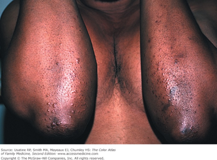
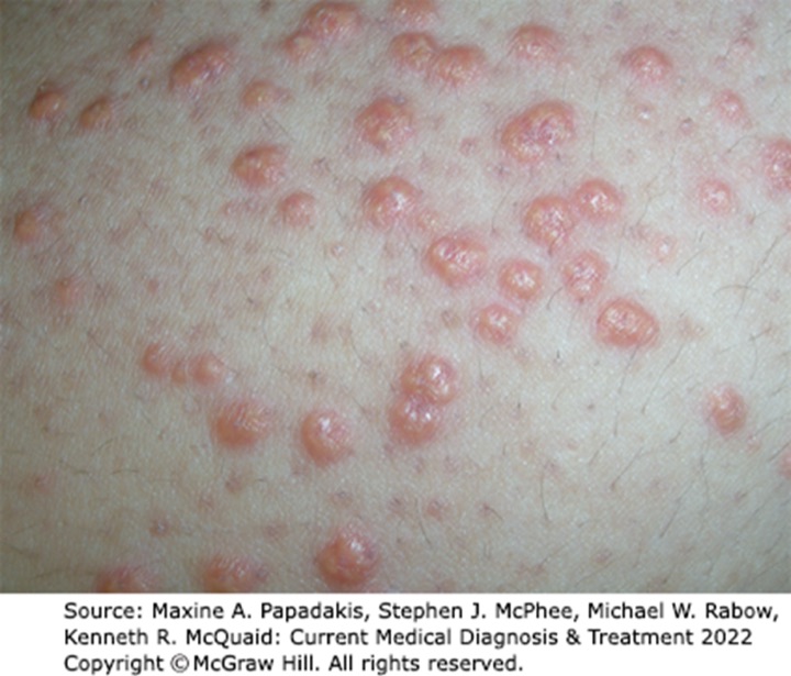
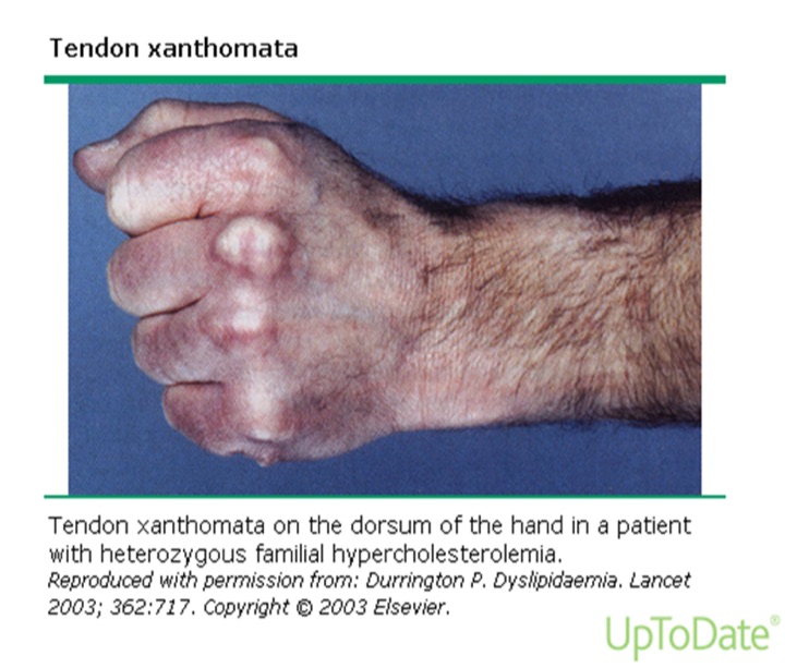
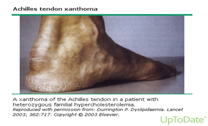
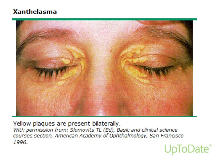
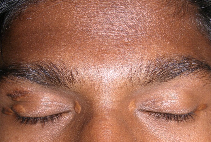
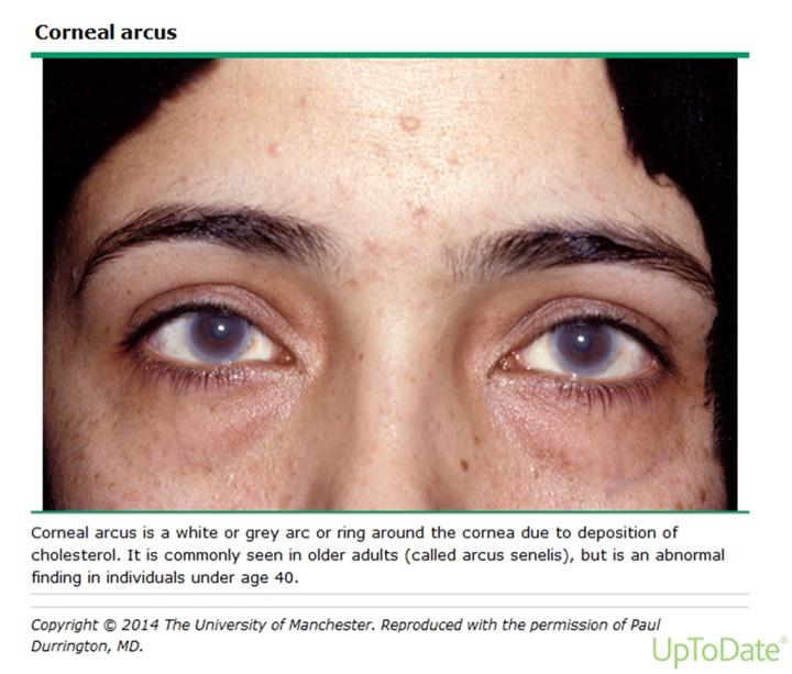
Carter on slide 32: eruptive xanthomas “present in different ways on different colored skin … very importantly, you pay attention.” The images are the deck’s own, credited on the slides to McGraw-Hill and UpToDate.
Also tested
- Xanthelasmas. They usually indicate an underlying genetic condition, and ruling out primary causes matters because they influence the patient's own risk and the management of family members.
- Corneal arcus. It is a white or gray ring around the cornea from cholesterol deposition; common in older adults (arcus senilis), before age 40 it usually indicates a genetic lipid disorder.
- Eruptive xanthomas. These clusters of small yellowish-white papules are usually seen with extremely high chylomicrons or very-low-density lipoprotein, the triglyceride carriers.
- Low high-density lipoprotein cholesterol. A level below 40 mg/dL is low and counts as a major coronary risk factor, while 60 mg/dL or more is high and removes one risk factor from the count.
Lab values (mg/dL)
| Test | Bands |
|---|---|
| Total cholesterol | <200 desirable · 200–239 borderline high · >240 high |
| LDL | <100 optimal · 100–129 near or above optimal · 130–159 borderline high · 160–189 high · >190 very high |
| HDL | <40 low · >60 high |
| Triglyceride | <150 normal · 150–199 borderline high · 200–499 high · >500 very high |
There are no “normal” cholesterol levels (LDL means vary across the globe), and there is no evidence adult cholesterol can be “too low”. Untreated values predict higher cardiovascular morbidity and mortality. Read the values as a decision, not a table: “this is not appropriate, I need to start you on a statin.”
Slides 31–40
3.5 · The conditions, one by one
Primary (genetic) causes: overproduction or defective clearance of triglyceride; overproduction or defective clearance of LDL; excessive clearance or underproduction of HDL. Genetic types are the most common cause of dyslipidemia in children. Secondary causes are in 3.6.
Dyslipidemia (overall) Routine
Gives itself away by: nothing, usually. Most patients are found from abnormal lab values.
- Definition
- A disorder of lipoprotein metabolism with raised total cholesterol, LDL, non-HDL cholesterol or triglyceride, and/or lowered HDL.
- Who gets it
- Primary (genetic) types are the most common cause in children; mixed dyslipidemia is the most common type in clinical practice.
- Risk factors
- Secondary causes: sedentary lifestyle with a diet high in carbohydrate and saturated or trans fat, diabetes, alcohol, chronic kidney disease, hypothyroidism, obstructive liver disease, medications (3.6).
- Classic signs and symptoms
- None in most patients. When present: xanthomas, arcus, premature coronary disease (30–50), pancreatitis.
- Physical exam findings
- Eruptive, tendinous or tuberous xanthomas; xanthelasma; corneal arcus before 40 (3.4).
- Diagnostics / tests
- Lipid panel read against the bands in 3.4. Critical first step: determine which lipoproteins are raised or lowered. Rule out secondary causes, then primary causes, before a statin (3.6). Screening in 3.7.
- First-line treatment
- Therapeutic lifestyle changes, drug therapy, and a clinician–patient risk discussion; LDL is the primary target (3.8).
- Complications
- Atherosclerotic cardiovascular disease; untreated values predict higher cardiovascular morbidity and mortality.
Slides 12, 16, 31–40, 42, 58–69, 72
Severe hypertriglyceridemia Urgent
Gives itself away by: triglyceride above 500 with high total and low HDL but usually no rise in LDL, and the risk of acute pancreatitis.
- Definition
- Triglyceride >500 mg/dL, raised total cholesterol, reduced HDL; usually no rise in LDL or apolipoprotein B. Cause: impaired lipolysis of triglyceride.
- Who gets it
- Usually patients with a polygenic disposition to secondary factors such as obesity or insulin resistance.
- Risk factors
- Obesity, insulin resistance, type 2 diabetes, high-carbohydrate diet, alcohol (alcohol history is important with raised triglyceride), chronic kidney disease, nephrotic syndrome; estrogen, glucocorticoids, beta blockers, isotretinoin, bexarotene.
- Classic signs and symptoms
- Acute pancreatitis.
- Physical exam findings
- Eruptive xanthomas with extremely high chylomicrons or VLDL.
- Diagnostics / tests
- Triglyceride band (>500 very high). At ≥500, rule out diabetes, chronic kidney disease, alcoholism, pregnancy and hypothyroidism before a statin. In hypertriglyceridemia, non-HDL cholesterol (total minus HDL) assesses heart risk better than LDL alone.
- First-line treatment
- Above 500: very low-fat diet (<15% of calories from fat), weight management, physical activity, and a fibrate to lower the pancreatitis risk (slide 118 adds niacin; the 2018 guideline does not use niacin).
- Complications
- Acute pancreatitis.
Slides 32, 40, 43, 59–62, 92, 115–118
Familial chylomicronemia syndrome Urgent
Gives itself away by: fasting triglyceride usually above 1000, abdominal pain from acute pancreatitis, eruptive xanthomas and lipemia retinalis. “This is important. This is one that can save somebody’s life.”
- Definition
- A disorder that causes severe hypertriglyceridemia: fasting triglyceride >500 and usually >1000 mg/dL.
- Who gets it
- Can present in childhood or adulthood. A primary (genetic) cause.
- Risk factors
- Not covered in the lecture.
- Classic signs and symptoms
- Severe abdominal pain due to acute pancreatitis.
- Physical exam findings
- Eruptive xanthomas (small yellowish-white papule clusters on the back, buttocks and extensor arms and legs); lipemia retinalis on fundoscopy (opalescence of the retinal blood vessels); hepatosplenomegaly may be present.
- Diagnostics / tests
- Fasting triglyceride >500, usually >1000; fundoscopic exam.
- First-line treatment
- Not covered specifically; the triglyceride >500 plan applies (very low-fat diet, weight management, activity, and a fibrate).
- Complications
- Acute pancreatitis.
Slides 43–44, 118
Hypercholesterolemia (raised LDL) Routine
Gives itself away by: raised LDL from impaired hepatic uptake of LDL (fewer or weaker LDL receptors).
- Definition
- Elevated LDL cholesterol.
- Who gets it
- Not covered in the lecture beyond its causes.
- Risk factors
- Anything that reduces LDL-receptor activity: diet high in saturated and trans fat, hypothyroidism, estrogen deficiency; also liver disorders, Cushing syndrome, nephrotic syndrome, glucocorticoids.
- Classic signs and symptoms
- Usually none; associated with premature atherosclerotic disease.
- Physical exam findings
- Tendinous xanthomas may be seen with high LDL.
- Diagnostics / tests
- LDL band. Rule out hypothyroidism, nephrotic syndrome, primary biliary cirrhosis, anorexia nervosa; screen raised-LDL patients for low thyroid.
- First-line treatment
- LDL is the primary target; statins first. LDL ≥190: high-intensity statin without calculating risk; add ezetimibe if LDL stays ≥100.
- Complications
- Premature atherosclerotic cardiovascular disease.
Slides 33, 40, 45, 59–61, 69, 92, 101
Familial hypercholesterolemia: homozygous versus heterozygous was the largest single disorder in the lecture (about 9 minutes). “Big thing I need you guys to understand is about like this comes from your parents. It’s important to do history.” His contrast: a child with hypercholesterolemia → think homozygous (skin biopsy); a 40-year-old with relatives who died young → think heterozygous (no definitive test).
Both forms: a codominant mutation of the LDL-receptor gene, reduced LDL clearance, LDL usually >190 with normal triglyceride, tendon xanthomas, premature atherosclerotic disease. Homozygous = two mutant alleles; heterozygous = one.
Familial hypercholesterolemia, homozygous Urgent
Gives itself away by: a child with cutaneous xanthomas, total cholesterol ~400 to >1000, and symptomatic atherosclerosis before puberty.
- Definition
- Two mutant LDL-receptor alleles. Receptor-negative (<2% of normal receptor activity) or receptor-defective (2–25%).
- Who gets it
- 1 in 1 million worldwide; presents in childhood.
- Risk factors
- Strong family history of raised lipids in parents and other first-degree relatives.
- Classic signs and symptoms
- Symptomatic atherosclerosis before puberty.
- Physical exam findings
- Cutaneous xanthomas on the hands, wrists, elbows, knees, heels or buttocks; tendon xanthomas.
- Diagnostics / tests
- Confirmed by skin biopsy with measurement of LDL-receptor activity. Total cholesterol ~400 to >1000.
- First-line treatment
- LDL apheresis is the treatment of choice (“like dialysis”: elective removal of LDL particles). Most need more than one drug: high-intensity statin, plus ezetimibe and nicotinic acid; refractory → PCSK9 inhibitor (evolocumab, alirocumab). Liver transplantation may help by providing LDL receptors. Evinacumab is an adjunct from age 12.
- Complications
- Accelerated atherosclerosis → disability and sudden death in childhood. Untreated receptor-negative patients rarely survive past the second decade; receptor-defective patients develop cardiovascular disease by 30.
Slides 46–50, 89, 109
Familial hypercholesterolemia, heterozygous Routine
Gives itself away by: an adult with LDL 200–400, normal triglyceride, Achilles tendon xanthomas and premature coronary disease in the family.
- Definition
- One mutant LDL-receptor allele; LDL 200–400 mg/dL.
- Who gets it
- 1 in 250 worldwide; up to 5% of premature myocardial infarctions (men <55, women <65). High cholesterol from birth, usually found incidentally on routine screening.
- Risk factors
- Inheritance; it is itself a high-risk condition for atherosclerotic events.
- Classic signs and symptoms
- Usually none until premature coronary disease.
- Physical exam findings
- Tendon xanthomas in ~75% (dorsum of hands, elbows, knees, mainly Achilles); corneal arcus common.
- Diagnostics / tests
- No definitive diagnostic test is available.
- First-line treatment
- Aggressive reduction of vascular risk factors (smoking, hypertension, diabetes); diet low in saturated fat and cholesterol; high-intensity statin, plus ezetimibe and nicotinic acid; refractory → PCSK9 inhibitor. “Don’t just jump straight to the PCSK9.” Inclisiran is an adjunct in adults.
- Complications
- Premature myocardial infarction and atherosclerotic disease.
Slides 27, 33, 46–47, 51–52, 90, 110
Familial defective apolipoprotein B-100 Routine
Gives itself away by: the familial hypercholesterolemia picture (raised LDL, tendon xanthomas, premature disease) from a different gene.
- Definition
- A disorder causing hypercholesterolemia: raised LDL.
- Who gets it
- 1 in 1,500 worldwide.
- Risk factors
- Not covered in the lecture.
- Classic signs and symptoms
- Premature atherosclerotic disease.
- Physical exam findings
- Tendon xanthomas.
- Diagnostics / tests
- Raised LDL.
- First-line treatment
- Not covered in the lecture.
- Complications
- Premature atherosclerotic cardiovascular disease.
Slides 9, 53
Mixed dyslipidemia Routine
Gives itself away by: triglyceride >150 and LDL >130 (or non-HDL >160). The most common type in clinical practice.
- Definition
- Raised triglyceride and LDL: triglyceride >150 with LDL >130 or non-HDL >160 mg/dL.
- Who gets it
- The most common dyslipidemia seen in practice.
- Risk factors
- Genetic predisposition plus medical conditions and environmental factors.
- Classic signs and symptoms
- Not covered in the lecture.
- Physical exam findings
- Not covered in the lecture.
- Diagnostics / tests
- The thresholds above.
- First-line treatment
- Not covered specifically; LDL first, then triglyceride (3.8).
- Complications
- Increased atherosclerotic risk.
Slide 54
Familial dysbetalipoproteinemia Routine
Gives itself away by: triglyceride and total cholesterol both 250–500, with palmar and tuberoeruptive xanthomas.
- Definition
- A rare disorder that causes mixed dyslipidemia: chylomicron and VLDL remnants accumulate, raising triglyceride and total cholesterol together.
- Who gets it
- Rare. Caused by an apolipoprotein E mutation, associated with apolipoprotein E2 homozygosity.
- Risk factors
- Needs environmental and/or other genetic factors on top of E2 homozygosity.
- Classic signs and symptoms
- Distinctive cutaneous xanthomas.
- Physical exam findings
- Palmar xanthomas; tuberoeruptive xanthomas.
- Diagnostics / tests
- Triglyceride and total cholesterol both 250–500 mg/dL.
- First-line treatment
- Not covered in the lecture.
- Complications
- Accelerated atherosclerosis.
Slides 11, 55–56
Low HDL cholesterol Routine
Gives itself away by: HDL <40. Best fixed by lifestyle.
- Definition
- HDL <40 mg/dL (metabolic-syndrome cut-off: men <40, women <50). HDL ≥60 is a negative risk factor.
- Who gets it
- Not covered in the lecture.
- Risk factors
- Genetic excessive clearance or underproduction of HDL; anabolic steroids. Rule out diabetes, cigarette smoking, obesity.
- Classic signs and symptoms
- None; it is a coronary risk factor.
- Physical exam findings
- Not covered in the lecture.
- Diagnostics / tests
- Lipid panel.
- First-line treatment
- Lifestyle: smoking cessation (up to +10%), weight loss (+1 mg/dL per 6 pounds lost), frequent aerobic exercise (+5%; brisk, 30 minutes a day, 5 days a week), healthier fats (olive, peanut, canola; saturated fat <7% of calories), limit alcohol to moderate. Drugs that raise HDL: niacin most (15–35%), fibrates (10–30%), statins (5–22%), ezetimibe (8%).
- Complications
- Loss of reverse cholesterol transport; raised coronary risk.
Slides 6, 21, 40, 42, 62, 79, 84–86, 92, 111–113
Elevated lipoprotein(a) Routine
Gives itself away by: nothing on exam; a causal, independent risk factor that calls for aggressive LDL lowering.
- Definition
- A lipoprotein like LDL that carries apolipoprotein(a) in addition to B-100.
- Who gets it
- Not covered in the lecture.
- Risk factors
- It is itself a risk factor; ≥50 mg/dL (or 125 nmol/L) is a 2018 risk enhancer.
- Classic signs and symptoms
- Not covered in the lecture.
- Physical exam findings
- Not covered in the lecture.
- Diagnostics / tests
- Measured in selected individuals (2018 guideline).
- First-line treatment
- If elevated, aggressive therapy to decrease LDL.
- Complications
- Atherosclerotic cardiovascular disease.
Slides 7, 11, 96, 105
Metabolic syndrome Routine
Gives itself away by: any 3 of 5 criteria, with obesity measured by waist circumference, not body mass index.
- Definition
- Any 3 of: waist >40 inches (men) or >35 inches (women); triglyceride >150; HDL <40 (men) or <50 (women); blood pressure >130/>85; fasting glucose >110.
- Who gets it
- Not covered in the lecture.
- Risk factors
- Not covered; the syndrome is itself a 2018 risk enhancer.
- Classic signs and symptoms
- The five criteria.
- Physical exam findings
- Abdominal obesity by waist circumference; raised blood pressure.
- Diagnostics / tests
- Any 3 of the 5 criteria.
- First-line treatment
- Lifestyle therapy is the primary intervention in all age groups.
- Complications
- Raised atherosclerotic risk (risk enhancer).
Slides 38, 96, 98, 105
Atherosclerosis (as the deck treats it) Routine
Gives itself away by: an ASCVD event, or a risk estimate. The deck never defines atherosclerosis or explains plaque.
- Definition
- Not covered in the lecture.
- Who gets it
- Coronary heart disease is the leading cause of death in United States men and women.
- Risk factors
- LDL (highest), VLDL (increased); the major risk factors and equivalents in 3.3; lipoprotein(a).
- Classic signs and symptoms
- Major events: acute coronary syndrome, myocardial infarction, ischemic stroke, symptomatic peripheral arterial disease.
- Physical exam findings
- Not covered in the lecture.
- Diagnostics / tests
- Risk estimation (Pooled Cohort Equation, 40–79); coronary artery calcium when a statin decision is uncertain.
- First-line treatment
- Statin-centered prevention per the 2018 guideline (3.8).
- Complications
- Atherosclerotic cardiovascular events; cardiovascular death.
Slides 13–15, 20–29, 96–106
Also tested
- Familial dysbetalipoproteinemia. An apolipoprotein E mutation, associated with apolipoprotein E2 homozygosity, lets chylomicron and very-low-density remnants accumulate, so triglyceride and total cholesterol both rise to 250 to 500 mg/dL.
- Severe hypertriglyceridemia. The pattern is triglyceride above 500 mg/dL with elevated total cholesterol and reduced high-density lipoprotein, usually without elevated low-density lipoprotein or apolipoprotein B.
- Apheresis. It is like dialysis: the elective removal of low-density particles from the circulation, and it is the treatment of choice for homozygous familial hypercholesterolemia.
- Familial chylomicronemia syndrome. Eruptive xanthomas, small yellowish-white papules, cluster on the back, buttocks and extensor surfaces of the arms and legs, alongside lipemia retinalis and pancreatitis.
- Diagnosing familial hypercholesterolemia. No definitive diagnostic test exists for the heterozygous form; skin biopsy with receptor activity confirms only the homozygous disorder.
- Raising low high-density lipoprotein. It is best improved by lifestyle modification: smoking cessation (up to 10%), weight loss (about 1 mg/dL per 6 pounds) and frequent aerobic exercise (about 5%).
- Heterozygous familial hypercholesterolemia. It causes premature atherosclerotic disease and accounts for up to 5% of premature heart attacks in men under 55 and women under 65.
- Familial dysbetalipoproteinemia. It is associated with accelerated atherosclerosis, the long-term danger of its remnant-driven rise in triglyceride and cholesterol.
3.6 · Secondary causes, and what to rule out before a statin
| Cause | Effect |
|---|---|
| High-carbohydrate diet | ↑ hepatic VLDL-triglyceride secretion → ↑ triglyceride |
| Saturated and trans fat | Down-regulates hepatic LDL receptors → ↑ LDL |
| Obesity, insulin resistance, type 2 diabetes | ↑ hepatic VLDL; insulin promotes hepatic fatty-acid synthesis → ↑ triglyceride |
| Alcohol (excess) | Inhibits hepatic free-fatty-acid oxidation → ↑ triglyceride. Take an alcohol history with raised triglyceride. |
| Chronic kidney disease | VLDL and remnant accumulation; reduced lipolysis and remnant clearance → ↑ triglyceride |
| Nephrotic syndrome | Excess VLDL production → ↑ triglyceride and LDL |
| Cushing syndrome | Glucocorticoid overproduction → ↑ VLDL synthesis, ↑ LDL |
| Hypothyroidism | Down-regulates hepatic LDL receptors → ↑ LDL. Screen raised-LDL patients for low thyroid. |
| Liver disorders | Down-regulate hepatic LDL receptors → ↑ LDL |
| Estrogen | ↑ triglyceride (↑ HDL). Monitor on birth control pills, new postmenopausal estrogen, or pregnancy. |
| Glucocorticoids | ↑ triglyceride and LDL |
| Beta blockers · isotretinoin · bexarotene | ↑ triglyceride |
| Anabolic steroids | ↓ HDL |
Slides 58–62, 92–94
Also tested
- Estrogen and lipids. Estrogen can elevate triglyceride (while increasing high-density lipoprotein), so patients starting birth control pills or postmenopausal estrogen, or who are pregnant, are monitored.
- Dietary carbohydrate and triglyceride. Excess carbohydrate increases hepatic very-low-density triglyceride secretion, raising triglyceride, so cutting it addresses a secondary cause.
- Anabolic steroids and lipids. Anabolic steroids decrease high-density lipoprotein, so they are a secondary cause of a low level, and stopping them addresses it.
- Thyroid testing before a statin. Decreased thyroid hormone down-regulates hepatic low-density lipoprotein receptors and raises low-density lipoprotein, so elevated levels call for screening for low thyroid before a statin.
- Primary causes of dyslipidemia. Even after secondary causes are excluded, primary causes are sought because they affect management of family members, who may carry the same disorder, as well as coronary risk and drug response.
- Family screening in familial hypercholesterolemia. Identifying a primary cause influences management of other family members, so lipid screening is appropriate for each child; elective screening is appropriate as young as 2 years with a family history of high cholesterol.
3.7 · Screening
- The decision to screen rests on overall cardiovascular risk, independent of lipid levels: age, sex, hypertension, smoking, family history of premature coronary disease (male relative <55, female <65), diabetes.
- Screen all adults 20 and older. (The United States Preventive Services Task Force line on the same slide says 20–35.)
- Children: a non-fasting lipid profile once at 9–11, and again in late adolescence. The deck gives that second window two ways (17–19 on slide 65, 17–21 on slide 66); the 2018 cholesterol guideline says 17–21. No question keys it. Elective screening from age 2 with a family history of heart disease or high cholesterol.
- Total cholesterol and HDL need no fasting; their ratio is the most efficient and cost-effective coronary risk predictor. The slide says triglyceride and calculated LDL need a 9–12 hour fast; under the 2018 guideline (the one tested) a non-fasting full panel is acceptable for routine screening, and a fasting sample is needed when triglyceride is 400 mg/dL or more. The quizzes key the 2018 version.
Slides 64–66
Also tested
- Fasting for lipid panels. Under the 2018 guideline a non-fasting panel, calculated low-density value included, is acceptable for routine screening; a fasting repeat is needed only when triglyceride is 400 mg/dL or more.
- Lipid screening in children. Elective lipid screening is appropriate for patients as young as 2 years old with a family history of heart disease or high cholesterol.
3.8 · Management
Three parts: therapeutic lifestyle changes, drug therapy, and a clinician–patient risk discussion.
LDL first, then triglyceride. “That’s what I need you to know. Address the LDLs, then address the triglycerides, and add your adjunctive therapy on top of it.”
- “The primary target of cholesterol-lowering therapy is elevated LDL-C.” (slide 69)
- “Primary aim of therapy is based on LDL goals, but if triglycerides are >200 mg/dL after LDL goal is reached additional treatment for lowering triglycerides is indicated.” (slide 116)
- Triglyceride 200–499: the slide says intensify the LDL drugs and/or add a triglyceride drug (fibrate, niacin). Above 500: very low-fat diet (<15% of calories from fat), weight management, physical activity, fibrate or niacin or both. (slide 118)
- In hypertriglyceridemia use non-HDL cholesterol = total − HDL, whose goal sits 30 above the LDL goal. (slides 115–117)
Also tested
- Non-high-density cholesterol. It is total cholesterol minus high-density cholesterol, and in hypertriglyceridemia it assesses heart-disease risk better than low-density lipoprotein alone.
- Weight goal in dyslipidemia. Therapeutic lifestyle changes aim for a 5% to 10% reduction of initial weight; for 200 pounds that is 10 to 20 pounds.
- Calcium score and statins. A coronary artery calcium score of 100 or more (or at or above the 75th percentile) indicates statin therapy unless the clinician-patient risk discussion defers it.
- Calculating non-high-density cholesterol. It is total cholesterol minus high-density cholesterol; for example, 250 minus 40 is 210 mg/dL.
- Coronary artery calcium scoring. In adults 40 to 75 without diabetes, with low-density lipoprotein of 70 to 189 and 10-year risk of 7.5% to 19.9%, it is considered when the statin decision is uncertain.
- Adding to a maximal statin. Ezetimibe is added before the injectable class such as evolocumab; adding evolocumab before ezetimibe is out of sequence and less cost-effective.
- Statin follow-up. Adherence and percentage response are assessed with a repeat lipid panel 4 to 12 weeks after starting a statin, then every 3 to 12 months as needed.
- Metabolic syndrome management. Lifestyle therapy is the primary intervention in all age groups, and a heart-healthy lifestyle is the foundation of risk reduction across the life course.
- Main treatment of metabolic syndrome. Lifestyle therapy is the primary intervention in all age groups, including weight reduction of 5% to 10% and daily moderate activity.
Therapeutic lifestyle changes
Saturated fat <7% of calories (the course's therapeutic-lifestyle figure; Carter once said “10%”, which is the looser Dietary Guidelines limit, not an error, but learn <7% here); more soluble fiber (20–30 g a day); consider plant stanols or sterols (in grains, vegetables, fruits, legumes, nuts and seeds); lose 5–10% of initial weight; moderate activity 30 minutes a day (brisk walking, biking, active yoga); stop smoking.
Which drug for which lipid. “Remember which medications go with which lipoprotein.” “The best one that’s increasing HDLs is niacin. And the one for lowering triglycerides is fibrates.” Generic names only.
| Class (generic names) | Main job | Effects |
|---|---|---|
| Statins: atorvastatin, fluvastatin, lovastatin, pravastatin, rosuvastatin, simvastatin, pitavastatin | Most effective for LDL | LDL ↓25–63%, maximum in 4–6 weeks; HDL ↑5–22%. Monitor lipids, liver function, creatine kinase. |
| Fibrates: gemfibrozil, fenofibrate | Most effective for triglyceride | Triglyceride ↓20–50%; HDL ↑10–30% |
| Niacin (nicotinic acid) | Raises HDL best | HDL ↑15–35%; triglyceride ↓20–50%; LDL ↓5–25% |
| Ezetimibe (cholesterol absorption inhibitor) | Adjunct LDL lowering | LDL ↓18%; HDL ↑8%; triglyceride ↓5–10% |
| Bile acid sequestrants: colestipol, cholestyramine, colesevelam | Adjunct LDL lowering | LDL ↓15–30% |
| Fish oil | — | Associated with lower coronary disease and coronary death; may lower thrombotic stroke; eat fish at least twice a week |
| PCSK9 (proprotein convertase subtilisin/kexin type 9) inhibitors: evolocumab, alirocumab | Refractory familial hypercholesterolemia; very high risk after statin + ezetimibe | Injectable monoclonal antibodies; approved for familial hypercholesterolemia; expensive |
| Bempedoic acid | Newer; oral | Up-regulates LDL receptors |
| Inclisiran | Adjunct to maximal statin in adults with heterozygous familial hypercholesterolemia or ASCVD | Subcutaneous injection |
| Evinacumab | Adjunct in homozygous familial hypercholesterolemia, age 12 and older | Intravenous; lowers LDL and triglyceride |
Carter called myopathy the “number one” statin side effect and said creatine kinase is “nephrotoxic”; neither is on a slide (and the second is wrong: in rhabdomyolysis the kidney injury comes from myoglobin). The slide fact is the monitoring list. Nystatin is not a statin (his slide 82 meme).
Statin intensity is defined by the LDL fall it produces: high about 50% or more, moderate 30% to under 50%, low under 30%.
The 2018 statin decisions (“For our testing … we are using the 2018”). Lifestyle is emphasized for everyone, at every age.
| Patient | 2018 recommendation |
|---|---|
| Clinical ASCVD, age ≤75 | High-intensity statin (lower LDL ≥50%); moderate if high is not tolerated |
| Clinical ASCVD, age >75 | Starting moderate- or high-intensity is reasonable; continuing high-intensity is reasonable |
| Very high-risk ASCVD, LDL ≥70 on maximal statin | Add ezetimibe first; then a PCSK9 inhibitor if LDL stays ≥70 (less cost-effective) |
| LDL ≥190 (severe primary hypercholesterolemia) | High-intensity statin without calculating 10-year risk; add ezetimibe if LDL stays ≥100 |
| Diabetes, age 40–75, LDL ≥70 | Moderate-intensity statin without calculating risk; high intensity reasonable with multiple risk factors or age 50–75 |
| Age 40–75, no diabetes, LDL 70–189 | Clinician–patient risk discussion, then by 10-year risk (3.3). If statins are indicated, lower LDL ≥30%; at ≥20% risk, lower LDL ≥50%. |
| Uncertain decision at intermediate risk | Coronary artery calcium: 0 → may withhold or delay (not in smokers, diabetes, or strong family history); 1–99 favors a statin (especially ≥55); ≥100 or ≥75th percentile → statin |
| Age 20–39 | Estimate lifetime risk to encourage lifestyle; consider a statin with a family history of premature ASCVD and LDL ≥160 |
| Age 0–19 | Lifestyle; a diagnosis of familial hypercholesterolemia → statin |
| Age >75, primary prevention | Clinical assessment and risk discussion |
Risk enhancers (favor a statin at borderline or intermediate risk): family history of premature ASCVD; LDL persistently ≥160; metabolic syndrome; chronic kidney disease; preeclampsia or premature menopause (<40); inflammatory disease (rheumatoid arthritis, psoriasis, HIV (human immunodeficiency virus)); South Asian ancestry; triglyceride persistently ≥175; and, if measured, apolipoprotein B ≥130, high-sensitivity C-reactive protein ≥2.0, ankle-brachial index <0.9, lipoprotein(a) ≥50 mg/dL.
Follow-up: repeat lipids 4–12 weeks after starting or changing a statin, then every 3–12 months; response is the percentage fall in LDL from baseline.
Slides 69, 72–90, 96–118
3.9 · Populations
| Population | What the deck gives |
|---|---|
| Infant | Nothing in the deck. |
| Child | Genetic types are the most common cause. Non-fasting screen once at 9–11; elective screening from age 2 with a family history. Homozygous familial hypercholesterolemia presents in childhood; confirm by skin biopsy; apheresis. Ages 0–19: lifestyle, and a statin once familial hypercholesterolemia is diagnosed. |
| Adolescent | Second screen in late adolescence (window stated two ways, see 3.7). Evinacumab is an adjunct in homozygous disease from age 12. |
| Adult | Screen from 20. Ages 20–39: lifetime risk and lifestyle; statin with premature family history and LDL ≥160. Ages 40–75: the 10-year risk pathway (3.3, 3.8). |
| Elderly | Age ≥65 is a high-risk condition. Above 75: clinical assessment and risk discussion in primary prevention; in clinical ASCVD, starting or continuing a statin is reasonable. |
Also tested
- Mechanical heart valve. It needs lifelong anticoagulation with a vitamin K antagonist, most commonly warfarin, monitored against an international normalized ratio target.
4 · Valvular Heart Disease
Instructional Objectives
VALVULAR HEART DISEASE — Grady G. Carter, MSHS, PA-C
- Compare and contrast the etiologies, epidemiology, risk factors, clinical manifestations, differential diagnosis, diagnostic testing (including ordering and interpretation), management (acute and chronic, including applicable rehabilitative and palliative care), appropriate referrals, patient education, prevention, and prognosis for valvular heart disease
- Acute rheumatic fever
- Aortic stenosis
- Aortic regurgitation
- Mitral stenosis
- Mitral regurgitation
- Mitral valve prolapse
- Tricuspid regurgitation
- Tricuspid stenosis
- Pulmonic stenosis
- Pulmonic regurgitation
- Compare and contrast anticoagulation therapy for prosthetic heart valves.
- Identify medical care strategies for valvular heart disease in the lecture topic list for the following populations.
The syllabus numbers the disease list (2–11) as though each were an objective, and objective 13 names no populations — the list is missing in the syllabus itself.
What is tested, in the lecturer's own words (lecture recording, 25 September, two parts):
- No audio. “There will be no audio clips on your test.” Murmurs arrive as words — “When you see the word harsh or rumble, it means stenosis … blowing, that's regurgitation.”
- Locations arrive anatomically. “They're not going to say at the pulmonic location … they're going to say the second intercostal space at the left sternal border. Then you have to know what valve is directly underneath there.”
- Maneuver → response. “You hear this sound, and then you have them do this test, and this is the response.”
- Every valve counts equally. “When it comes to the test, you will just as likely have a tricuspid valve question as you will an aortic valve question.” Aortic stenosis got the most time because it matters most in practice, not because it is weighted more on the exam.
- Severity grading is asked only for aortic stenosis and mitral stenosis (“I'm not asking about the lesser valves”). For aortic and mitral regurgitation learn only severe = regurgitant fraction of 50% or more.
- Not tested: the suffusion sign on slide 67 (“I'm not going to test you on it”), and the low-flow / low-gradient “pseudo-severe” aortic stenosis named on slide 21 (aimed at future cardiology PAs).
- Electrocardiogram findings are recognition only — the electrocardiogram lecture has not happened yet.
- Program rules: no dosing, no mechanism-of-action questions, generic names only, no acronyms in questions. INR (international normalized ratio) targets are laboratory targets, not doses, and are learned.
Built from the slides. The recording adds weight only: ★ marks show what he emphasized. Where a slide, his words and standard references disagree, this guide teaches what is medically true and says what the slide or the lecture said instead (the boxes below).
Erratum from the lecture — he said these slides are wrong
| Slide | The slide says | His correction (learn this) |
|---|---|---|
| 28 (aortic regurgitation exam) | “High-Pitch BLOWING DIASTOLIC | The murmur is a high-pitched blowing DIASTOLIC murmur. “It's not holosystolic because it's diastolic. So cross that out.” Holosystolic belongs to mitral and tricuspid regurgitation. |
| 29 (aortic regurgitation echo) | “Used to determine cause of | Echocardiography determines the cause of aortic regurgitation — aortic root dilation, aortic dissection. “That should be an A.” |
Deck error — copy-paste slips he did not mention
- Slide 44: the mitral regurgitation definition is labeled “Mitral Stenosis:”. The definition itself — incomplete mitral valve closure with backflow into the left atrium — is mitral regurgitation.
- Slide 52: “Children & young adults – Percutaneous Aortic Balloon Valvuloplasty” is repeated under mitral regurgitation surgery, copied from the aortic stenosis slide (21). It belongs to aortic stenosis; it is not learned for mitral regurgitation.
- Slide 53: the mitral regurgitation “When to refer” slide says “MS” throughout (copied from slide 43). It is not used here.
- Slide 65: tricuspid stenosis is defined as inflow obstruction “from the LA and SVC/IVC”. The tricuspid valve sits between the right atrium and the right ventricle — slide 66 has it right (“obstruction of inflow from RA to RV”).
- Slide 31: “vasodilators (nitroprusside & dobutamine)”. Dobutamine is an inotrope, not a vasodilator. For acute severe aortic regurgitation awaiting surgery, learn intravenous diuretics + nitroprusside.
Where the slide or the lecture is not accurate — learn the true version
- Slide 29, which leaflet flutters on echo. The slide is right: the regurgitant jet makes the anterior mitral leaflet flutter in diastole. Asked in class, he first said mitral, then said “It's the aortic leaflet” and asked for it to be crossed out; that correction is not accurate. Learn anterior mitral leaflet.
- Slide 74, valve type by age (“TAVR/TMVR … <55”, “Mechanical … <70”) has it backwards. What he taught earlier is the standard (2020 ACC/AHA valvular guideline): a younger patient gets a mechanical valve because it lasts; an older patient gets a bioprosthetic or transcatheter valve. No quiz question keys an age cut-off.
- Slide 38, mitral stenosis “Increased with Valsalva” is not accurate: Valsalva reduces venous return and softens the mitral stenosis murmur, as it does every murmur except hypertrophic cardiomyopathy and mitral valve prolapse. No quiz question asks a mitral stenosis maneuver.
- Pulmonic regurgitation, most common cause. Slide 61 and the lecture say iatrogenic (“the most common overall reason is iatrogenic”); slide 64 says “almost always congenital”. The accurate version: significant pulmonic regurgitation is most often iatrogenic (after outflow-tract surgery such as tetralogy of Fallot repair), while mild regurgitation from pulmonary hypertension is the commonest overall (Merck Manual). The quizzes ask it as “significant”.
- Slide 40, mitral stenosis bands. Mild (1.5–2.5) and moderate (1.0–1.5) overlap at 1.5. Learn severe only.
Spoken slips — the slide is right: his pulmonic regurgitation example put the balloon in a mitral valve (slide 61: surgery on the right ventricular outflow tract); carcinoid disease was called “cancer of the valve” (slides 55, 61, 65 say carcinoid disease); a transcatheter valve was called “plastic … rubber” (not on any slide). His “pulmonary hypertension is our number one cause” of pulmonic regurgitation, which he overrode himself 40 seconds later, is true of pulmonic regurgitation overall (see above).
Where each objective is answered
| Objective | Answered in |
|---|---|
| 1. Etiology, epidemiology, risk factors, manifestations, testing, management, referral, education, prevention, prognosis | 4.1 (murmur foundations), then each lesion's subsection and its eight-point block in 4.13, 4.12 (test tips) · the deck has no prevention content for any lesion |
| 2. Acute rheumatic fever | No section in the deck. It appears only as an etiology — rheumatic heart disease, an autoimmune reaction to group A beta-hemolytic streptococci (slides 12, 24, 34, 45, 65): see 4.2, 4.3, 4.4, 4.5 and 4.9 |
| 3. Aortic stenosis | 4.2 |
| 4. Aortic regurgitation | 4.3 |
| 5. Mitral stenosis | 4.4 |
| 6. Mitral regurgitation | 4.5 |
| 7. Mitral valve prolapse | 4.6 |
| 8. Tricuspid regurgitation | 4.10 |
| 9. Tricuspid stenosis | 4.9 |
| 10. Pulmonic stenosis | 4.7 |
| 11. Pulmonic regurgitation | 4.8 |
| 12. Anticoagulation for prosthetic heart valves | 4.11 |
| 13. Populations | 4.14 · only percutaneous aortic balloon valvuloplasty for children and young adults (slide 21), mitral valve prolapse in women 15–30 (slide 54) and the aortic stenosis age cut-offs (slides 12, 13, 16) |
4.1 · Anatomy, heart sounds and the murmur words
“This is how I passed this test in PA school.” The testing hints appear twice in the deck (slides 8 and 73, opening and closing):
| Word in the stem | Means | Flow | Overload |
|---|---|---|---|
| Harsh or rumble | Stenosis | Abnormal forward flow | Pressure overload |
| Blowing | Regurgitation | Abnormal back flow | Volume overload |
| Timing | Lesions |
|---|---|
| Systolic | Aortic stenosis, mitral regurgitation · pulmonic stenosis, tricuspid regurgitation |
| Diastolic | Aortic regurgitation, mitral stenosis · pulmonic regurgitation, tricuspid stenosis — “ARMS rest because they are PRetty TiredS” |
“What do we know about the word harsh? Stenosis. Systolic. We only have two systolic stenotic diseases … you just went from a 25% chance to a 50-50.” (Aortic stenosis and pulmonic stenosis.)
Also tested
- Pulmonic stenosis murmur and breathing. The murmur gets louder with inspiration, which raises venous return to the right heart, and it comes with a split second heart sound.
Where you hear it: A-P-E-T-M (the picture on slide 8)
| Site | Location given in a stem |
|---|---|
| Aortic | 2nd intercostal space, right sternal border (right upper sternal border) |
| Pulmonic | 2nd intercostal space, left sternal border (left upper sternal border) |
| Erb's point | 3rd intercostal space, left sternal border |
| Tricuspid | 5th intercostal space, lower left sternal border |
| Mitral | Apex — point of maximal impulse, 5th intercostal space at the midclavicular line |
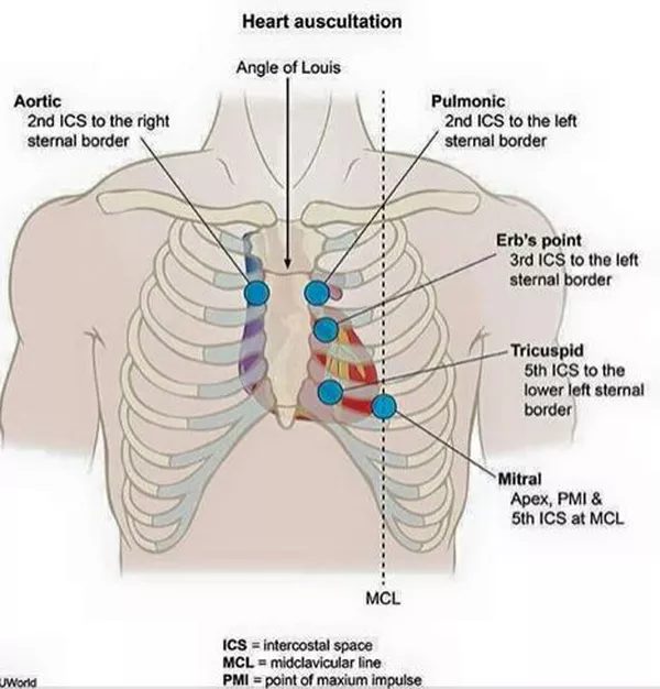
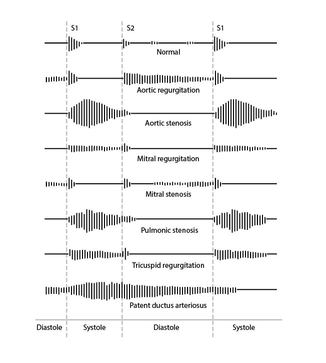
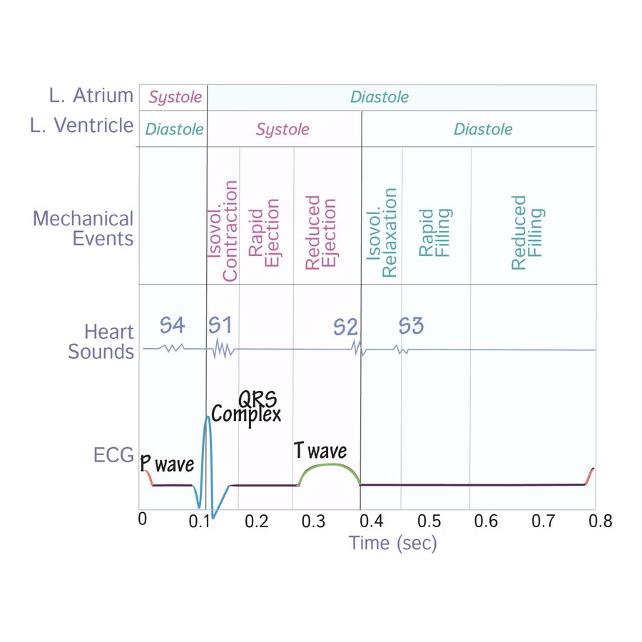
Heart sounds (slide 7)
- S1 (systolic): the atrioventricular valves close — tricuspid, mitral.
- S2: the semilunar valves close — pulmonic, aortic.
- S3 (diastolic): high volume into a dilated left ventricle (systolic heart failure); the slide lists aortic regurgitation (back flow) and mitral stenosis (late opening).
- S4: high pressure against a hypertrophied left ventricle (diastolic heart failure), secondary to aortic regurgitation or aortic stenosis.
Respiration: “RILE” (slides 9–10)
| Inspiration | Expiration | |
|---|---|---|
| Intrathoracic pressure | Negative | Increased |
| Venous return | Increased to the right heart (more right-sided preload) | Reduced to the right heart; blood forced out of the lungs into the left atrium (more left-sided preload) |
| Louder murmurs | Right-sided: tricuspid regurgitation, pulmonic stenosis, tricuspid stenosis, pulmonic regurgitation | Left-sided: mitral regurgitation, aortic stenosis, aortic regurgitation, mitral stenosis |
Carvallo sign = a right-sided murmur that gets louder with inspiration — in the deck, the tool that separates tricuspid regurgitation from mitral regurgitation (slide 71).
Slides 5–10, 73
4.2 · Aortic stenosis
“That was the big heavy one … That's the one I want you to remember.” Obstruction of left ventricular outflow across the aortic valve — the most common valvular disease.
| Severe aortic stenosis — “4, 40 and 1” | Value |
|---|---|
| Valve area | <1.0 cm |
| Mean gradient | >40 mmHg |
| Jet velocity | >4.0 m/s |
(Slide 19 also gives mild >1.5 cm / <25 mmHg / <3.0 m/s and moderate 1.0–1.5 cm / 25–40 / 3.0–4.0; he taught severe only.)
| Cardinal symptom | Average survival once it appears |
|---|---|
| Angina (usually exertional) | 5 years |
| Syncope (usually exertional) | 3 years |
| Congestive heart failure / exertional dyspnea — the most common symptom | 2 years — the worst prognosis |
Symptomatic aortic stenosis has a 3-year mortality of 75%. “50% of people with severe aortic stenosis will be dead in two years if it is untreated … can't miss this one.”
The murmur: HARSH SYSTOLIC ejection crescendo-decrescendo murmur at the 2nd right intercostal space (right upper sternal border), radiating to the carotids. Decreased with Valsalva, standing, handgrip (more afterload or less volume → less flow through the valve); increased with squatting and sitting forward (more preload; sitting forward brings the valve closer to the chest wall).
Simultaneous palpation of a forceful left ventricular apex beat and a delayed, weakened carotid pulse is a clue that severe aortic stenosis is present — “a clinical way of knowing … moderate versus severe.”
Patients are PRELOAD dependent. Medical treatment is not effective (mild and moderate only), and the rules are what to avoid:
- Strenuous physical activity and competitive sports
- Dehydration and hypovolemia
- With hypertension: beta blockers and calcium channel blockers
- With angina: nitrates (“What does nitrates do to your preload? Drops it.”)
Also tested
- Aortic stenosis and nitrates. Patients are preload dependent and nitrates drop preload, so nitrates are avoided if angina is present; the same logic removes dehydration and strenuous exertion.
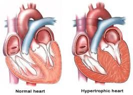
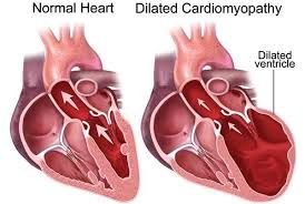
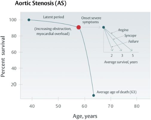
Causes, who gets it, and the mechanism
- Degenerative (atherosclerosis: hyperlipidemia, diabetes, smoking; chronic wear and tear): the most common cause over 70.
- Congenital bicuspid aortic valve: the most common cause under 70.
- Acquired: rheumatic heart disease (an autoimmune reaction to group A beta-hemolytic streptococci); endocarditis — 50% of endocarditis is mitral and 20% aortic, and untreated endocarditis carries an 80% one-year mortality.
- Epidemiology: 80% of symptomatic patients are men; present in 25% of patients older than 65 and 35% older than 70; 10–20% progress to significant stenosis within 10–15 years. Risk factors: hypertension, hyperlipidemia, smoking.
- Mechanism: outflow obstruction → left ventricular pressure overload → myocyte remodeling → concentric left ventricular hypertrophy → diastolic heart failure (heart failure with preserved ejection fraction).
- Symptoms are rare until the orifice is about 1 cm (normal 3–4 cm), usually at ages 60–80. Progression is insidious because resting cardiac output holds until late; once symptomatic, the prognosis is severely worsened.
Diagnosis, surgery and referral
- Electrocardiogram: left ventricular hypertrophy in severe disease (low sensitivity).
- Transthoracic echocardiogram: thickened, calcified leaflets, reduced systolic opening, left ventricular hypertrophy in advanced disease. Transthoracic for degenerative; transesophageal useful for congenital.
- Definitive: left heart catheterization.
- Any symptom suggestive of aortic stenosis (exertional chest pressure, shortness of breath, syncope) → echocardiogram.
- Surgery when severe (<1, >40, >4) and symptomatic left ventricular systolic dysfunction, or a bicuspid valve: TAVR (transcatheter aortic valve replacement); mechanical valve (titanium and carbon — lifelong anticoagulation) vs biological (pig or cow tissue — no anticoagulation); children and young adults → percutaneous aortic balloon valvuloplasty.
- Refer: all patients with aortic stenosis on echocardiogram get a cardiology consult to evaluate and set follow-up frequency.
4.3 · Aortic regurgitation
Incomplete aortic valve closure lets blood flow back into the left ventricle. “AR and AI [aortic regurgitation and aortic insufficiency] are the same thing” — a stem may say aortic insufficiency.
The murmur (corrected, see the erratum): a high-pitched BLOWING DIASTOLIC murmur at the 3rd intercostal space / left upper sternal border, radiating to the apex. Severity is proportional to the murmur's duration. Decreased with Valsalva and standing; increased with squatting, sitting forward and handgrip. Other findings: bounding pulses, wide pulse pressure, thrills, displaced point of maximal impulse.
Corrigan's pulse (“water hammer” or “collapsing” pulse: a very rapid upstroke followed by a rapid collapse) goes with aortic regurgitation. “The PANCE (Physician Assistant National Certifying Exam) did it to me … Corrigan's pulse was noted, and they didn't tell you what it was.” de Musset sign = head bobbing.
Severe = a regurgitant fraction of 50% or more — “50% of your cardiac output passed that valve going back.” (The orifice-area and volume columns of slide 30 are not taught; its echo picture shows stenosis-type numbers and is not used.)
Also tested
- Dilated aortic root. Patients with a dilated aortic root should be monitored by a cardiologist. A patient with an audible regurgitant murmur should have a cardiology consult to decide whether follow-up is needed.
- Chronic aortic regurgitation with reduced ejection fraction. With a mildly reduced or reduced ejection fraction, with or without symptoms, the next step is evaluation as a surgical candidate; untreated, survival is only about 2 to 3 years.
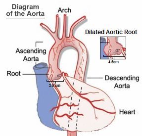
- Causes — valve disease, aortic root disease or both. Valvular: congenital bicuspid valve, endocarditis, rheumatic fever, myxomatous (collagen) disease, trauma, syphilis, ankylosing spondylitis. Root: aortic dissection, cystic medial degeneration, Marfan syndrome, nonsyndromic familial aneurysm, aortitis, hypertension.
- Mechanism: diastolic backflow → eccentric hypertrophy and dilation → left ventricular dilation (systolic heart failure) → heart failure with reduced ejection fraction.
- Acute (myocardial infarction, endocarditis, aortic dissection, trauma): cardiogenic shock, pulmonary edema. Chronic: asymptomatic for 10–15 years, then congestive heart failure (exertional sinus tachycardia, exertional dyspnea, orthopnea, paroxysmal nocturnal dyspnea, excessive sweating, anginal chest pain).
- Diagnosis: electrocardiogram — left ventricular hypertrophy in chronic disease (non-specific); echocardiogram determines the cause (aortic root dilation, aortic dissection) and shows diastolic fluttering of the anterior mitral leaflet where the jet strikes it; catheterization is definitive but not necessary.
- Treatment: medical = afterload reduction (ACE (angiotensin-converting enzyme) inhibitor or angiotensin receptor blocker, dihydropyridine calcium channel blocker, hydralazine) for aortic regurgitation with preserved ejection fraction, or asymptomatic. Acute severe: emergency aortic valve replacement or repair, ideally within 24 hours; if surgery is delayed, stabilize in intensive care with intravenous diuretics and nitroprusside. Chronic with mildly reduced or reduced ejection fraction, symptomatic or not: surgical candidate — untreated, survival is only 2–3 years.
- Refer: any audible murmur → cardiology consult; a dilated aortic root is monitored by cardiology.
4.4 · Mitral stenosis
Obstruction of left atrial outflow across the mitral valve. Rheumatic heart disease is the MOST COMMON cause. 80% of mitral stenosis from rheumatic fever is in women; it is usually diagnosed about 20 years after the rheumatic fever, and it is rare in developed countries because rheumatic fever has declined.
Left atrial enlargement → atrial fibrillation → thromboembolism. “Your mitral stenosis patients that have already affected their left atrium, most of them will develop AFib [atrial fibrillation].”
Also tested
- Mitral stenosis follow-up. All patients with evidence of mitral stenosis on echocardiogram should have a cardiology consult to evaluate them and set the frequency of follow-up. Asymptomatic disease in sinus rhythm needs no therapy yet.
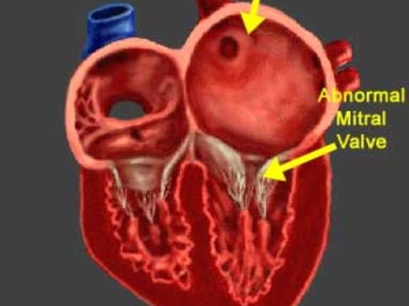
- Mechanism: thickened, immobile leaflets narrow the orifice → left atrial size and pressure rise → pulmonary vasoconstriction, raised pulmonary artery pressure, right ventricular pressure overload → right-sided heart failure; heart failure with preserved ejection fraction.
- Symptoms are rare until the orifice is a third of normal, then left-sided heart failure: dyspnea on exertion, cough, orthopnea, paroxysmal nocturnal dyspnea, fatigue. Plus hemoptysis (high left atrial pressure ruptures small bronchial vein anastomoses), hoarseness — Ortner syndrome (the enlarged left atrium presses on the left recurrent laryngeal nerve) and dysphagia (the left atrium presses on the esophagus).
- The murmur: low-pitched decrescendo-crescendo RUMBLING DIASTOLIC murmur at the apex, starting with an opening snap after S2; listen with the bell, patient in the left lateral decubitus position (lying on the left side). Like most murmurs it softens with Valsalva (slide 38's\n “increased” is not accurate); no quiz question asks a mitral stenosis maneuver.
- Electrocardiogram: left atrial abnormality (the P wave); atrial fibrillation is common in severe disease; right ventricular hypertrophy with pulmonary hypertension. Transthoracic echocardiogram: large left atrium, stenotic valve. Catheterization is not necessary (usually done for an ischemic evaluation).
- Severe mitral stenosis: area <1.0 cm, mean gradient >10 mmHg, peak velocity >3.0 m/s, proximal flow always present.
- Treatment: asymptomatic in sinus rhythm → no therapy; dyspnea and orthopnea → loop diuretics (furosemide, bumetanide). Surgery when severe and symptomatic dyspnea or pulmonary hypertension: percutaneous balloon valvotomy; valve replacement with moderate symptoms or pulmonary hypertension.
- Refer everyone with mitral stenosis on echocardiogram; any symptoms suggestive (dyspnea on exertion, orthopnea) → echocardiogram.
4.5 · Mitral regurgitation
Incomplete mitral valve closure lets blood flow back into the left atrium during systole (the slide mislabels this line “Mitral Stenosis” — see the deck-error box).
- The murmur: HOLOSYSTOLIC BLOWING murmur at the apex, best heard in the left lateral decubitus position, radiating to the axilla; ± S3, ± thrill. Increased with squatting and handgrip (more resistance to ejection pushes more blood back into the left atrium); decreased with standing and Valsalva.
- Acute causes: acute myocardial infarction with papillary muscle rupture, infective endocarditis, rheumatic fever, rupture of the chordae tendineae, acute left ventricular dilation (myocarditis, ischemia), mechanical failure of a prosthetic mitral valve. Chronic causes: mitral valve prolapse, infective endocarditis, rheumatic fever, hypertrophic obstructive cardiomyopathy, dilated cardiomyopathy.
- Mechanism: left atrial dilation with pressure and volume overload → left ventricular dilation with eccentric hypertrophy (systolic heart failure). In acute mitral regurgitation the reduced left-sided pressures avoid right heart failure; chronic disease leads to right heart failure and then combined right and left failure.
- Symptoms: mild-to-moderate isolated disease is usually asymptomatic; palpitations; acute → left-sided failure (dyspnea, fatigue, orthopnea, paroxysmal nocturnal dyspnea, rales, elevated jugular venous pressure, leg edema).
- Diagnosis: electrocardiogram — left atrial enlargement, left ventricular hypertrophy in severe disease (low sensitivity); transthoracic echocardiogram — regurgitant flow, left ventricular hypertrophy, left atrial enlargement; left heart catheterization not required. Severe = regurgitant fraction 50% or more (“Again, 50% regurge, that's a lot”).
- Medical treatment: increase forward cardiac output while reducing regurgitant volume; treat the comorbid atrial fibrillation and pulmonary hypertension.
- Surgery: severe disease, symptomatic left ventricular systolic dysfunction, ejection fraction <60%, left ventricular end-systolic dimension >40 mm. Options: mitral valve clip, TMVR (transcatheter mitral valve replacement), mechanical (lifelong anticoagulation) or biological (none) valve replacement.
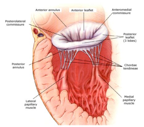
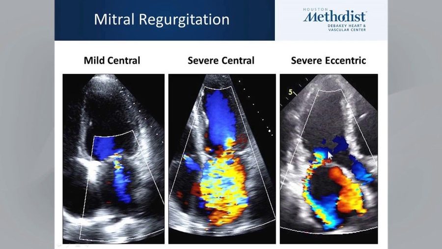
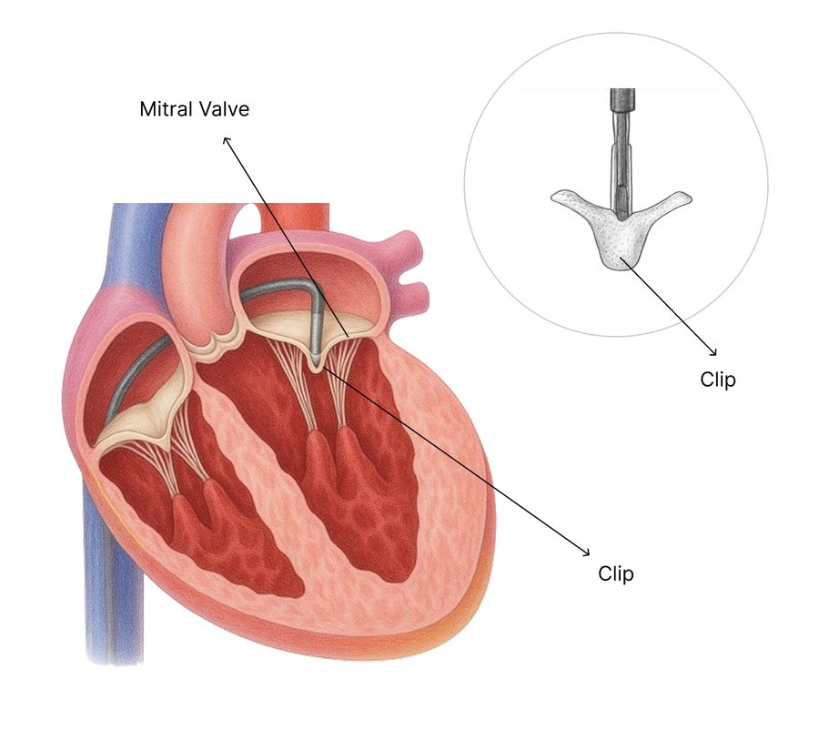
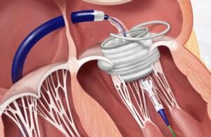
Also tested
- Chronic mitral regurgitation. It dilates the left ventricle with eccentric hypertrophy, causing left heart failure, then pulmonary hypertension, and finally combined right and left heart failure.
- Medical management of mitral regurgitation. Treatment aims to increase forward cardiac output while reducing regurgitant volume, and to treat comorbidities such as atrial fibrillation and pulmonary hypertension.
4.6 · Mitral valve prolapse
“It's a different instructional objective, so you have to know it, but basically, it's just mitral regurg. The reason is just different.”
| Field | Slide 54 |
|---|---|
| Definition | Flaring of the mitral valve leaflets into the left atrium during systole |
| Cause | Myxomatous degeneration of the mitral valve; the slide calls it a congenital condition |
| Who | Usually healthy women 15–30 years old |
| Presentation | Usually benign and asymptomatic; mid-systolic click at the apex; may develop mitral regurgitation symptoms and signs |
| Diagnosis | Echocardiogram: thickened, redundant mitral leaflets (>5 mm) |
| Treatment | Usually none. If it progresses to mitral regurgitation: clip, transcatheter mitral valve replacement, biological or mechanical replacement |
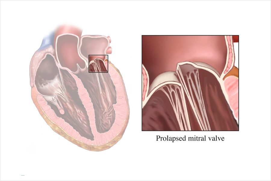
4.7 · Pulmonic stenosis
- Definition: right ventricular outflow obstruction of blood to the lungs.
- Cause: almost always congenital; carcinoid disease; commonly part of tetralogy of Fallot.
- Mechanism: raised right ventricular pressure → right ventricular hypertrophy (diastolic failure); volume overload → dilated right heart failure (systolic failure).
- Symptoms: mild-moderate usually asymptomatic; severe → right heart failure symptoms: exertional dyspnea, fatigue, angina, syncope. Late: right heart failure, hepatomegaly, ascites, edema.
- The murmur: “Harsh, high pitched, crescendo decrescendo, mid systolic ejection. Look at your words.” At the left 2nd–4th intercostal space / left upper sternal border; increases with inspiration; split S2.
- Diagnosis: electrocardiogram — right axis deviation, right ventricular hypertrophy; echocardiogram — abnormal valve motion, increased jet on Doppler. (The grading table is not tested — “not the lesser valves”.)
- Treatment: mild-moderate → none, with a 94% 20-year survival. Severe with symptoms → diuretics for right heart failure symptoms, pulmonic balloon valvuloplasty; severe congenital disease may need replacement.
- Refer to cardiology if present on echocardiogram, whether congenital or heart-failure related.
4.8 · Pulmonic regurgitation
Incomplete pulmonic valve closure lets blood flow back into the right ventricle. Iatrogenic is the MOST COMMON cause of significant regurgitation — after surgical valvotomy, valvectomy or valvuloplasty to relieve right ventricular outflow tract obstruction. He said “the most common overall reason is iatrogenic”; overall, mild regurgitation from pulmonary hypertension is commoner.
Also tested
- Pulmonic regurgitation cause. Significant pulmonic regurgitation is most often iatrogenic: surgical valvotomy, valvectomy or valvuloplasty done to relieve right ventricular outflow tract obstruction leaves the valve leaking.
- Pulmonic regurgitation discovery. It is usually an incidental finding on an echocardiogram or physical examination; management is to treat the underlying condition.
- Pulmonic regurgitation referral. Pulmonic regurgitation is referred to cardiology when it is seen on echocardiogram or when there are right heart failure signs and symptoms.
- Pulmonic regurgitation. It usually comes to attention as an incidental echo or exam finding, on echocardiogram or physical examination; the murmur is soft and hard to hear.
- Pulmonic regurgitation murmur maneuvers. Anything that increases venous return makes the murmur louder: inspiration, squatting and lying supine. Expiration and standing decrease it.
- Causes: high-pressure — pulmonary hypertension; low-pressure — congenital (bicuspid), carcinoid disease, iatrogenic (most common cause of significant\n regurgitation).
- Mechanism and symptoms: retrograde flow from the pulmonary artery → right ventricular overload → right-sided heart failure: fatigue, peripheral edema, jugular venous distension, ascites.
- The murmur: brief, high-pitched, decrescendo early DIASTOLIC murmur at the 2nd left intercostal space, radiating to the mid right sternal border with full inspiration — the Graham Steell murmur. Louder with more venous return (inspiration, squatting, supine); softer with less (expiration, standing).
- Diagnosis: usually an incidental finding on echocardiogram or physical exam.
- Treatment: treat the underlying condition. Refer when seen on echocardiogram or with right heart failure symptoms and signs.
Also tested
- Pulmonic stenosis referral. Whether congenital or related to heart failure, pulmonic stenosis present on echocardiogram should be referred to a cardiologist.
4.9 · Tricuspid stenosis
- Definition: right ventricular inflow obstruction — the narrowed tricuspid valve obstructs inflow from the right atrium to the right ventricle (slide 66; slide 65's “from the LA” is a deck error).
- Cause: most commonly rheumatic fever, usually alongside rheumatic mitral disease; carcinoid disease is the other notable cause. Slide 65 says that in the United States it is “almost always congenital”; that is not accurate, since congenital tricuspid stenosis is rare. Most common in women.
- Mechanism: right atrial dilation and hypertrophy, low cardiac output, portal congestion → right heart failure without right ventricular dysfunction.
- Symptoms: mild-moderate usually asymptomatic; severe → a fluttering discomfort in the neck, cold skin (low cardiac output), right upper quadrant pain (enlarged liver).
- The murmur: soft opening snap and mid-DIASTOLIC RUMBLE at the left lower sternal border near the xiphoid; increases with inspiration; jugular venous distension.
- Diagnosis: electrocardiogram — right atrial enlargement out of proportion to right ventricular hypertrophy; echocardiogram — abnormal valve motion, increased jet on Doppler (grading not tested; the slide's example picture grades mild by its own table and is not used).
- Treatment (severe with symptoms): low salt, diuretics, aldosterone antagonist; bioprosthetic valve replacement is preferred; balloon valvotomy and valve repair are reserved for hepatic congestion that may lead to cirrhosis or severe systemic venous congestion. The “Mild to Moderate” heading on slide 69 has nothing under it.
- Refer when seen on echocardiogram or with heart-failure symptoms.
4.10 · Tricuspid regurgitation
Incomplete closure lets blood flow from the right ventricle back into the right atrium.
- Most common cause: dilation of the tricuspid annulus from right ventricular dilation in the setting of pulmonary hypertension.
- Primary cause: infective endocarditis in intravenous drug users — “when it's on the tricuspid valve, it's usually IV [intravenous] drug use.”
- The murmur: HOLOSYSTOLIC at the left mid or lower sternal border near the epigastrium; increases with inspiration (Carvallo sign), which distinguishes it from mitral regurgitation.
Also tested
- Carvallo sign. Inspiration increases venous return to the right heart, so the tricuspid regurgitation murmur grows louder while mitral regurgitation does not; deep inspiration distinguishes them.
- Tricuspid regurgitation referral. It is referred when identified on echocardiogram or when there are right heart failure symptoms; pedal edema and ascites mark severe disease.
- Tricuspid regurgitation neck findings. Raised jugular venous pressure with neck pulsations and a venous thrill over the right jugular vein, as blood is driven back into the right atrium.
- Tricuspid regurgitation murmur. It is a holosystolic murmur at the left lower sternal border that grows louder on inspiration, and it accompanies pulmonary arterial hypertension in right-sided failure.
- Other findings: elevated jugular venous pressure with neck pulsations; a venous thrill over the right jugular vein. Mild-moderate usually asymptomatic; severe → pedal edema, ascites.
- Diagnosis: echocardiogram with Doppler shows the regurgitation and prolapsed, scarred or displaced leaflets.
- Treatment: treat the underlying condition; annuloplasty if the regurgitation is from annular dilation; valve repair or replacement if from a primary cause.
- Refer when identified on echocardiogram or with right heart failure symptoms.
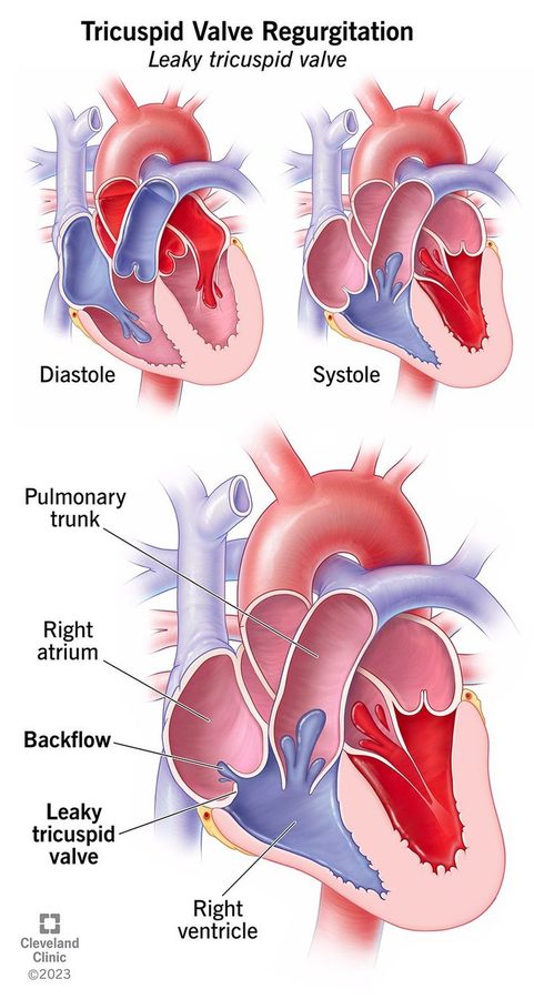
4.11 · Prosthetic valves and anticoagulation
| Mechanical (titanium and carbon) | Biological / bioprosthetic (pig or cow tissue) | |
|---|---|---|
| Anticoagulation | Lifelong | None beyond the immediate postoperative period |
| Preferred when | — | Life expectancy is shorter than the valve's expected longevity, or any contraindication to anticoagulation |
For mechanical valves, a vitamin K antagonist is recommended over no vitamin K antagonist and over antiplatelet agents. The most commonly used one is warfarin. “It's the only one that's going to work well with mechanical.”
| Mechanical valve position | Target INR (international normalized ratio) |
|---|---|
| Aortic | 2.5 (range 2.0–3.0) |
| Mitral | 3.0 (range 2.5–3.5) |
| Both aortic and mitral | 3.0 (range 2.5–3.5) — “stick with the higher” |
Bridging: if surgery requires the switch to heparin, warfarin must be BRIDGED until the international normalized ratio is therapeutic. “The word we use is bridge … the answer is heparin.”
Also tested
- Mechanical valve and warfarin interruption. If surgery requires interrupting warfarin, bridge with heparin until the international normalized ratio is therapeutic; stopping warfarin with no bridge, leaving the valve uncovered, is to be avoided.
- Choice of valve with contraindication to anticoagulation. A bioprosthetic valve is preferred when there is any contraindication to anticoagulation; it does not need anticoagulation beyond the immediate postoperative period.
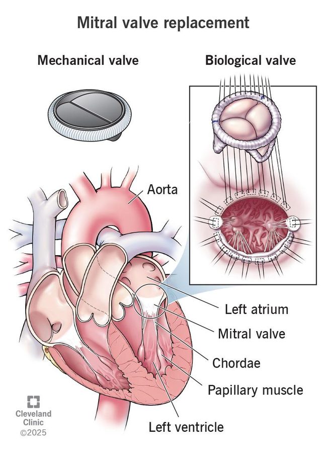
By age: slide 74's cut-offs are reversed; the standard is mechanical for a younger patient and bioprosthetic or transcatheter for an older one (see the box above 4.1). No quiz question keys an age cut-off.
4.12 · Test tips: the nine murmurs side by side
“This is the best way and easiest way to identify them on your test. It is not the best and easiest way to identify them in real life.”
| Lesion | Timing | Words | Where | Radiates / respiration | The clue |
|---|---|---|---|---|---|
| Aortic stenosis | Systolic ejection | Harsh, crescendo-decrescendo | 2nd right intercostal space | Carotids; louder on expiration | Angina / syncope / heart failure; forceful apex + delayed weak carotid = severe |
| Aortic regurgitation | Diastolic | High-pitched blowing | 3rd intercostal space / left upper sternal border | Apex; louder on expiration | Corrigan's pulse, de Musset sign, wide pulse pressure |
| Mitral stenosis | Diastolic | Low-pitched rumble after an opening snap | Apex (bell, left lateral decubitus) | Louder on expiration | Rheumatic history; atrial fibrillation; hoarseness, hemoptysis, dysphagia |
| Mitral regurgitation | Holosystolic | Blowing | Apex (left lateral decubitus) | Axilla; louder on expiration | Acute after infarction (papillary muscle) or chordal rupture |
| Mitral valve prolapse | Mid-systolic | Click | Apex | — | Healthy woman 15–30 |
| Pulmonic stenosis | Mid-systolic ejection | Harsh, high-pitched, crescendo-decrescendo | Left 2nd–4th intercostal space | Louder on inspiration; split S2 | Congenital; tetralogy of Fallot |
| Pulmonic regurgitation | Early diastolic | Brief, high-pitched, decrescendo | 2nd left intercostal space | Mid right sternal border on full inspiration (Graham Steell) | Prior valvotomy / valvuloplasty (iatrogenic) |
| Tricuspid stenosis | Mid-diastolic | Rumble, soft opening snap | Left lower sternal border near the xiphoid | Louder on inspiration | Right atrial enlargement out of proportion; right upper quadrant pain |
| Tricuspid regurgitation | Holosystolic | — | Left mid/lower sternal border near the epigastrium | Louder on inspiration (Carvallo) | Intravenous drug use; pulmonary hypertension |
Also tested
- Mitral regurgitation murmur with handgrip. It becomes louder with squatting and handgrip, because raised peripheral vascular resistance to ventricular ejection pushes more regurgitation back into the left atrium.
- Mitral regurgitation murmur with standing. It becomes softer with standing or the Valsalva maneuver and increases with squatting and handgrip, which raise resistance to ventricular ejection.
- Symptoms of pulmonic stenosis. Mild to moderate disease is usually asymptomatic; severe stenosis produces right heart failure symptoms such as exertional dyspnea, fatigue, angina and syncope.
Slides 8–10, 17, 27–28, 38, 48, 54, 57, 63, 67, 71, 73
Also tested
- Tricuspid stenosis referral. It is referred when seen on an echocardiogram or when there are heart failure related symptoms.
- Tricuspid stenosis management. Severe symptomatic tricuspid stenosis is managed with a low-salt diet together with diuretics and an aldosterone antagonist, which limits the systemic venous congestion.
4.13 · The conditions, point by point
Aortic stenosis
Defining feature: harsh systolic crescendo-decrescendo murmur at the 2nd right intercostal space radiating to the carotids; severe = <1.0 cm, >40 mmHg, >4.0 m/s
- Definition
- Obstruction of left ventricular outflow across the aortic valve; the most common valvular disease.
- Who gets it
- 80% of symptomatic patients are men; 25% of patients older than 65 and 35% older than 70; symptoms usually at 60–80. Degenerative is the most common cause over 70, bicuspid valve under 70.
- Risk factors
- Hypertension, hyperlipidemia, smoking; rheumatic heart disease; endocarditis.
- Classic signs & symptoms
- Exertional angina (5-year survival), exertional syncope (3 years), congestive heart failure / exertional dyspnea (2 years — most common, worst prognosis).
- Physical exam findings
- Harsh systolic ejection murmur, right upper sternal border, to the carotids; softer with Valsalva, standing, handgrip; louder with squatting, sitting forward. Forceful apex beat with a delayed, weak carotid pulse = severe.
- Diagnostics / tests
- Echocardiogram for any suggestive symptom: thick, calcified leaflets, reduced opening, left ventricular hypertrophy. Electrocardiogram: left ventricular hypertrophy (low sensitivity). Left heart catheterization is definitive.
- First-line treatment
- Medical treatment is not effective; preload dependent — avoid strenuous activity, dehydration, beta blockers and calcium channel blockers, nitrates. Severe + symptomatic systolic dysfunction or bicuspid valve → valve replacement (transcatheter, mechanical or biological); children and young adults → balloon valvuloplasty. Refer all to cardiology.
- Complications
- Concentric hypertrophy → diastolic heart failure; symptomatic 3-year mortality 75%.
Slides 11–22
Aortic regurgitation
Defining feature: high-pitched blowing DIASTOLIC murmur at the left upper sternal border with Corrigan's (water hammer) pulse
- Definition
- Incomplete aortic valve closure with backflow into the left ventricle (aortic insufficiency).
- Who gets it
- Not covered in the lecture.
- Risk factors
- Causes: bicuspid valve, endocarditis, rheumatic fever, myxomatous disease, trauma, syphilis, ankylosing spondylitis; aortic dissection, cystic medial degeneration, Marfan syndrome, familial aneurysm, aortitis, hypertension.
- Classic signs & symptoms
- Acute: cardiogenic shock, pulmonary edema. Chronic: silent 10–15 years, then congestive heart failure symptoms and anginal pain.
- Physical exam findings
- Diastolic blowing murmur to the apex, displaced point of maximal impulse, de Musset sign (head bobbing), Corrigan's pulse (rapid upstroke, rapid collapse), bounding pulses, wide pulse pressure, thrills.
- Diagnostics / tests
- Echocardiogram determines the cause (root dilation, dissection) and shows fluttering of the anterior mitral leaflet; severe = regurgitant fraction 50% or more. Electrocardiogram: left ventricular hypertrophy (non-specific). Catheterization definitive, not necessary.
- First-line treatment
- Afterload reduction (ACE inhibitor / angiotensin receptor blocker, dihydropyridine, hydralazine). Acute severe: emergency valve replacement or repair within 24 hours (bridge with intravenous diuretics + nitroprusside). Chronic with reduced function: surgery. Refer any audible murmur.
- Complications
- Eccentric hypertrophy → heart failure with reduced ejection fraction; untreated severe disease with heart failure: survival 2–3 years.
Slides 23–32
Mitral stenosis
Defining feature: low-pitched rumbling diastolic murmur at the apex after an opening snap, in a patient with rheumatic fever years before
- Definition
- Obstruction of left atrial outflow across the mitral valve.
- Who gets it
- 80% of rheumatic mitral stenosis is in women; diagnosed about 20 years after rheumatic fever; rare in developed countries.
- Risk factors
- Rheumatic heart disease — the most common cause.
- Classic signs & symptoms
- Left-sided heart failure (dyspnea on exertion, cough, orthopnea, paroxysmal nocturnal dyspnea, fatigue); hemoptysis; hoarseness (Ortner syndrome); dysphagia.
- Physical exam findings
- Opening snap then a decrescendo-crescendo rumble at the apex, bell, left lateral decubitus.
- Diagnostics / tests
- Echocardiogram (large left atrium, stenotic valve) for dyspnea or orthopnea. Electrocardiogram: left atrial abnormality, atrial fibrillation, right ventricular hypertrophy. Severe: <1.0 cm, >10 mmHg, >3.0 m/s. Catheterization not necessary.
- First-line treatment
- Asymptomatic in sinus rhythm: none. Dyspnea / orthopnea: loop diuretics. Severe + symptoms or pulmonary hypertension: percutaneous balloon valvotomy; valve replacement. Refer everyone on echo.
- Complications
- Left atrial enlargement → atrial fibrillation → thromboembolism; pulmonary hypertension → right heart failure.
Slides 33–43
Mitral regurgitation
Defining feature: holosystolic blowing murmur at the apex radiating to the axilla
- Definition
- Incomplete mitral valve closure with backflow into the left atrium during systole.
- Who gets it
- Not covered in the lecture.
- Risk factors
- Acute: myocardial infarction with papillary muscle rupture, endocarditis, rheumatic fever, chordal rupture, acute left ventricular dilation, prosthetic valve failure. Chronic: mitral valve prolapse, endocarditis, rheumatic fever, hypertrophic obstructive cardiomyopathy, dilated cardiomyopathy.
- Classic signs & symptoms
- Mild-moderate asymptomatic; palpitations; acute left-sided failure (dyspnea, orthopnea, rales, edema).
- Physical exam findings
- Holosystolic blowing murmur, apex to axilla, ± S3, ± thrill; louder with squatting and handgrip, softer with standing and Valsalva.
- Diagnostics / tests
- Transthoracic echocardiogram (regurgitant flow, left atrial enlargement, hypertrophy); severe = regurgitant fraction 50% or more. Electrocardiogram: left atrial enlargement. Catheterization not required.
- First-line treatment
- Increase forward output, reduce regurgitant volume; treat atrial fibrillation and pulmonary hypertension. Surgery for severe disease, ejection fraction <60% or end-systolic dimension >40 mm: clip, transcatheter replacement, mechanical or biological valve.
- Complications
- Eccentric hypertrophy → systolic heart failure; chronic → pulmonary hypertension → combined right and left failure.
Slides 44–52
Mitral valve prolapse
Defining feature: mid-systolic click at the apex in a healthy young woman
- Definition
- Flaring of the mitral leaflets into the left atrium during systole.
- Who gets it
- Usually healthy women 15–30.
- Risk factors
- Not covered in the lecture (cause: myxomatous degeneration; called congenital).
- Classic signs & symptoms
- Usually benign and asymptomatic.
- Physical exam findings
- Mid-systolic click at the apex.
- Diagnostics / tests
- Echocardiogram: thickened, redundant leaflets (>5 mm).
- First-line treatment
- Usually none; if mitral regurgitation develops, treat as mitral regurgitation.
- Complications
- Progression to mitral regurgitation.
Slide 54
Pulmonic stenosis
Defining feature: harsh mid-systolic ejection murmur at the left upper sternal border that increases with inspiration, with a split S2
- Definition
- Right ventricular outflow obstruction of blood to the lungs.
- Who gets it
- Not covered in the lecture (almost always congenital).
- Risk factors
- Congenital disease; tetralogy of Fallot; carcinoid disease.
- Classic signs & symptoms
- Mild-moderate asymptomatic; severe: exertional dyspnea, fatigue, angina, syncope.
- Physical exam findings
- Harsh crescendo-decrescendo murmur, left 2nd–4th intercostal space, louder on inspiration, split S2; late hepatomegaly, ascites, edema.
- Diagnostics / tests
- Electrocardiogram: right axis deviation, right ventricular hypertrophy. Echocardiogram: abnormal valve motion, increased jet.
- First-line treatment
- Mild-moderate: none. Severe with symptoms: diuretics, pulmonic balloon valvuloplasty; replacement if severe congenital. Refer if on echo.
- Complications
- Right ventricular hypertrophy, then dilated right heart failure. Mild-moderate: 94% 20-year survival.
Slides 55–57, 59
Pulmonic regurgitation
Defining feature: brief early diastolic decrescendo murmur at the 2nd left intercostal space (Graham Steell) after prior outflow-tract surgery — iatrogenic, the most common cause of significant regurgitation
- Definition
- Incomplete pulmonic valve closure with backflow into the right ventricle.
- Who gets it
- Not covered in the lecture.
- Risk factors
- Iatrogenic (most common cause of significant regurgitation); pulmonary hypertension (commonest overall, usually mild); congenital bicuspid valve; carcinoid disease.
- Classic signs & symptoms
- Right-sided heart failure: fatigue, peripheral edema, jugular venous distension, ascites.
- Physical exam findings
- Graham Steell murmur; louder with inspiration, squatting, supine; softer with expiration, standing.
- Diagnostics / tests
- Usually an incidental finding on echocardiogram or exam.
- First-line treatment
- Treat the underlying condition; refer when seen on echo or with right heart failure.
- Complications
- Right ventricular overload → right heart failure.
Slides 60–64
Tricuspid stenosis
Defining feature: mid-diastolic rumble at the left lower sternal border near the xiphoid that increases with inspiration; right atrial enlargement out of proportion
- Definition
- Right ventricular inflow obstruction from the right atrium to the right ventricle.
- Who gets it
- Most common in women.
- Risk factors
- Rheumatic fever (most common); carcinoid disease. (Slide 65's “congenital in the United States” is not accurate.)
- Classic signs & symptoms
- Severe: fluttering discomfort in the neck, cold skin, right upper quadrant pain.
- Physical exam findings
- Soft opening snap, mid-diastolic rumble, louder on inspiration; jugular venous distension.
- Diagnostics / tests
- Electrocardiogram: right atrial enlargement out of proportion to right ventricular hypertrophy. Echocardiogram: abnormal valve motion, increased jet.
- First-line treatment
- Severe with symptoms: low salt, diuretics, aldosterone antagonist; bioprosthetic replacement preferred; balloon valvotomy only for hepatic congestion or severe venous congestion. Refer if on echo.
- Complications
- Portal congestion (cirrhosis risk); right heart failure without right ventricular dysfunction; low cardiac output.
Slides 65–69
Tricuspid regurgitation
Defining feature: holosystolic murmur at the left lower sternal border that increases with inspiration (Carvallo sign); think intravenous drug use
- Definition
- Incomplete closure with backflow from the right ventricle into the right atrium.
- Who gets it
- Intravenous drug users (primary disease from endocarditis).
- Risk factors
- Pulmonary hypertension (annular dilation — most common); infective endocarditis in intravenous drug use.
- Classic signs & symptoms
- Mild-moderate asymptomatic; severe: pedal edema, ascites.
- Physical exam findings
- Holosystolic murmur near the epigastrium, louder on inspiration; elevated jugular venous pressure with neck pulsations; venous thrill over the right jugular vein.
- Diagnostics / tests
- Echocardiogram with Doppler: regurgitation; prolapsed, scarred or displaced leaflets.
- First-line treatment
- Treat the underlying condition; annuloplasty for annular dilation; repair or replacement for a primary cause. Refer when on echo or with right heart failure.
- Complications
- Right-sided congestion (edema, ascites).
Slides 9, 70–72
Prosthetic heart valves
Defining feature: mechanical = lifelong warfarin (a vitamin K antagonist, never antiplatelets alone); biological = no long-term anticoagulation
- Definition
- Mechanical (titanium and carbon) or biological (pig or cow tissue) replacement valves; transcatheter aortic or mitral valve replacement.
- Who gets it
- Bioprosthetic preferred with a short life expectancy or a contraindication to anticoagulation; mechanical for a younger patient, bioprosthetic or transcatheter for an older one (slide 74's age cut-offs are reversed).
- Risk factors
- Not covered in the lecture.
- Classic signs & symptoms
- Not covered in the lecture (mechanical prosthetic mitral failure is an acute cause of mitral regurgitation).
- Physical exam findings
- Not covered in the lecture.
- Diagnostics / tests
- International normalized ratio targets: aortic 2.5 (2.0–3.0); mitral 3.0 (2.5–3.5); both 3.0 (2.5–3.5).
- First-line treatment
- Mechanical: lifelong vitamin K antagonist (warfarin). Around surgery: bridge with heparin until the ratio is therapeutic. Bioprosthetic: none beyond the immediate postoperative period.
- Complications
- Not covered in the lecture.
Slides 21, 45, 52, 74–75
Also tested
- Rheumatic mitral stenosis. It is rare in developed countries because rheumatic fever, its most common cause, has become less common there.
4.14 · Populations
| Population | What the deck gives |
|---|---|
| Infant, child, adolescent | Children and young adults with aortic stenosis → percutaneous aortic balloon valvuloplasty (slide 21). Pulmonic stenosis is almost always congenital, often with tetralogy of Fallot (slide 55). No other pediatric strategy. |
| Adult | Mitral valve prolapse: usually healthy women 15–30, usually no treatment (slide 54). Bicuspid valve is the most common cause of aortic stenosis under 70 (slide 12). Rheumatic mitral stenosis usually diagnosed about 20 years after rheumatic fever (slide 34). |
| Elderly | Aortic stenosis is present in 25% of patients older than 65 and 35% older than 70; degenerative disease is the most common cause over 70; symptoms usually appear at 60–80 (slides 12, 13, 16). |
5 · Coronary Artery Disease
Instructional Objectives
CORONARY ARTERY DISEASE — Grady G. Carter, MSHS, PA-C
- Compare and contrast the etiologies, epidemiology, risk factors, clinical manifestations, differential diagnosis, diagnostic testing (including ordering and interpretation), management (acute and chronic, including applicable rehabilitative and palliative care), appropriate referrals, patient education, and prognosis of the following coronary artery disease:
-
- Angina pectoris
- Stable
- Unstable
- Vasospastic (Prinzmetal variant)
- Acute coronary syndrome
- Acute myocardial infraction
- Non-ST segment elevation
- ST segment elevation
- Identify medical care strategies for coronary artery disease in the lecture topic list for the
following populations.
- infant
- child
- adolescent
- adult
- elderly
Quoted as the syllabus prints it, including its own lettering (a lone “b.” before the angina list, then b, c and d again) and its typo “infraction” for infarction. There are two real objectives: a, the compare-and-contrast across the disease list, and d, populations.
What is tested, in the lecturer's own words (lecture recording, 25 September, two parts):
- Troponin numbers. The one “for testing” statement of the lecture: “a person is considered to have non-abnormal troponin if it is 0.04 or less … With high sensitivity troponin, a 12 is an abnormal number for a woman, a 20 is an abnormal value for a man … Those are the numbers that you need to know. The other part you need to know is we use troponin to do what? Rule it out.” These come from a slide shown in class that is not in the posted deck (5.5).
- Risk scores: only one cut-off. “I don't need you to know all the cut-offs on the scores or the percentages, just know that this is what we use to justify … A risk score of anything three or higher” — TIMI (Thrombolysis in Myocardial Infarction) 3 or higher justifies an early invasive approach. The HEART score's parts are learned; its bands and percentages are not.
- Your words have meaning. Heart catheterization, coronary angiography, percutaneous coronary intervention and plain old balloon angioplasty are different things, and so are unstable angina, non-ST-elevation and ST-elevation infarction (5.7, 5.10).
- The new name: “Anterior interventricular artery … That is the new name for the LAD … you need to know that's the name.”
- Three main coronary arteries is the answer (“That's not gonna be on the test … three is the answer”); the extra grafts of a quadruple bypass are not.
- Self-study: the big localization chart on slide 62 (“feel free to review”). Learn the lead groups of slide 61 plus the posterior row he taught.
- Program rules: no dosing, no mechanism-of-action questions, generic names only, no acronyms in questions. Drug classes may be named (“P2Y12 receptor antagonist”). No electrocardiogram tracings are used in the quizzes; findings are given as words.
Built from the slides. The recording adds weight only: ★ marks show what he emphasized. There is no slide erratum this lecture (his one “that's wrong” withdrew his own spoken claim that V4 is a lateral lead, so slide 61 stands). Where the slide, his words and standard references disagree, this guide teaches what is medically true and says what was said instead (the lists below).
Deck error — slips he did not mention
- Slide 86: non-ST-elevation ischemia is written “ST inversion (> 1mm)”. There is no such thing: the ST segment is depressed or elevated, and T waves invert. Learn slide 47's wording — ST depression >1 mm and/or T-wave inversion in two or more consecutive leads, with elevated troponin. (He read 86 aloud as “usually it's an ST depression”.)
- Typos: “Forsinopril” (slide 82) is fosinopril; “Antistreplase” (slide 74) is anistreplase; “Drug Eluding Stent” (slide 50) is drug-eluting; “suppled” (slide 12) is supplied; “PCTA” (slides 69–72) is usually written PTCA (percutaneous transluminal coronary angioplasty).
- Slide 52, “Noncompliance with DAPT in the first 6 weeks → 50% risk of Sudden Re-InStent Stenosis” (he called it “a flip of the coin”). The event is stent thrombosis — a clot that suddenly occludes the stent — not restenosis, which is slow scar-tissue narrowing over months. Stopping dual antiplatelet therapy early is its strongest predictor, and stent thrombosis is often fatal, but no source gives a 50% risk. Learn stopping early → sudden stent thrombosis; no quiz keys a percentage.
- Slide 54 cites the unstable angina classification as “APACHE II”; it is the Braunwald classification. The table was brushed past and is not used here.
- Slide 18's caption (“compare thickness of cap with previous picture”) points at the thin-cap picture, which comes after it on slide 19.
Where the slides disagree — what is true, and what is not keyed
- Slide 65, “Elevated cardiac markers must be present” for ST-elevation infarction. In class he said the opposite (“Do I need an elevated troponin? No, because I may have caught it before I can catch it in the blood”), and he is right about practice: ST-elevation infarction is recognized from the symptoms and the electrocardiogram, and reperfusion never waits for troponin, which rises later and confirms the infarct. No question asks whether troponin is required.
- Vasospastic angina, slide 91's “T-wave inversion; maybe ST depression”. Slide 90 says transient ST-segment elevation during an episode, and he described only “ST segment changes” that resolve. Learn slide 90; slide 91's line is left alone.
- Nitroglycerin in right ventricular infarct: slide 63 says “DO NOT GIVE”; slide 73 says “Caution in inferior MIs (right sided) with evidence of CHF”. He said “do not give” three times and never read 73's line — learn do not give. He also generalized it to every inferior infarct, but the right ventricle is involved in only 15–30% of right coronary occlusions: the rule is for a right ventricular infarct.
- Dual antiplatelet durations: slide 52 says clopidogrel for a minimum of 12 months and aspirin for a minimum of 6; slide 83 says clopidogrel “usually for a full year”. The accurate version (2016 ACC/AHA dual antiplatelet update): after an acute coronary syndrome the P2Y12 antagonist is given for about 12 months, and aspirin is continued indefinitely — the “minimum of 6 months” for aspirin is not accurate. He gave no durations. Not keyed.
- Coronary dominance numbers: slides 11/13 say the right coronary artery gives the posterior interventricular branch in 67% (left 15–33%); slide 62's chart says right dominant in ~70–85%. If you need one, it is 67%, but no question asks it.
- Microvascular angina and MINOCA. Slide 94 lists “Myocardial Infarction with Non Obstructive Coronary Arteries (MINOCA)” under microvascular angina, and he equated them (“primary care … microvascular angina … a cardiology report … MINOCA”). That is not accurate: MINOCA is an infarction (troponin rise with ischemia) with non-obstructed coronaries, while microvascular angina (cardiac syndrome X) is angina with normal coronaries and no infarction. Slide 94's “possibly even with elevated cardiac biomarkers” belongs to MINOCA. No question asks you to tell them apart.
- Stress-test numbers (slide 35, “Abn Sensitivity 90%, Nrml Specificity 77%”) and the HEART score's bands, points and percentages (slide 34) and TIMI's percentage table (slide 72) are not asked.
- Slide 72a extras — the 1-hour and 3-hour change values and “as early as 90 min” — were never spoken. Not keyed.
Spoken slips — the slide is right: smoking is a strong predictor in women under 45 (he said “over 45”); vasospastic angina is often under age 50 (he said “under 15”); the troponin cut-offs are the 99th percentile (he said “90th”, and read the 40 row as “99% of people”); nitroglycerin is not a phosphodiesterase inhibitor (it is primarily a venodilator, slide 40); troponins are proteins, not enzymes; avoid nonselective beta blockers in vasospasm (he said “the beta blockers”); bypass uses an artery first (he said “usually a vein from the leg”); subendocardial ischemia gives ST depression and transmural ischemia ST elevation (a non-ST-elevation infarct is still an infarct). His two morphine remarks cancel out and are not learned.
Where each objective is answered
| Objective | Answered in |
|---|---|
| a. Etiology, epidemiology, risk factors, manifestations, differential, testing, management, referral, education, prognosis | 5.1–5.5 (the shared foundation), then each disease's subsection and its eight-point block in 5.14 · rehabilitative care appears only as risk-factor control (slide 79) and “exercise as tolerated” (slide 53); no palliative content |
| a. Angina pectoris | 5.3 |
| b. Stable | 5.6 |
| c. Unstable | 5.7 |
| d. Vasospastic (Prinzmetal variant) | 5.12 |
| b. Acute coronary syndrome | 5.4, 5.5, 5.7 |
| c. Acute myocardial infarction — 1. non-ST segment elevation | 5.7, 5.8 |
| c. Acute myocardial infarction — 2. ST segment elevation | 5.8, 5.9, 5.10, 5.11 |
| d. Populations | 5.15 · the deck narrows this to adult and elderly; it has no infant, child or adolescent content |
Also in the deck but not in the syllabus list: right ventricular infarct (5.9), sudden cardiac death (5.11) and microvascular angina (5.13, named in the deck's own objectives).
5.1 · Ischemia and the coronary arteries
Ischemia is a mismatch between the myocardium's oxygen demand and its supply (“writing checks that its vasculature can't cash”).
| ↓ Oxygen supply | ↑ Oxygen demand |
|---|---|
| Atherosclerosis, thrombus/embolus, anemia, spasm, dissection, carboxyhemoglobin, hypoxia | Exercise, emotional stress, left ventricular hypertrophy/cardiomyopathy, hyperthyroidism, hyperthermia, cocaine |
- The heart is 0.3% of body weight but uses 7% of the body's oxygen. Irreversible injury occurs in 40–60 minutes.
- Ischemia shifts the cell to anaerobic metabolism (↑ lactic acid); damage comes from free radicals, lactic acid and raised extracellular calcium.
- Regional wall motion abnormalities, seen on an echocardiogram: hypokinesia = reduced movement, akinesia = no movement, dyskinesia = bulging wall movement.
Also tested
- Cardiogenic shock after infarction. It is more common in left-sided myocardial infarctions, and left ventricular involvement carries a higher mortality.
- Angina and exertion. Exercise and emotional stress raise myocardial oxygen demand; a narrowed artery cannot raise supply to match, and the mismatch produces pain.
- Hyperthyroidism and ischemia. Myocardial ischemia is a mismatch between oxygen demand and supply, and hyperthyroidism raises the heart's oxygen demand, as do exercise, emotional stress, left ventricular hypertrophy, hyperthermia and cocaine.
- ST elevation in V1 through V4. Leads V1-V4 are the anterior and septal leads, supplied by the anterior interventricular artery, formerly the left anterior descending artery, the most common culprit in acute infarction.
The arteries and what they feed
- Three main coronary arteries: right coronary artery, left anterior descending and circumflex.
- The right coronary artery supplies the atrioventricular node — so a right coronary infarct is more likely to cause dysrhythmias, and a patient who develops complete heart block during an infarct has a right coronary lesion. The sinoatrial node is supplied by the right coronary artery in 60% and by the circumflex in 40% (“so you can't know for sure”).
- Anterior interventricular artery (AIV) = the left anterior descending (LAD) — the new name. Culprit vessel in acute infarction: AIV/LAD 40–50% (most common), right coronary artery 30–40%, left circumflex 15–20%.
- The left ventricle is supplied by the anterior interventricular, circumflex, left marginal and diagonal branches; left ventricular involvement carries a higher mortality. The interventricular septum is mostly left coronary. Anterolateral papillary muscle: anterior interventricular + circumflex; posteromedial papillary muscle: posterior interventricular branch.
- “Everybody's heart is not the same” — coronary distribution varies between people (slide 13), which is why the posterior interventricular branch matters for nitroglycerin use.
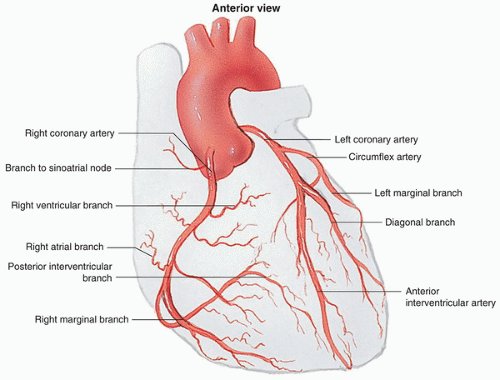
Slides 5–14
5.2 · Atherosclerosis and risk factors
- Atherosclerosis = fat, smooth muscle, fibroblasts and intracellular matrix in the wall; macrophages become foam cells under a fibrous cap.
- A thick fibrous cap covers the necrotic core of a stable atheroma; a thin-cap fibroatheroma has only a thread-like cap between the core and the lumen.
- Plaque rupture → platelet activation, and tissue factor → clotting cascade → fibrin deposition (the thrombus of an acute coronary syndrome).
| Nonmodifiable | Modifiable |
|---|---|
| Age ≥45 in men, ≥55 in women Male > premenopausal female; the gap narrows with age (“at 80”) Family history: sudden death or infarction in a male first-degree relative <55 or a female first-degree relative <65 |
Obesity · dyslipidemia · diabetes mellitus · smoking — a strong predictor, especially in women <45 · hypertension · excessive alcohol · sedentary lifestyle · ineffective stress management |
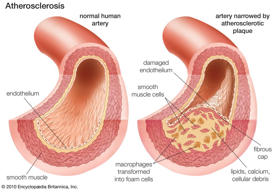
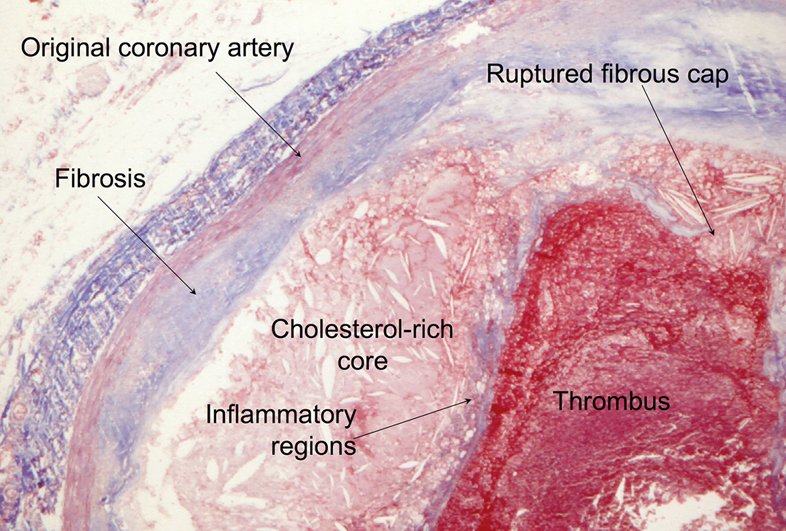
Slides 15–23
5.3 · Angina pectoris: the pain, silent ischemia and referred pain
Angina pectoris = transient myocardial ischemia, not long or severe enough to cause infarction; the symptoms recur. The pain is not affected by position and typically worsens with exertion (“the more you work, the higher your demand goes”).
- “Typical” cardiac chest pain: substernal chest pressure, worse with exertion, radiating to the neck or shoulder, with shortness of breath and diaphoresis, relieved with nitroglycerin.
- Words patients use: squeezing, tightness, pressure, constriction, burning, heartburn, fullness, band-like — and toothache.
- Levine's sign: a clenched fist held over the sternum.
- Silent ischemia — chest discomfort may be absent in diabetes mellitus, peripheral neuropathy, a transplanted heart, afferent (pain) dysfunction and different pain thresholds. It presents instead as shortness of breath, cough, nausea/vomiting, abdominal pain, diaphoresis, dizziness/lightheadedness or fatigue (“Every one of these questions is important.”)
| Referred pain (slide 31) | Goes to |
|---|---|
| Acute coronary syndrome | Jaw, tooth, arm or teeth most commonly; can be back, scapular region or epigastric/left upper quadrant |
| Thoracic aortic dissection | Left / interscapular region |
| Pancreas | Straight to the back |
| Gallbladder | Right scapular region |
| Aneurysm | Back |
Also tested
- Angina pectoris. It is transient myocardial ischemia that is not long or severe enough to cause infarction, and its symptoms are recurrent.
- Position and angina. The pain of angina pectoris is not affected by position and typically worsens with exertion; pain that changes with position lowers the suspicion that it is cardiac.
- Silent ischemia. Chest discomfort may be absent with diabetes, peripheral neuropathy and a transplanted heart; dyspnea, nausea and fatigue can be the presentation. Every chest pain workup starts with a stat electrocardiogram.
- Levine sign. A clenched fist held over the sternum while describing the discomfort is the classic gesture of ischemic, anginal chest pain, suggesting myocardial ischemia.

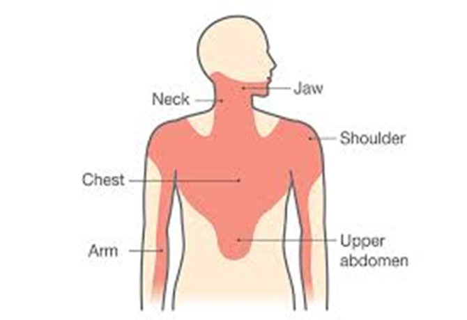
Slides 24–31
5.4 · The chest pain workup and risk scores
- All chest pain patients get a chest radiograph and a STAT electrocardiogram.
- Definitely: complete blood count, metabolic panel, prothrombin/partial thromboplastin time, troponin, lipids (later). Maybe: drug screen, D-dimer, lipase, pro-B-type natriuretic peptide.
- Risk scores predict the risk of an adverse outcome: GRACE, TIMI, PURSUIT, HEART and others.
- HEART score = History, EKG (electrocardiogram), Age, Risk factors (diabetes, hypertension, hyperlipidemia, obesity, smoking, family history of coronary disease; known coronary disease counts more), initial Troponin. “All goes out the window when your T is elevated.” Its bands and percentages are not tested.
- TIMI (Thrombolysis in Myocardial Infarction) score — for unstable angina and non-ST-elevation infarction — 3 or higher justifies an early invasive approach (“will need ICU or emergent revascularization”). It is the interventionalist's score. Its seven one-point criteria (slide 71) are for recognition; the percentage table (slide 72) is not tested.
Also tested
- Serial troponin I. Troponin is drawn serially: when one is drawn, a second follows, because the change over time is what matters; a rising troponin I would turn the presentation into an infarction.
- Coronary calcium score. A cardiac computed tomography calcium score (0 to 400 or more, higher meaning higher risk) stratifies future risk; it is not a test for acute coronary syndrome.
- HEART score components. The score rates history, the electrocardiogram, age, risk factors such as diabetes and smoking, and the initial troponin.
Diagnostics (slide 35)
- Cardiac computed tomography calcium score = risk stratification, not acute coronary syndrome (0 to 400+; higher = more risk). “It's not used for diagnostics or treatment … with ACS.”
- Stress testing: treadmill electrocardiogram; nuclear stress test — if abnormal ⇒ cardiac catheterization.
- Echocardiogram; coronary computed tomography angiography.
Slides 32–35, 71–72
5.5 · Troponin — the numbers you need to know
From a slide shown in class between posted slides 71 and 72 that is not in the posted deck; a student asked, and he answered “Because this is so important.” The photograph below is its source.
- Troponins are protein molecules of cardiac and skeletal muscle; smooth muscle has none. Three types: I, T and C. Troponin I is specific to the heart.
| Assay | Abnormal (99th percentile) |
|---|---|
| Standard troponin I | Above 0.04 ng/mL — “0.04 or less … non-abnormal. If it is 0.05 or higher, we consider it abnormal.” |
| High-sensitivity troponin I | 12 ng/L in women · 20 ng/L in men |
- Troponin rules OUT. “A tool that says who can safely go home, not a predicting value of who definitely is having a heart attack … this rules people out better, not ruling people in.” (The high-sensitivity assay is why a woman with a value the old assay called normal is no longer sent home.)
- One means two. “When you get one, you have to get two … because we're watching the change … if the second one is higher, then you order another one.”
- Troponin is what separates unstable angina (normal) from non-ST-elevation infarction (elevated) (slide 47).
- Say “not abnormal”, not “negative”.
Not learned from this slide: the 1-hour and 3-hour change values and “as early as 90 min” (never spoken), the limit-of-quantitation and highest-reportable rows, and the 40 ng/L row (“Traditional Tn-I 0.04 = hs-Tn 40”), which he misread as “99% of people”. Other course: Principles of Diagnostic Medicine gives high-sensitivity troponin T limits and a conventional troponin I normal of <0.03 ng/mL — a different assay; keep each course's numbers with that course.
Also tested
- Troponin and disposition. Troponin is a tool that says who can safely go home: two high-sensitivity troponin I values of 4 ng/L, far below the men's 20 ng/L, with a normal electrocardiogram rule out an infarct.
- Serial troponin. When one troponin is drawn, two are needed: the change between draws is what matters, and a rising second value leads to a third as more cells die.
- Interpreting serial troponin. Serial troponins are drawn to watch the change; a second high-sensitivity troponin I above the women's 99th percentile of 12 ng/L and higher than the first is rising and abnormal, so another is ordered.
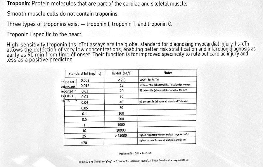
Slide 72a (supplement), slide 47
5.6 · Stable angina (stable ischemic heart disease)
- The most common form of angina pectoris. Usually precipitated by activity; relieved by rest and nitroglycerin — which is what separates it from unstable angina (pain at rest).
- Diagnosis: stress-test-induced electrocardiogram changes (T-wave inversion usually) on exercise treadmill, nuclear stress or cardiac computed tomography; coronary angiography is the gold standard for coronary artery disease (“where they actually do a heart cath and then spray dye”).
- Treatment: sublingual nitroglycerin; lifestyle modification; beta blockers (calcium channel blockers if beta blockers are contraindicated); statins; aspirin.
- Sublingual nitroglycerin is the cornerstone of angina therapy — primarily a venodilator (lowers preload, wall stress and oxygen demand; also dilates arteries). It treats the pain, not the disease.
- Patient education: call emergency medical services if the pain is unimproved or worse 5 minutes after one dose, or if 3 doses have failed (“if the third one didn't work, then you need to call 911”).
- Prevention of future attacks: aspirin (chronic stable angina and survivors of an acute coronary syndrome) and clopidogrel — more effective together.
Also tested
- Antiplatelet therapy in chronic stable angina. Aspirin plus clopidogrel both inhibit platelets, and together they are more effective than either one alone; aspirin is used in chronic stable angina and after a survived acute coronary syndrome.
Slides 36–41
5.7 · Unstable angina and non-ST-elevation infarction
Acute coronary syndromes = unstable angina, non-ST-elevation myocardial infarction (NSTEMI) and ST-elevation myocardial infarction (STEMI).
Unstable angina = a change in the patient's stable angina: it occurs at rest, is new-onset and limits activity, or is increasing (more discomfort, more often). Nitroglycerin usually, but not always, helps.
| Electrocardiogram | Troponin | |
|---|---|---|
| Stable angina | Changes on a stress test (usually T-wave inversion) | — |
| Unstable angina | Non-specific changes, usually inverted T waves; pain at rest | Normal |
| Non-ST-elevation infarction | ST depression >1 mm and/or T-wave inversion in two or more consecutive leads | Elevated — “this is what differentiates unstable from an NSTEMI” |
| ST-elevation infarction | ST elevation >1 mm in two or more consecutive leads (5.9) | Elevated |
Nuclear stress test: reversible perfusion defect = ischemia; non-reversible = infarction. A stress test can show ST depression or just T-wave inversion.
Also tested
- Change in stable angina. A change occurring at rest, new and limiting, or increasing in frequency and discomfort is unstable angina, an acute coronary syndrome and a medical emergency.
Treatment — non-ST-elevation infarction is treated the same as unstable angina
- A medical emergency. Aspirin (chewable); optimize hemodynamics with a beta blocker (to reduce oxygen demand) and intravenous nitrates if appropriate; antithrombotic therapy — typically dual antiplatelet therapy (aspirin + clopidogrel) ± anticoagulation.
- “MONA” — she's old now (morphine, oxygen, nitroglycerin, aspirin): give oxygen only if SpO2 is <91% (“if you hyper-oxygenate these people, sometimes it makes it worse”; with normal oxygen it causes peripheral vasoconstriction).
- After initial stabilization: left heart catheterization, coronary angiography, and angioplasty or percutaneous coronary intervention (5.10).
- Then: exercise as tolerated, a low-salt, low-fat diet, hydration and scheduled follow-up.
Slides 42–53, 86
5.8 · The electrocardiogram: ischemia, infarction and where the lesion is
- Subendocardial ischemia → ST depression over the region; transmural ischemia → ST elevation.
- Evolution of an ST-elevation infarction: hyperacute T waves and ST elevation (minutes–hours) → ST elevation with T inversion and a Q wave (hours–1 day) → Q wave with a “coronary T” (1 week) → a persistent Q wave (months). Pathologic Q waves mean the infarction has already happened.
| Leads | Territory | Culprit artery |
|---|---|---|
| II, III, aVF | Inferior | Right coronary artery (and/or circumflex) |
| I, aVL, V5–V6 | Lateral | Circumflex (or a diagonal of the LAD) |
| V1–V4 (V1–V2 septal, V3–V4 anterior) | Anterior / septal | Left anterior descending |
| ST depression in V1–V3 | Posterior | Right coronary or circumflex — confirm with posterior leads V7–V9 (a 15-lead) |
| V1 and V4R | Right ventricle | Right coronary artery |
His memory aids (they match the chart): “Inferior = RCA” — the right coronary artery is as common as the home appliance of the same initials; “my patients are ILL” — more I's than L's in the limb leads (II, III, aVF inferior; I, aVL lateral); and for V1→V6 “A-S-S up, A-L-L down” (V3, V2, V1 = anterior, septal, septal; V4, V5, V6 = anterior, lateral, lateral).
Posterior infarction is “super, super, super easy to miss”: ST depression in V1–V3 is the reciprocal of ST elevation on the back. Flip the tracing upside down and it looks like an ST-elevation infarct (the “Upside down” slide, 60). Get posterior leads V7–V9.
Self-study, not tested: slide 62's high-lateral and right-atrial rows, its V2–V3 millimeter cut-offs by age and sex, and its dominance percentages.
Also tested
- Right ventricular infarct and nitroglycerin. Nitroglycerin is not given because it lowers preload, reducing the Frank-Starling effect on the right ventricle, which can fail into right-sided heart failure, dropping output and precipitating shock.
- Right ventricular infarct and nitroglycerin. Avoid nitroglycerin in a right ventricular infarct (ST elevation in the right-sided lead V4R): it lowers preload, the failing right ventricle loses its Frank-Starling support, output falls further and the patient can go into shock.
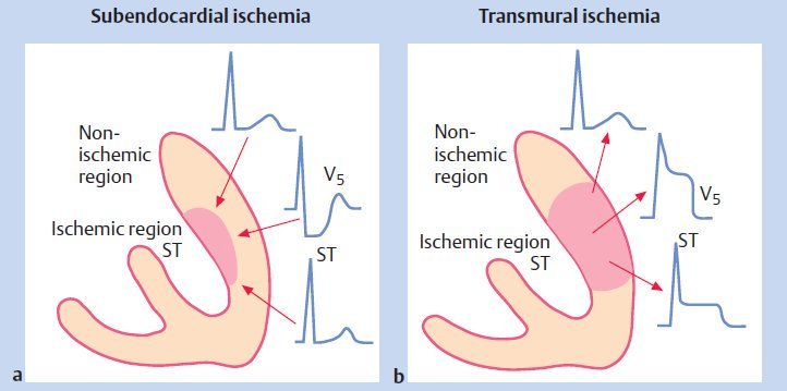
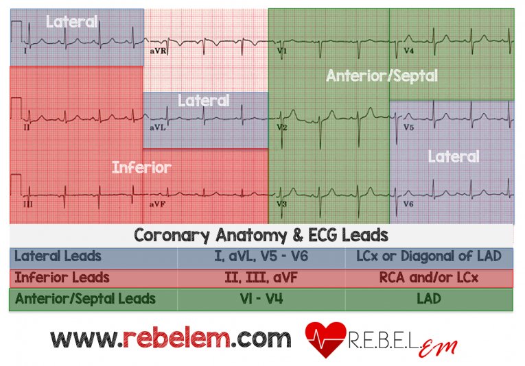
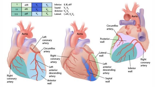
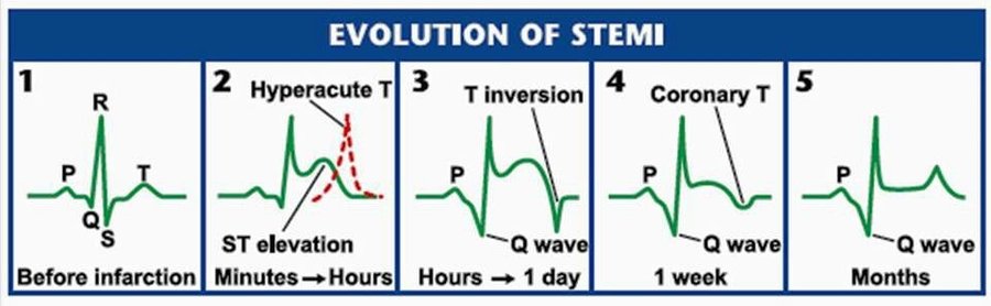
Slides 55, 61–62, 64 (tracings on 56–60 are unannotated)
5.9 · ST-elevation infarction and right ventricular infarct
ST-elevation myocardial infarction (transmural infarction): myocardial necrosis with ST elevation not quickly reversed by nitroglycerin in two or more (consecutive) leads, with reciprocal ST depression on the opposite side of the heart, and Q waves. Slide 65 lists a new left bundle branch block, and he taught it as a STEMI equivalent (“you don't have anything to show they didn't have it before … then we consider that a STEMI”). Current guidelines no longer treat a new or presumed-new block alone as a STEMI equivalent, because the block hides the ST segment: with ongoing ischemic pain, hypotension or heart failure it is managed like a STEMI (catheterization team); otherwise it is judged on the rest of the picture. The quizzes key that version.
Also tested
- Nitroglycerin in right ventricular infarction. Nitroglycerin reduces preload, removing the Frank-Starling effect the injured right ventricle depends on; output drops further, the right ventricle fails and shock follows.
- Right ventricular infarct and nitroglycerin. The infarcted right ventricle is preload dependent; nitroglycerin decreases preload, reducing the Frank-Starling effect, so it fails into right-sided heart failure, output drops and shock follows.
Time is muscle
- Electrocardiogram within 10 minutes of arrival; goal door-to-balloon time <90 minutes.
- Aspirin (chewable) and call the cardiac catheterization (percutaneous coronary intervention) team. Once catheterization is confirmed: a P2Y12 receptor antagonist (ticagrelor, prasugrel or clopidogrel) and a heparin drip.
- Nitroglycerin (sublingual every 5 minutes, paste or infusion) and a beta blocker if there is no bradycardia. “Did I mention aspirin?”
- If the closest percutaneous intervention unit is 2+ hours away, give thrombolytics, started within 30 minutes of arrival. Tenecteplase is the most common today (also alteplase, reteplase, streptokinase, anistreplase, lanoteplase). Risk: intracerebral hemorrhage. Glycoprotein IIb/IIIa inhibitors have fallen out of favor.
| Absolute contraindications to thrombolytics | Relative |
|---|---|
| Active bleeding or known bleeding disorder · major surgery or trauma in the last 4 weeks · hemorrhagic stroke in the previous year · stroke of unknown type within 6 weeks · pregnancy · uncontrolled hypertension (>190/100 mmHg) | Potential serious bleeding · severe diabetic retinopathy · current oral anticoagulation · recent non-compressible vascular puncture · prolonged (>5 min) or traumatic cardiopulmonary resuscitation |
Medication caveats (slide 73)
- Oxygen only if SpO2 <91%; with normal oxygen do not give it (peripheral vasoconstriction).
- Nitroglycerin is contraindicated within 24 hours of a phosphodiesterase inhibitor for erectile dysfunction — and ask women too, because the same drugs treat pulmonary arterial hypertension.
Right ventricular infarct
Right coronary occlusion affects the inferior and posterior left ventricular wall and the septum, and extends into the right ventricle in 15–30% (ST elevation in V1 and V4R). DO NOT GIVE nitroglycerin: dropping the preload removes the Frank-Starling push the right ventricle depends on, so output falls further and the patient goes into shock / right-sided heart failure (“you will kill somebody”). The rule is for a right ventricular infarct, not for every inferior infarct.
Slides 62–68, 73–75
5.10 · Catheterization, stents and bypass — words have meaning
| Term | What it is |
|---|---|
| Left heart catheterization | A guidewire introduced to reach the chambers of the heart and/or the coronary arteries — “the tube that goes in” |
| Coronary angiography | Dye to identify the lesion — “we sprayed dye and took pictures”; the gold standard for diagnosing coronary disease |
| Coronary angioplasty | A device to “balloon” open a narrowed artery |
| Plain old balloon angioplasty | Opening a narrowed artery without a stent |
| Percutaneous coronary intervention | Placement of a drug-eluting (or non-drug-eluting) stent to keep the artery open — “if we do anything while we're in there … it's a PCI” |
During the intervention: balloon angioplasty (dilates the vessel), atherectomy (physical removal of atheroma, calcium and excess cellular material) and stent placement (after balloon pre-dilation; direct stenting may lead to better outcomes).
Also tested
- Stopping dual antiplatelet therapy after a stent. Stopping a P2Y12 receptor antagonist such as clopidogrel plus aspirin in the first 6 weeks is the strongest predictor of stent thrombosis, a sudden clot in the stent that can cause a large, often fatal infarction.
After a stent
- Medical optimization: dual antiplatelet therapy — a P2Y12 receptor antagonist (clopidogrel) plus a cyclooxygenase inhibitor (aspirin) — a statin, an ACE (angiotensin-converting enzyme) inhibitor, and anticoagulation if indicated.
- Stopping dual antiplatelet therapy in the first 6 weeks risks sudden stent thrombosis, a clot that occludes the stent. You have to take your meds. The slide calls it a “50% risk of sudden in-stent restenosis” and he called it “a flip of the coin”; the danger is real, but the event is thrombosis and the 50% figure has no source. (In a patient who will not take them, think of bypass or balloon angioplasty rather than a stent.)
Coronary artery bypass graft
Anastomosis of one or both mammary arteries or a radial artery to the coronary artery distal to the obstruction. A vein is used only if an artery cannot be — usually the great saphenous vein, connecting the aorta to the coronary artery beyond the obstruction.
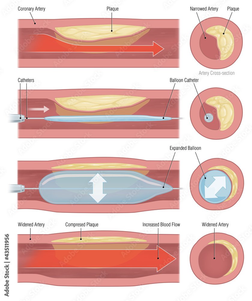
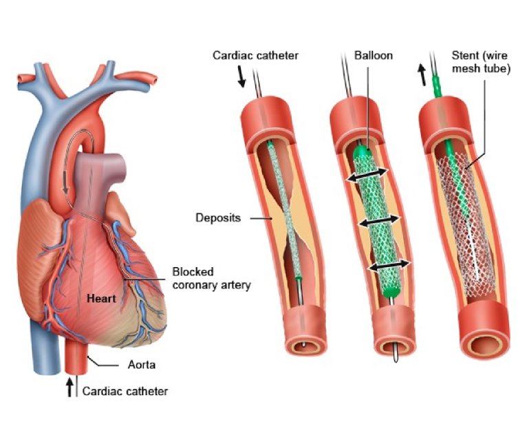
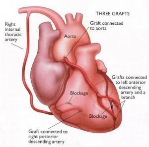
Slides 38, 50–52, 69–70, 74, 76–77
5.11 · After the infarct: complications, secondary prevention, sudden cardiac death
| Mechanical complications | Electrical |
|---|---|
| Post-infarction ischemia · congestive heart failure · myocardial rupture · ventricular aneurysm · thromboembolism · pericarditis (Dressler syndrome) · cardiogenic shock — more common in left-sided infarcts | Arrhythmias |
- Control the risk factors: blood pressure, lipids and glucose; weight loss; alcohol restriction; exercise; smoking cessation; stress.
- Beta blockers improve survival when the ejection fraction is reduced; calcium channel blockers do not improve prognosis. The deck says beta blockers improve survival “across the board”; that is no longer the position: the benefit is clear with a reduced ejection fraction and not established with a preserved one (REDUCE-AMI; 2025 ACC/AHA acute coronary syndrome guideline). The deck also adds that diltiazem and verapamil reduce mortality with preserved left ventricular function; that is not accurate. They have no mortality benefit after infarction: they relieve angina or control rate when a beta blocker cannot be used, and they are avoided with reduced ejection fraction or heart failure.
- High-intensity statin for every acute coronary syndrome, regardless of baseline low-density lipoprotein, started before discharge (atorvastatin or rosuvastatin).
- ACE inhibitors reduce remodeling (scarring); indicated with reduced left ventricular function to prevent dilation and heart failure; most patients go home on one (captopril, enalapril, fosinopril, quinapril).
- Aspirin recommended; clopidogrel is necessary after a stent. Slide 83 says warfarin is recommended for large anterior wall infarcts; current guidance adds warfarin only when the anterior infarct leaves an akinetic apex (may be considered) or a left ventricular thrombus, not routinely. The quizzes key that version.
Sudden cardiac death: the most common cause is ventricular fibrillation, and it is the first manifestation of coronary disease in over 20% (“one in five”). Other causes: left ventricular hypertrophy, hypertrophic cardiomyopathy, hypoxia, electrolyte abnormalities.
Also tested
- Diltiazem after myocardial infarction. Diltiazem and verapamil do not lower mortality; when a beta blocker cannot be used they relieve angina or control rate, provided function is preserved and there is no heart failure.
- Pericarditis after myocardial infarction. Pericarditis (Dressler syndrome) is listed among the mechanical complications of infarction; pleuritic chest pain with a pericardial friction rub weeks after the infarct points to the pericardium rather than the ventricle.
Slides 78–85
5.12 · Vasospastic (Prinzmetal) angina
Also called Prinzmetal, variant or coronary vasospasm angina. Relatively uncommon, often under age 50; caused by coronary artery vasospasm in normal or diseased arteries (“The arteries themselves are not blocked up, but they will spasm shut”). It occurs at rest and responds to nitroglycerin.
- Cigarette smoking is a major risk factor. Drugs may precipitate it: cocaine, ephedrine, marijuana, alcohol, amphetamine, triptans.
- Timing: a chronic pattern of recurrent rest pain, most often between midnight and 8 in the morning, gradual on/off, lasting 5 to 15 minutes (episodes may last longer); “typical” chest pain.
- Electrocardiogram: transient ST-segment elevation at the time of an episode — transient ischemic ST changes in multiple leads that resolve, without a cause for increased oxygen demand.
- No high-grade coronary stenoses at arteriography.
- Gold standard: coronary spasm provoked during angiography by ergonovine or acetylcholine, then relieved with nitroglycerin through the catheter (“a little squirt of nitro, and it goes away. Guess what? You got diagnosed.”).
- Treatment: stop smoking; nitrates for symptoms; first-line calcium channel blockers (diltiazem, amlodipine). AVOID nonselective beta blockers, aspirin and triptans.
Also tested
- Vasospastic angina timing. Pain most often occurs from midnight to early morning, more often between midnight and 8 in the morning, at rest, coming on and off gradually over 5 to 15 minutes.
- Vasospastic angina diagnosis. The gold standard is coronary artery spasm provoked during angiography by ergonovine or acetylcholine, then resolved with nitroglycerin given through the catheter.
- Aspirin in vasospastic angina. Aspirin is on the avoid list, together with nonselective beta blockers and triptans; prevention is a calcium channel blocker plus stopping smoking.
- Triptans in vasospastic angina. Triptans such as sumatriptan can precipitate coronary vasospasm and are on the avoid list, along with nonselective beta blockers and aspirin.
- Modifiable coronary risk factors. These are obesity, dyslipidemia, diabetes, smoking, hypertension, excessive alcohol, sedentary lifestyle and ineffective management of stress.
Slides 87–92
Also tested
- Family history as a coronary risk factor. It counts when a male first-degree relative had sudden death or myocardial infarction before 55, or a female first-degree relative before 65.
- Age as a coronary risk factor. Age becomes a nonmodifiable coronary risk factor at 45 or older for men and 55 or older for women.
- Nonmodifiable coronary risk factors. Age, sex and genetics (family history) cannot be changed, whereas hypertension, obesity and a sedentary lifestyle are modifiable.
- Age as a coronary risk factor. Age is a nonmodifiable risk factor from 45 years in men and from 55 years in women; men carry more risk than premenopausal women, a gap that narrows with age.
5.13 · Microvascular angina (formerly “cardiac syndrome X”)
- Angina with ST changes but normal coronary arteries on the angiogram. (Slide 94 adds “possibly even with elevated cardiac biomarkers”; a biomarker rise means infarction, which is MINOCA, not microvascular angina.)
- More frequent in females, often associated with stress or anxiety.
- A diagnosis of exclusion that requires angiography to rule out coronary artery disease.
- Proposed mechanisms: endothelial dysfunction, myocardial ischemia, insulin resistance/diabetes, abnormal autonomic control, altered cardiac sensitivity, estrogen deficiency, altered pain perception.
- Treatment is very similar to coronary disease: lifestyle and risk-factor reduction, calcium channel blocker, beta blocker, ACE inhibitor, statin.
Not the same thing: the slide also names “Myocardial Infarction with Non Obstructive Coronary Arteries (MINOCA)” here, and he treated the two as the same thing seen from primary care and from cardiology. They are not: MINOCA is an infarction (troponin rise) with non-obstructed coronaries; microvascular angina has no infarction. No question asks you to separate them.
Slides 93–96
Also tested
- Microvascular angina. It was formerly called cardiac syndrome X: angina with ST changes but normal coronary arteries on angiography.
5.14 · The conditions, point by point
Stable angina
Defining feature: chest pressure with exertion, relieved by rest and nitroglycerin
- Definition
- Transient myocardial ischemia short of infarction, usually precipitated by activity (stable ischemic heart disease); the most common form of angina pectoris.
- Who gets it
- Men ≥45, women ≥55; male > premenopausal female.
- Risk factors
- Family history (male relative <55, female <65), obesity, dyslipidemia, diabetes, smoking, hypertension, excessive alcohol, sedentary lifestyle, stress.
- Classic signs & symptoms
- Substernal pressure worse with exertion, radiating to the neck or shoulder, with shortness of breath and diaphoresis; not positional; Levine's sign (clenched fist over the sternum).
- Physical exam findings
- Not covered in the lecture.
- Diagnostics / tests
- Stress test (treadmill, nuclear, cardiac computed tomography): changes, usually T-wave inversion. Coronary angiography is the gold standard.
- First-line treatment
- Sublingual nitroglycerin (call emergency medical services if unimproved 5 minutes after one dose or after 3 doses); lifestyle; beta blocker (calcium channel blocker if contraindicated); statin; aspirin ± clopidogrel.
- Complications
- Progression along the spectrum to unstable angina and infarction.
Slides 22–27, 36–41
Unstable angina
Defining feature: pain at rest or a changed pattern, with troponin not abnormal
- Definition
- A change in stable angina — at rest, new-onset and activity-limiting, or increasing; an acute coronary syndrome.
- Who gets it
- Patients with coronary artery disease; not otherwise covered in the lecture.
- Risk factors
- The coronary risk factors; plaque rupture.
- Classic signs & symptoms
- Rest pain; nitroglycerin usually, but not always, helps.
- Physical exam findings
- Not covered in the lecture.
- Diagnostics / tests
- Non-specific electrocardiogram changes, usually inverted T waves; normal troponin; TIMI 3 or higher → early invasive approach.
- First-line treatment
- Medical emergency: chewable aspirin, beta blocker, intravenous nitrates, dual antiplatelet therapy ± anticoagulation; oxygen only if SpO2 <91%; then catheterization and angiography.
- Complications
- Progression to non-ST-elevation or ST-elevation infarction.
Slides 44–53, 71–72, 86
Non-ST-elevation myocardial infarction
Defining feature: ST depression >1 mm and/or T-wave inversion in two or more consecutive leads with elevated troponin
- Definition
- Myocardial infarction diagnosed by electrocardiogram changes, signs and symptoms, and elevated troponin / high-sensitivity troponin.
- Who gets it
- Not covered in the lecture.
- Risk factors
- The coronary risk factors; TIMI criteria.
- Classic signs & symptoms
- Ischemic chest pain; nitroglycerin may help.
- Physical exam findings
- Not covered in the lecture.
- Diagnostics / tests
- Subendocardial ischemia → ST depression; troponin I above 0.04 ng/mL (high-sensitivity above 12 ng/L in women, 20 ng/L in men); serial troponins.
- First-line treatment
- Treated the same as unstable angina.
- Complications
- As for infarction (5.11).
Slides 47–53, 55, 72a
ST-elevation myocardial infarction
Defining feature: ST elevation >1 mm in two or more consecutive leads with reciprocal depression — or a new left bundle branch block with ongoing ischemia or instability (the block alone is no longer enough)
- Definition
- Transmural myocardial necrosis with ST elevation not quickly reversed by nitroglycerin.
- Who gets it
- Culprit: anterior interventricular (LAD) 40–50%, right coronary 30–40%, circumflex 15–20%.
- Risk factors
- The coronary risk factors.
- Classic signs & symptoms
- Typical or referred pain (jaw, tooth, arm); may be silent in diabetes, neuropathy or a transplanted heart.
- Physical exam findings
- Not covered in the lecture.
- Diagnostics / tests
- Electrocardiogram within 10 minutes: the lead group names the artery (5.8); posterior = ST depression V1–V3 → V7–V9; Q waves = completed infarct.
- First-line treatment
- Chewable aspirin, call the catheterization team (door-to-balloon <90 minutes), P2Y12 receptor antagonist + heparin, nitroglycerin, beta blocker if no bradycardia; thrombolytics within 30 minutes if intervention is 2+ hours away; bypass (artery first).
- Complications
- Heart failure, rupture, aneurysm, thromboembolism, Dressler pericarditis, cardiogenic shock (left-sided), arrhythmias; sudden cardiac death.
Slides 14, 55–78
Right ventricular infarct
Defining feature: inferior infarct with ST elevation in V1 / V4R — no nitroglycerin
- Definition
- Right coronary occlusion extending into the right ventricle.
- Who gets it
- 15–30% of right coronary occlusions.
- Risk factors
- Not covered in the lecture.
- Classic signs & symptoms
- Not covered in the lecture beyond shock / right-sided failure if preload is dropped.
- Physical exam findings
- Not covered in the lecture.
- Diagnostics / tests
- ST elevation in V1 and V4R (reciprocal I, aVL); the right coronary artery also supplies the atrioventricular node (heart block).
- First-line treatment
- Do not give nitroglycerin — the right ventricle is preload dependent.
- Complications
- Shock and right-sided heart failure if preload falls.
Slides 11, 62–63
Vasospastic (Prinzmetal) angina
Defining feature: rest pain after midnight in a young smoker, transient ST elevation, normal coronaries
- Definition
- Angina from coronary artery vasospasm, at rest, responding to nitroglycerin.
- Who gets it
- Relatively uncommon; often <50.
- Risk factors
- Cigarette smoking (major); cocaine, ephedrine, marijuana, alcohol, amphetamine, triptans.
- Classic signs & symptoms
- Recurrent rest pain, midnight to 8 a.m., gradual on/off, 5 to 15 minutes.
- Physical exam findings
- Not covered in the lecture.
- Diagnostics / tests
- Transient ST elevation during an episode; no high-grade stenoses; gold standard = ergonovine or acetylcholine provocation at angiography, relieved by nitroglycerin.
- First-line treatment
- Stop smoking; nitrates; calcium channel blocker first-line; avoid nonselective beta blockers, aspirin, triptans.
- Complications
- Not covered in the lecture.
Slides 87–92
Microvascular angina
Defining feature: angina with ST changes but normal coronary arteries on angiogram, usually in a woman
- Definition
- Formerly cardiac syndrome X: angina with ST changes, normal coronaries, no infarction (a biomarker rise would mean MINOCA, which slide 94 lists here).
- Who gets it
- More frequent in females; stress or anxiety.
- Risk factors
- Proposed: insulin resistance/diabetes, estrogen deficiency, endothelial dysfunction, abnormal autonomic control.
- Classic signs & symptoms
- Angina.
- Physical exam findings
- Not covered in the lecture.
- Diagnostics / tests
- Diagnosis of exclusion; angiography required to rule out coronary artery disease.
- First-line treatment
- Very similar to coronary disease: lifestyle, calcium channel blocker, beta blocker, ACE inhibitor, statin.
- Complications
- Not covered in the lecture.
Slides 93–96
Sudden cardiac death
Defining feature: ventricular fibrillation; the first sign of coronary disease in over 20%
- Definition
- Not covered in the lecture.
- Who gets it
- Over 20% of patients with coronary disease present this way first.
- Risk factors
- Sudden death in a first-degree relative is a coronary risk factor.
- Classic signs & symptoms
- Not covered in the lecture.
- Physical exam findings
- Not covered in the lecture.
- Diagnostics / tests
- Not covered in the lecture.
- First-line treatment
- Not covered in the lecture.
- Complications
- Other causes: left ventricular hypertrophy, hypertrophic cardiomyopathy, hypoxia, electrolyte abnormalities.
Slides 22, 85
Also tested
- Nuclear stress test defects. A reversible perfusion defect indicates ischemia, while a non-reversible (fixed) defect indicates infarction.
- Unstable angina versus non-ST-elevation myocardial infarction. Elevated troponin or high-sensitivity troponin differentiates the infarction from unstable angina, in which troponin is normal; both can occur at rest.
- Infarction without ST elevation. ST-segment depression above 1 mm and/or T-wave inversion in two or more consecutive leads, with symptoms and an elevated troponin I (standard troponin I above 0.04 ng/mL), indicates non-ST-elevation infarction.
5.15 · Populations
| Population | What the deck gives |
|---|---|
| Infant, child, adolescent | Nothing — the deck's own objective narrows this to adult and elderly. |
| Adult | Risk from age 45 in men and 55 in women (slide 22); smoking a strong predictor in women under 45 (slide 23); vasospastic angina often under 50 (slide 88); microvascular angina more frequent in females (slide 94); ask women about phosphodiesterase inhibitors for pulmonary arterial hypertension (slide 73). |
| Elderly | The sex gap in risk narrows with age (“at 80”, slide 22); age 65 or older is a TIMI criterion (slide 71). |
6 · Heart Failure
Instructional Objectives
HEART FAILURE — Grady G. Carter, MSHS, PA-C
- Compare and contrast the etiologies, epidemiology, risk factors, clinical manifestations, differential diagnosis, diagnostic testing (including ordering and interpretation), management (acute and chronic, including applicable rehabilitative and palliative care), appropriate referrals, patient education, prevention, and prognosis for heart failure.
- Differentiate between systolic dysfunction and diastolic dysfunction..
- Define each class of the New York Heart Association (NYHA) symptomatic and functional classification of heart failure.
- Define the following diagnostic and therapeutic groups of HF patients, and the treatment
implications/recommendations for each group:
- HF with reduced EF (HFrEF)
- HF with mildly reduced EF (HFmrEF)
- HF with preserved EF (HFpEF)
- Differentiate each stage of the American College of Cardiology/American Heart Association (ACC/AHA), natural history classification of heart failure, and associated treatment recommendations of each stage.
- Compare and contrast precipitating factors, clinical manifestations, diagnosis (including role of B-type natriuretic peptide, BNP), and treatment of acute decompensated heart failure.
- Identify medical care strategies for heart failure in the lecture topic list for the
following populations.
- infant
- child
- adolescent
- adult
- elderly
What is tested, in the lecturer's own words. Heart failure is tested on the 2022 AHA/ACC/HFSA (American Heart Association / American College of Cardiology / Heart Failure Society of America) guideline: “This will not be on your test. You'll be tested on the 2022 guidelines.” The final slide's 2026 changes (slide 57) are for rotations only, so heart failure with mildly reduced ejection fraction stays a live group here.
Scope caps he stated: no dosing and no mechanism-of-action questions (the ten mechanism pictures on slides 41–50 are background); generic drug names only; no acronyms in questions; the right heart catheterization pressure table on slide 10 is not tested; the small-font “other cardiomyopathies” block on slide 16 is not tested here (it belongs to the cardiomyopathy lecture); the ejection fraction and stroke volume formulas on slide 22 are not tested, though the concepts and normal values are. Murmurs and heart sounds are tested as written descriptions, not audio.
Built from the slides. The recording (24 September, three parts) adds weight only: ★ marks show what he emphasized. Where he misspoke against his own slide, the slide is right and the slip is listed below; where the deck strays from the tested 2022 guideline, the 2022 wording is taught (the table below).
2022 guideline vs the deck — which wording is tested
| Topic | The deck says | Learn this (2022) |
|---|---|---|
| Name of the 41–49% group | “moderately reduced” (slide 23) | Mildly reduced ejection fraction — also the syllabus word |
| Reduced and preserved cut-offs | <40% and >50% (slide 23), leaving 40 and 50 unassigned | ≤40% reduced · 41–49% mildly reduced · ≥50% preserved (slide 52, the 2022 figure) |
| “Ejection fraction >40%” | The diastolic chain (slides 25, 39, 56) | That is the chain shorthand, not the definition of preserved ejection fraction (≥50%) |
| Stage D | “Decompensated heart failure” (slide 24) | Advanced heart failure (slide 52). “Decompensated” elsewhere means an acute exacerbation |
| Calcium channel blockers | Dihydropyridines “contraindicate”, verapamil and diltiazem “avoid” (slide 40) | Avoid calcium channel blockers in heart failure. The subclass ranking contradicts 2022, so learn only “avoid” |
| Beta blockers | Examples: metoprolol, atenolol, propranolol (slide 40) | Learn the class (“beta blocker”); 2022 recommends only specific evidence-based agents, so no individual agent is keyed |
| Digitalis | Listed under inotropes, “ICU use ONLY” (slide 51) | 2022 allows digoxin as an outpatient consideration in selected patients. Learn “inotropes (dobutamine, milrinone) are intensive care only”; digitalis is not keyed |
| Slide 57 | “Main 2026 Changes”: HFmrEF is out, foundational + additional therapy, an ESC 2026 poster | Not tested. Rotations only |
Two slips on the recording (both transcripts agree, so these are real): he said asymptomatic left ventricular hypertrophy is stage A — slide 24 says stage B (structural disease without symptoms). He said a defibrillator goes in when life expectancy is under a year — slide 53 says at least one year. The slide is right both times, and matches the 2022 guideline.
Where each objective is answered
| Objective | Answered in |
|---|---|
| a. Etiology, epidemiology, risk factors, manifestations, testing, management, prognosis | 6.1, 6.5–6.7 and the condition blocks in 6.8 · prevention appears only as stage A therapy; the deck names no referral or education content beyond cardiac rehabilitation |
| b. Systolic vs diastolic dysfunction | 6.2 |
| c. NYHA classes | 6.4 |
| d. Reduced / mildly reduced / preserved ejection fraction and their treatment | 6.3 (definitions) and 6.7 (therapy by group) |
| e. ACC/AHA stages and treatment per stage | 6.4 |
| f. Acute decompensated heart failure, role of BNP | 6.6 (natriuretic peptides) and the acute block in 6.8 · the deck has no precipitating-factors slide; its only list is “MI, sepsis, ARF, etc” (slide 53) |
| g. Populations | 6.9 · adult and elderly only; the deck has no infant, child or adolescent content |
6.1 · Definition, epidemiology and causes
Heart failure is the inability of the heart to pump blood at a rate that meets the body's metabolic demands at rest and during effort, or doing so only with abnormally high filling pressures. It is a complex clinical syndrome (a constellation of signs and symptoms) that can result from any structural or functional disorder that impairs the ventricle's ability to fill with or eject blood, with effort intolerance, fluid retention and reduced longevity.
The body answers increased demand three ways: a faster heart rate (neurohormonal input), stronger contraction (catecholamines, autonomic input) and more preload (venous capacitance, renal compensation). Four basic mechanisms produce failure: excessive preload (increased blood volume), excessive afterload (increased resistance), decreased contractility and decreased filling. Acute damage or chronic conditions then drive cardiac remodeling — myocyte hypertrophy and collagen fibrosis. In the remodeling picture, the sympathetic and renin-angiotensin-aldosterone systems worsen the damage while the natriuretic peptides are the beneficial compensation.
Also tested
- Modifiable heart failure risk factors. Tobacco, excessive alcohol and obesity are modifiable, whereas family history cannot be changed, so counseling targets the modifiable ones.
- Response to increased metabolic demand in heart failure. The body raises heart rate, contractility and preload; these help at first and later drive damage.
- Modifiable heart failure risk factors. Tobacco, excess alcohol and obesity can be changed; age, family history and genetic abnormality cannot.
- Heart failure defined. The heart cannot pump blood at a rate that meets the body's metabolic needs at rest and during effort, or manages it only at abnormally high filling pressures.
- Ischemic cardiomyopathy. Its causes are infarction (myocardial infarction), ongoing ischemia (persistent or chronic intermittent myocardial ischemia) or shock (hypoperfusion); everything else is non-ischemic cardiomyopathy.
- Valvular causes of heart failure. Aortic stenosis is the most common valvular cause; mitral stenosis appears instead as a cause of right-sided failure.
Epidemiology
- 6.7 million Americans older than 20 have heart failure (2025).
- 60% of patients with heart failure are older than 65, and in that group one-year mortality after a hospitalization is 35%.
- 10–15% of people older than 80 have heart failure. Lifetime risk is 24% (1 in 4).
- Heart failure accounts for 45% of cardiovascular deaths; in 2022, 47% of cardiovascular deaths were caused by heart failure against 29% by heart attack.
Risk factors
Age · hypertension · coronary artery disease · diabetes · chronic tachyarrhythmias · obesity / metabolic syndrome · excessive alcohol · tobacco · family history · genetic abnormality · cardiotoxic medications.
Causes
“All heart failure has a cardiomyopathy etiology.” Causes are sorted as ischemic cardiomyopathy (myocardial infarction, persistent or chronic intermittent ischemia, hypoperfusion from shock) versus non-ischemic cardiomyopathy (everything else): hypertrophic cardiomyopathy, hypertension, valvular disease (most commonly aortic stenosis), dilated cardiomyopathy, congenital heart disease, toxins (alcohol, chemotherapy), autoimmune or metabolic disease (diabetes, thyroid disease, rheumatoid arthritis, lupus).
The deck's list of heart failure “types” (slide 7): left vs right, systolic vs diastolic, high- vs low-output, dilated vs hypertrophic vs restrictive, compensated vs decompensated, acute vs chronic.
6.2 · Systolic vs diastolic heart failure
Systole is the SQUEEZE — systolic heart failure is dysfunction of contraction. Diastole is the REST — diastolic heart failure is dysfunction of relaxation.
| Hypertrophic cardiomyopathy | Dilated cardiomyopathy | |
|---|---|---|
| Tissue | Enlarged (thick) cardiac tissue | Thin, dilated (ballooned) cardiac tissue |
| Muscle | Increased muscle = increased contraction / poor relaxation | Decreased muscle = poor contraction / loss of elasticity |
| Leads to | Diastolic failure | Systolic failure (with ischemic cardiomyopathy) |
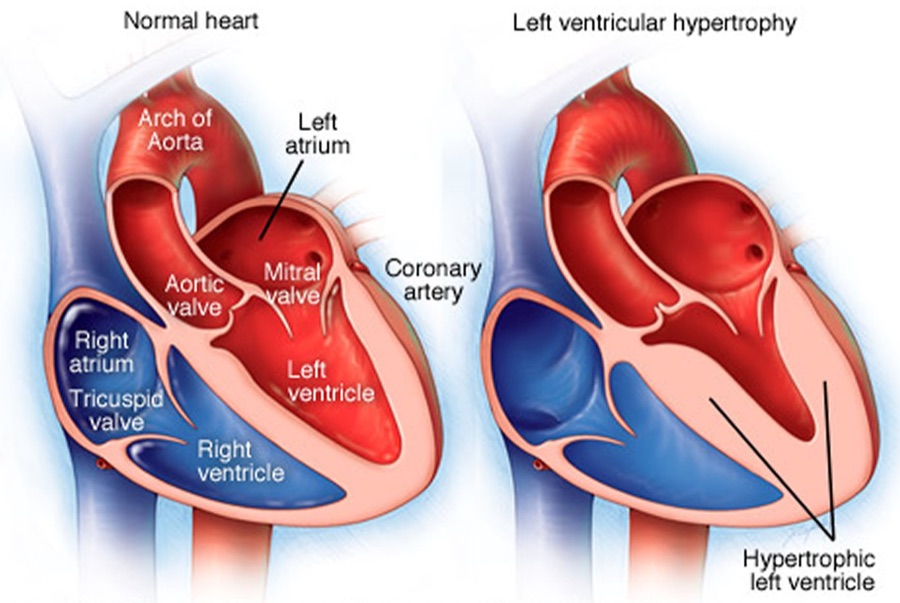
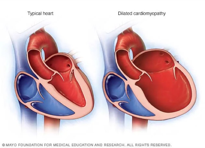
“Burn this in your memory.” The two chains are the lecture's spine (about 15 minutes, returned to at the end of each part). He described scanning a test question for one clue — a third heart sound, a normal or low pressure — and filling in the rest of the chain.
| Left ventricular… | Cause | Ejection fraction | Blood pressure | Heart sound | Edema |
|---|---|---|---|---|---|
| Systolic failure | Ischemic or dilated cardiomyopathy | Reduced | Normal or low | S3 (third heart sound) — rapid filling of a dilated left ventricle | Pulmonary |
| Diastolic failure | Hypertrophic cardiomyopathy | Preserved | Hypertension | S4 (fourth heart sound) — strong left atrial contraction against a hypertrophic left ventricle | Peripheral, then pulmonary |
“Add an elevated BNP and…” — the slam-dunk diagnosis. Most patients with systolic failure you will be asked about have ischemic or dilated cardiomyopathy.
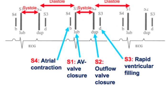
Also tested
- Titrating therapy in reduced ejection fraction. Guideline-directed therapy is prioritized for the patient's comorbidities and optimized as hemodynamically stable, so each class is increased as blood pressure and tolerance allow.
- Reduced ejection fraction therapy. Core therapy is a beta blocker, a renin-angiotensin blocker such as lisinopril, a mineralocorticoid receptor antagonist such as spironolactone and a sodium-glucose cotransporter 2 inhibitor such as dapagliflozin, with diuretics as needed.
- Dilated versus hypertrophic cardiomyopathy. Dilated is thin, ballooned muscle that contracts poorly with loss of elasticity; hypertrophic is increased muscle that contracts strongly but relaxes poorly.
- Ejection fraction in dilated cardiomyopathy. Poor contraction (the systolic chain) gives a reduced ejection fraction of 40% or less, such as about 25%, with low-normal pressure, a third heart sound and pulmonary edema.
6.3 · Ejection fraction and the treatment groups
Ejection fraction is the percentage of the filled (end-diastolic) volume that the ventricle ejects with each beat. Cardiac output is stroke volume × heart rate. (The formulas themselves are not tested.)
Normal cardiac output is 5–6 liters per minute (“know it, know it, learn it, love it”). Normal ejection fraction is 50% or more (“cannot express that enough”) — the “giant line down the middle” of treatment is 50% and above versus 49% and below.
| Group | Left ventricular ejection fraction (2022) |
|---|---|
| Heart failure with reduced ejection fraction (HFrEF) | ≤40% |
| Heart failure with mildly reduced ejection fraction (HFmrEF) | 41–49% — “those numbers are very specific” |
| Heart failure with preserved ejection fraction (HFpEF) | ≥50% |
| Heart failure with improved ejection fraction (HFimpEF) | Was ≤40%, now >40% — “these are important”: someone who has recovered is treated differently from someone who was never reduced |
6.4 · NYHA classes and ACC/AHA stages
The NYHA (New York Heart Association) class grades symptoms. The ACC/AHA (American College of Cardiology / American Heart Association) stage is natural history and does not move backward.
| NYHA class | Symptoms | ACC/AHA stage | Definition (slide 24; names from slide 52) |
|---|---|---|---|
| I | No symptoms | A — at risk | Risk factors, normal heart function, no symptoms |
| II | Symptoms with exertion | B — pre-heart failure | Asymptomatic structural heart disease: ischemic cardiomyopathy, systolic dysfunction, diastolic dysfunction, left ventricular hypertrophy |
| III | Symptoms with slight exertion | C — symptomatic | Symptomatic structural heart disease, past or current — resolved symptoms stay stage C |
| IV | Symptoms at rest | D — advanced | Advanced heart failure (slide 24 says “decompensated”; see the 2022 box) |
2022 therapy by stage (slide 52). “It'll be on your test: what is the number one first medication … if you're high risk, especially if you're diabetic? SGLT2, not a beta blocker.”
| Stage | Therapy (strength of recommendation) |
|---|---|
| A — at risk | Sodium-glucose cotransporter 2 inhibitor in patients with diabetes (strong). No beta blocker yet. |
| B — pre-heart failure | Sodium-glucose cotransporter 2 inhibitor in diabetes (strong) · ACE (angiotensin-converting enzyme) inhibitor (strong) · angiotensin receptor blocker if the ACE inhibitor is not tolerated (strong) · beta blocker (strong) |
| C and D | By ejection fraction group — see 4.7 |
Also tested
- Heart failure prevention at stage A. An at-risk patient with diabetes and a normal heart is stage A, where the first recommended drug is a sodium-glucose cotransporter 2 inhibitor such as dapagliflozin or empagliflozin. A beta blocker enters at stage B.
- Heart failure stage C. Stage C is symptomatic structural heart disease, past or current; once symptoms have occurred, resolution on treatment does not move the patient backward, so the patient remains in stage C.
- Heart failure drug recommendations. In mildly reduced ejection fraction, diuretics as needed are strong, the sodium-glucose cotransporter 2 inhibitor moderate, and the beta blocker, antagonist and renin-angiotensin drugs weak. In reduced ejection fraction, the beta blocker, renin-angiotensin blockade, mineralocorticoid antagonist and sodium-glucose cotransporter 2 inhibitor are all strong.
- Fluid overload in heart failure. Diuretics are used as needed for congestion in every ejection fraction group; a loop diuretic such as furosemide or bumetanide is the potassium-wasting diuretic that clears fluid.
- Renin-angiotensin drugs by symptom class. Symptoms at rest are class IV. In reduced ejection fraction, an angiotensin-converting enzyme inhibitor or receptor blocker covers class II to IV, while the angiotensin receptor-neprilysin inhibitor is recommended for class II to III.
- Preserved ejection fraction drug recommendations. Diuretics as needed are strong, and the sodium-glucose cotransporter 2 inhibitor is next with a moderate recommendation; the neprilysin combination, the mineralocorticoid antagonist and the receptor blocker are weak.
- Stage B (pre-heart failure) therapy. An angiotensin-converting enzyme inhibitor is listed, with an angiotensin receptor blocker such as losartan if the inhibitor is not tolerated, alongside a beta blocker.
6.5 · Clinical manifestations
Clinical heart failure = signs and symptoms of heart failure from a structural or functional cardiac abnormality, plus one of: an elevated BNP (B-type natriuretic peptide), or objective evidence of cardiogenic pulmonary or systemic congestion.
- Volume overload: dyspnea on exertion, paroxysmal nocturnal dyspnea (waking breathless), orthopnea (breathless lying flat), pulmonary or peripheral edema, elevated jugular venous pressure, tachycardia, S3 and S4.
- Inadequate tissue perfusion: weakness, fatigue, lethargy, malaise; syncope, near-syncope, obtundation.
- Renal dysfunction: acute kidney injury, decreased urine output.
Also tested
- Renal complication of heart failure. Renal dysfunction presents as acute kidney injury with decreased (low) urine output, one of the signs of inadequate tissue perfusion.
Bedside hemodynamic profiles
Congestion (wet vs dry) crossed with perfusion (warm vs cold): warm and dry is normal, warm and wet is congestion, cold and dry is hypoperfusion, cold and wet is both. Low-perfusion signs: narrow pulse pressure, sleepy or obtunded, low serum sodium, cool extremities, hypotension with an ACE inhibitor, renal dysfunction. Congestion signs: orthopnea / paroxysmal nocturnal dyspnea, jugular venous distension, hepatomegaly, edema, rales (rare in chronic heart failure).
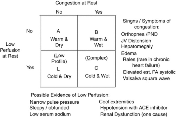
6.6 · Diagnosis and natriuretic peptides
Congestive heart failure is a CLINICAL DIAGNOSIS: signs and symptoms of heart failure + an elevated BNP or cardiogenic pulmonary or systemic congestion. “Clinical diagnosis requires congestion; congestion = edema” (pulmonary and/or peripheral). Fatigue and exertional dyspnea without any edema do not make it congestive.
| Test | What it answers |
|---|---|
| Echocardiogram | Most accurate for structures (valves); classifies by ejection fraction; the definitive test for right-sided failure |
| MUGA (multigated acquisition scan) | Most accurate ejection fraction and perfusion — when it disagrees with the echo on the ejection fraction, trust this one |
| Chest radiograph | Pleural effusion, cephalization of vessels, Kerley B lines, increased cardiothoracic ratio |
| Complete blood count | The differential diagnosis (for example anemia) |
| Complete metabolic panel | Electrolytes, potassium above all; renal function (blood urea nitrogen, creatinine, filtration rate) |
| Troponin | Only with concern for a non-ST-elevation infarction or heart failure secondary to ischemic cardiomyopathy |
| Electrocardiogram | Ischemic changes (ST elevation, T-wave inversion) · left ventricular hypertrophy: tall R in V5–V6, deep S in V1–V2 · right heart strain: right axis deviation, right bundle branch block, T-wave inversion in V1–V3 |
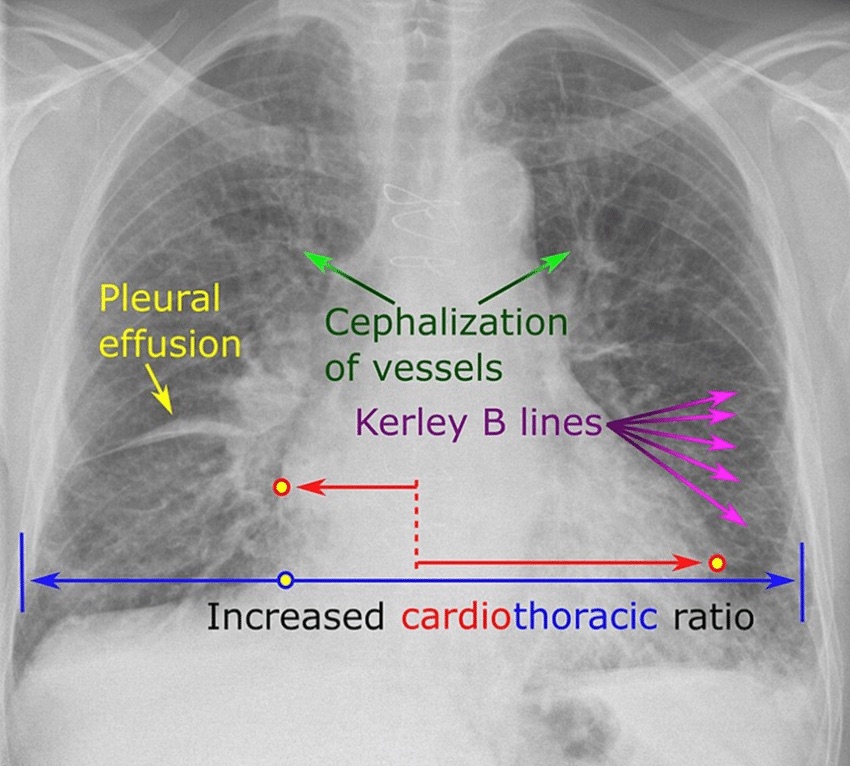
“Make sure you know the difference between whether your test was a BNP or the new pro-BNP” — and “what you do need to remember is what your cutoff levels are.”
| BNP (B-type natriuretic peptide) | NT-proBNP (N-terminal pro-B-type natriuretic peptide) | |
|---|---|---|
| What it is | Bioactive hormone secreted mainly by the ventricular myocardium in response to wall stress from volume or pressure overload | Inactive precursor fragment; requires enzymatic activation |
| Timing | Rises 1–3 hours, peaks at 12 hours | More stable: peaks at 24 hours, detected earlier and detectable longer |
| Caveat | Degraded by neprilysin, so altered by sacubitril-valsartan | Not the one degraded by neprilysin |
| Rule out acute heart failure | <100 (negative predictive value above 90%) | <300 (negative predictive value above 90%) |
| Gray zone | 100–400: no useful sensitivity or specificity | — |
| Likely heart failure | >400 | >125 if younger than 75 · >450 if older than 75 |
| Likely acute heart failure | >900 (“over 900, they're not going home”) | — |
The two have similar sensitivity and specificity for ruling heart failure in and out. Prognosis: each BNP rise of 100 means a 35% higher relative risk of death, and a predischarge BNP below 430 means a lower risk of 30-day readmission.
Also tested
- High-output heart failure treatment. It is treated by correcting the underlying cause of increased demand, such as hyperthyroidism, with a diuretic for congestion.
- High-output heart failure. It is elevated cardiac output from increased heart rate and contractility, meeting an increased peripheral demand; over time it may lead to ischemic cardiomyopathy or remodeling.
- Diagnosing congestive heart failure. It is a clinical diagnosis: signs and symptoms of heart failure plus an elevated B-type natriuretic peptide or cardiogenic pulmonary or systemic congestion; no single test makes the diagnosis alone.
- High-output heart failure treatment. It is driven by peripheral demand, so treatment targets the cause, such as anemia, hyperthyroidism or an arteriovenous fistula, with a diuretic for congestion.
- B-type natriuretic peptide rule-out. A level below 100 rules out acute heart failure with a negative predictive value above 90%, so breathlessness then needs another explanation.
- Complete metabolic panel in heart failure. It follows potassium and renal function (blood urea nitrogen, creatinine and filtration rate), because diuretics and several therapies move potassium and affect the kidneys.
- Monitoring on a loop diuretic. Potassium and kidney function are followed on the metabolic panel, because loop diuretics waste potassium.
- Workup for high-output heart failure. It is an echocardiogram, complete blood count, complete metabolic panel and B-type natriuretic peptide; the blood count matters because anemia is one of its causes.
6.7 · Treatment
Goals: decrease symptoms · increase survival · limit disease progression — which is why the regimen continues after the patient feels better.
| Class | Generic examples (slide 40) | Note |
|---|---|---|
| Potassium-wasting diuretics | Thiazide-like: hydrochlorothiazide, metolazone · loop: furosemide, bumetanide | Monitor potassium and renal function |
| ACE inhibitor / angiotensin receptor blocker / angiotensin receptor-neprilysin inhibitor | Lisinopril, enalapril / losartan, valsartan, irbesartan / sacubitril-valsartan | |
| Beta blocker | Learn the class | See the 2022 box |
| Sodium-glucose cotransporter 2 inhibitor | Dapagliflozin, empagliflozin | |
| Mineralocorticoid receptor antagonist | Spironolactone, eplerenone | Potassium-sparing diuretic |
| Nitrates | Isosorbide dinitrate (with hydralazine) | For the selected group below |
“The other part I want you to remember is GDMT” (guideline-directed medical therapy). “GDMT for HFpEF and HFmrEF are very similar. It's different for HFrEF.” Strength: strong / moderate / weak (slide 52, the 2022 figure).
| Reduced (≤40%) | Mildly reduced (41–49%) | Preserved (≥50%) |
|---|---|---|
| Angiotensin receptor-neprilysin inhibitor in NYHA II–III, or ACE inhibitor / angiotensin receptor blocker in NYHA II–IV (strong) Beta blocker (strong) Mineralocorticoid receptor antagonist (strong) Sodium-glucose cotransporter 2 inhibitor (strong) Diuretics as needed (strong) Hydralazine + isosorbide dinitrate for African American patients in NYHA III–IV (strong) |
Diuretics as needed (strong) Sodium-glucose cotransporter 2 inhibitor (moderate) ACE inhibitor, angiotensin receptor blocker, angiotensin receptor-neprilysin inhibitor (weak) Mineralocorticoid receptor antagonist (weak) Beta blocker (weak) |
Diuretics as needed (strong) Sodium-glucose cotransporter 2 inhibitor (moderate) Angiotensin receptor-neprilysin inhibitor (weak) Mineralocorticoid receptor antagonist (weak) Angiotensin receptor blocker (weak) |
Reduced ejection fraction therapy is prioritized for comorbidities and optimized as hemodynamically stable — each class is raised as blood pressure and tolerance allow (slide 56).
Also tested
- Furosemide monitoring. Furosemide is a potassium-wasting loop diuretic, so potassium and renal function are followed on the complete metabolic panel.
- Calcium channel blockers in heart failure. Verapamil is a calcium channel blocker, and calcium channel blockers are avoided in heart failure; pressure control comes from optimizing the recommended classes as tolerated.
- Implantable defibrillator criteria. The device is indicated only with a life expectancy of at least one year, together with ejection fraction 35% or less, class II or III and optimal therapy.
- Goals of heart failure treatment. The goals are to decrease symptoms, increase survival and limit disease progression; feeling better meets only the first of the three.
- Defibrillator vest. The vest covers the period in which guideline-directed therapy is optimized and cardiac rehabilitation is completed; the echocardiogram is then repeated to decide whether an implantable device is needed.
- Defibrillator after infarction. The implantable defibrillator is placed only if the repeat echocardiogram shows no improvement; with an ejection fraction well above the 35% threshold, continue therapy without a device.
- Defibrillator after acute heart failure from infarction. The pathway is a wearable defibrillator, optimized therapy and cardiac rehabilitation, then a repeat echocardiogram; implanting a defibrillator now is premature, as the implantable device follows only if there is no improvement.
- Spironolactone in heart failure. A mineralocorticoid receptor antagonist, it is a potassium-sparing diuretic: it spares potassium, so levels are checked, with renal function, alongside the potassium-wasting loop diuretic.
- Diltiazem in heart failure. Diltiazem is a calcium channel blocker, and calcium channel blockers are avoided in heart failure, for example when planning rate control in atrial fibrillation; a beta blocker belongs to core therapy.
Sudden death protection (slide 53)
- Implantable cardioverter-defibrillator for ischemic or non-ischemic cardiomyopathy with ejection fraction ≤35%, NYHA class II–III, on optimal guideline-directed therapy, with a life expectancy of at least one year.
- 52% of cardiac arrests in hospitalized patients with reduced ejection fraction are due to ventricular arrhythmias.
- After acute heart failure (from infarction, sepsis and similar) with ejection fraction <35%: wearable defibrillator vest → optimize therapy + cardiac rehabilitation → recheck echocardiogram → implantable device only if not improved.
6.8 · The conditions, point by point
Heart failure — the syndrome overall
Defining feature: output that cannot meet demand, or meets it only at high filling pressures — clinically, symptoms plus an elevated BNP or congestion
- Definition
- Inability to pump blood at a rate that meets metabolic demand at rest and with effort, or doing so only with abnormally high filling pressures. A syndrome from any structural or functional disorder that impairs filling or ejection.
- Who gets it
- 6.7 million Americans older than 20; 60% of patients are older than 65; 10–15% of people older than 80; lifetime risk 1 in 4.
- Risk factors
- Age, hypertension, coronary artery disease, diabetes, chronic tachyarrhythmias, obesity / metabolic syndrome, excessive alcohol, tobacco, family history, genetic abnormality, cardiotoxic medications.
- Classic signs & symptoms
- Dyspnea on exertion, paroxysmal nocturnal dyspnea, orthopnea, edema; fatigue, weakness, near-syncope from poor perfusion.
- Physical exam findings
- Pulmonary or peripheral edema, S3 and/or S4, elevated jugular venous pressure, tachycardia, crackles; low-perfusion signs (narrow pulse pressure, cool extremities, obtundation).
- Diagnostics / tests
- Clinical diagnosis + BNP or congestion. Echocardiogram (structures, ejection fraction group), MUGA (most accurate ejection fraction), chest radiograph, complete blood count, complete metabolic panel, troponin if ischemia is a concern, electrocardiogram.
- First-line treatment
- By stage and ejection fraction group (6.4, 6.7). Goals: fewer symptoms, longer survival, slower progression. Avoid calcium channel blockers.
- Complications
- Renal dysfunction (acute kidney injury, low urine output); ventricular arrhythmias; reduced longevity — 35% one-year mortality after hospitalization in those older than 65; 45% of cardiovascular deaths.
Slides 4–6, 16, 18, 26, 32–33, 38, 40, 52–53
Left-sided heart failure
Defining feature: the most common by far — 70–80%; pulmonary congestion
- Definition
- Left ventricular involvement: systolic or diastolic failure; dilated, hypertrophic or restrictive cardiomyopathy; ischemic or non-ischemic; preserved or reduced ejection fraction.
- Who gets it
- 70–80% of all heart failure.
- Risk factors
- Not covered separately (the general list applies).
- Classic signs & symptoms
- Systolic: normal or low blood pressure, S3, pulmonary edema. Diastolic: hypertension, S4, peripheral then pulmonary edema. Orthopnea, paroxysmal nocturnal dyspnea.
- Physical exam findings
- S3 (rapid filling of a dilated left ventricle) or S4 (atrial contraction against a hypertrophic left ventricle); crackles.
- Diagnostics / tests
- Not covered separately from heart failure overall.
- First-line treatment
- Not covered separately — therapy follows the ejection fraction group.
- Complications
- Chronic left-sided failure is the most common cause of right-sided failure.
Slides 8, 9, 26–27, 39, 56
Right-sided heart failure
Defining feature: fluid backs up — edema, distended neck veins, an enlarged liver; most often caused by chronic left-sided failure
- Definition
- Heart failure of the right ventricle (systolic or diastolic; any cardiomyopathy; ischemic from the right coronary artery or non-ischemic).
- Who gets it
- Epidemiology, risk factors and pathology are the same as left-sided.
- Risk factors
- Causes: chronic left-sided heart failure (most common: left failure → right hypertrophic → right dilated cardiomyopathy), mitral valve stenosis, pulmonary arterial hypertension.
- Classic signs & symptoms
- Systemic overload: peripheral edema, jugular venous distension. Portal overload: nausea, loss of appetite, hepatomegaly (congestion, ascites).
- Physical exam findings
- With mitral stenosis: rumbling diastolic murmur at the apex with an opening snap. With tricuspid regurgitation in pulmonary arterial hypertension: holosystolic murmur at the left lower sternal border, louder on inspiration.
- Diagnostics / tests
- Clinical heart failure with jugular venous distension raises suspicion; echocardiogram gives the definitive diagnosis (by ejection fraction); right heart catheterization grades hemodynamic severity (its pressure values are not tested). Electrocardiogram: right heart strain.
- First-line treatment
- Same as left-sided: symptom management, the underlying cause, heart transplant, palliative care.
- Complications
- Prognosis much worse than left-sided failure.
Slides 9–10, 27, 32–34
Combined right and left heart failure
Defining feature: both ventricles have failed — management moves to comfort and transplant
- Definition
- Not covered in the lecture (title only).
- Who gets it
- Not covered in the lecture.
- Risk factors
- Not covered in the lecture (chronic left failure is the usual route to the right side).
- Classic signs & symptoms
- Not covered in the lecture.
- Physical exam findings
- Not covered in the lecture.
- Diagnostics / tests
- Not covered in the lecture.
- First-line treatment
- Symptom management, comfort care, transplant.
- Complications
- Not covered in the lecture.
Slide 11
Reduced ejection fraction (systolic) — including improved ejection fraction
Defining feature: the weak squeeze — ejection fraction ≤40%, normal or low pressure, S3, pulmonary edema
- Definition
- Failure of systolic function (contraction). Ejection fraction ≤40%. Improved ejection fraction: was ≤40%, now >40%.
- Who gets it
- Not covered in the lecture; the causes are ischemic or dilated cardiomyopathy.
- Risk factors
- Ischemic cardiomyopathy (infarction, ischemia, shock); dilated cardiomyopathy.
- Classic signs & symptoms
- Normal or low blood pressure, pulmonary edema, dyspnea.
- Physical exam findings
- S3 — rapid filling of a dilated left ventricle; crackles.
- Diagnostics / tests
- Echocardiogram (ejection fraction ≤40%; MUGA most accurate for the number) + elevated BNP.
- First-line treatment
- Angiotensin receptor-neprilysin inhibitor (NYHA II–III) or ACE inhibitor / angiotensin receptor blocker (II–IV), beta blocker, mineralocorticoid receptor antagonist, sodium-glucose cotransporter 2 inhibitor, diuretics as needed — all strong; hydralazine + isosorbide dinitrate for African American patients in NYHA III–IV.
- Complications
- Ventricular arrhythmias cause 52% of cardiac arrests in hospitalized patients — defibrillator if ejection fraction ≤35%, NYHA II–III, on optimal therapy, life expectancy ≥1 year.
Slides 12–13, 17, 22–23, 25, 32, 39, 52–53, 56
Preserved ejection fraction (diastolic)
Defining feature: the stiff, thick ventricle that cannot relax — hypertension, S4, edema peripheral first; ejection fraction ≥50%
- Definition
- Failure of diastolic function (relaxation). Ejection fraction ≥50%.
- Who gets it
- Not covered in the lecture; the cause is hypertrophic cardiomyopathy, driven by hypertension.
- Risk factors
- Hypertension → hypertrophic cardiomyopathy.
- Classic signs & symptoms
- Hypertension; peripheral edema, then pulmonary edema.
- Physical exam findings
- S4 — strong left atrial contraction against a hypertrophic left ventricle.
- Diagnostics / tests
- Echocardiogram (preserved fraction; left ventricular wall >10 mm = hypertrophy) + elevated BNP. Electrocardiogram: tall R in V5–V6, deep S in V1–V2.
- First-line treatment
- Diuretics as needed (strong); sodium-glucose cotransporter 2 inhibitor (moderate); angiotensin receptor-neprilysin inhibitor, mineralocorticoid receptor antagonist, angiotensin receptor blocker (weak). Other classes for comorbidities.
- Complications
- Not covered in the lecture.
Slides 12–14, 17, 23, 25, 32, 39, 52, 56
Mildly reduced ejection fraction
Defining feature: ejection fraction 41–49% that has never been ≤40%
- Definition
- Ejection fraction 41–49% (the deck says “moderately”; learn “mildly”).
- Who gets it
- Not covered in the lecture.
- Risk factors
- Not covered in the lecture.
- Classic signs & symptoms
- Not covered in the lecture.
- Physical exam findings
- Not covered in the lecture.
- Diagnostics / tests
- Echocardiogram ejection fraction 41–49%.
- First-line treatment
- Diuretics as needed (strong); sodium-glucose cotransporter 2 inhibitor (moderate); ACE inhibitor, angiotensin receptor blocker, angiotensin receptor-neprilysin inhibitor, mineralocorticoid receptor antagonist, beta blocker (weak) — “very similar” to preserved.
- Complications
- Not covered in the lecture.
Slides 23, 30, 52, 56
Acute and acute decompensated heart failure
Defining feature: sudden onset — new, or an exacerbation (“acute on chronic” once disease has lasted over a year)
- Definition
- Acute = sudden, new onset or exacerbation. Chronic = more than 1 year. Sequence: pre-heart failure (asymptomatic) → acute new-onset (symptomatic) → resolution → exacerbation → chronic → acute on chronic.
- Who gets it
- Not covered in the lecture.
- Risk factors
- Precipitants named: myocardial infarction, sepsis, “ARF” (unexpanded on the slide), “etc”.
- Classic signs & symptoms
- Congestion = edema (pulmonary and/or peripheral); dyspnea.
- Physical exam findings
- Crackles; hemodynamic profile (warm/cold, wet/dry).
- Diagnostics / tests
- BNP <100 rules out acute heart failure; >900 makes it likely. NT-proBNP <300 rules it out.
- First-line treatment
- Diuretics for congestion; inotropes (dobutamine, milrinone and others) in intensive care only; wearable defibrillator pathway if ejection fraction <35% after the event.
- Complications
- One-year mortality after hospitalization 35% (older than 65); each BNP rise of 100 = 35% higher relative risk of death; predischarge BNP <430 = lower 30-day readmission.
Slides 6, 28–31, 38, 51, 53
High-output heart failure
Defining feature: elevated cardiac output driven by increased peripheral demand
- Definition
- Elevated cardiac output from increased heart rate and contractility.
- Who gets it
- Not covered in the lecture. Causes: anemia, hyperthyroidism, arteriovenous fistula.
- Risk factors
- Obesity, chronic obstructive pulmonary disease, cirrhosis, Paget disease (osteitis deformans).
- Classic signs & symptoms
- The heart failure syndrome: fatigue, dyspnea, edema, palpitations.
- Physical exam findings
- Not covered in the lecture beyond the syndrome.
- Diagnostics / tests
- Echocardiogram, complete blood count, complete metabolic panel, BNP.
- First-line treatment
- Treat the underlying cause; diuretic.
- Complications
- Increased demand may lead to ischemic cardiomyopathy or remodeling.
Slide 54
Also tested
- Congestive heart failure requires congestion. A clinical diagnosis requires congestion, meaning pulmonary or peripheral edema; symptoms and a mildly reduced ejection fraction without edema do not meet the congestive label.
- Combined right and left heart failure. It is managed with symptom control, comfort care and heart transplant; when optimized therapy no longer controls symptoms, transplant evaluation or comfort care is discussed.
- Mitral stenosis murmur. A rumbling diastolic murmur at the apex with an opening snap; mitral stenosis is one of three causes of right-sided failure, with chronic left-sided failure and pulmonary arterial hypertension.
6.9 · Populations
| Population | What the deck gives |
|---|---|
| Infant, child, adolescent | Nothing — the deck's own objectives slide limits it to adult and elderly patients. |
| Adult | NT-proBNP: heart failure likely above 125 when younger than 75. |
| Elderly | 60% of patients are older than 65, with 35% one-year mortality after hospitalization; 10–15% of people older than 80 have heart failure. NT-proBNP threshold rises to 450 when older than 75. |
★ Quick reference
Secondary cause → the one clue that names it
| Clue | Cause |
|---|---|
| Unexplained hypokalemia with resistant pressure | Primary aldosteronism |
| Abdominal bruit, asymmetric kidneys, flash pulmonary edema | Renovascular disease |
| Marked creatinine rise after starting an ACE inhibitor | Bilateral renal artery stenosis |
| Loud snoring, witnessed apneas, daytime sleepiness | Obstructive sleep apnea |
| Proximal weakness, bruising, thin skin, purple striae | Cushing syndrome |
| Paroxysmal pressure, perspiration, palpitations, pallor, tremor | Pheochromocytoma |
| Radial-femoral delay; leg pressure lower than arm | Coarctation of the aorta |
| Systolic elevation with tachycardia / diastolic elevation with cold intolerance | Hyper- / hypothyroidism |
Adverse effect → the class it belongs to
| Effect | Class |
|---|---|
| Dry cough, hyperkalemia, angioedema | ACE inhibitor |
| Dependent ankle edema, flushing, gingival enlargement | Dihydropyridine calcium channel blocker |
| Bradycardia and heart block (worse with a beta blocker) | Non-dihydropyridine (verapamil, diltiazem) |
| Hyponatremia, hypokalemia, hyperuricemia, volume depletion | Thiazide-type diuretic |
| Reflex tachycardia and fluid retention | Hydralazine |
| Sedation, dry mouth, rebound hypertension on withdrawal | Central alpha-2 agonist (clonidine) |
The things that are NOT what they look like
| It looks like… | …but |
|---|---|
| A very high reading means an emergency | No numeric cutoff exists — acute organ injury defines it |
| A normal electrocardiogram excludes hypertrophy | The criteria are specific but not sensitive |
| A normal chest radiograph excludes dissection | It does not — and neither do normal pulses |
| A normal ejection fraction excludes heart failure | Preserved-fraction failure is a recognized endpoint |
| Normal office readings mean normal pressure | Masked hypertension carries real risk |
| Resistant pressure means more drugs are needed | Nonadherence is the commonest cause; check technique too |
| ACE inhibitor + ARB is a logical combination | Never — harm without benefit |
| Lowering the pressure fast in an emergency is safest | Autoregulation has adapted; rapid reduction causes ischemia |