1 · Review of General Microbiology
Instructional Objectives
- Describe an overview of molecular mechanisms related to microbiology that influence health and disease
- Describe the types of infectious pathogens.
- Compare and contrast standard and specialized structures of a bacterial cell.
- Describe bacterial cell morphologies, including Gram-staining characteristics.
- Review the methods utilized in bacterial identification.
- Review the types of culture media used to identify pathogens.
- Review the phases of the bacterial growth curve.
- Differentiate the different processes of phage replication.
- Compare and contrast phage replication to animal virus replication.
- Describe the cytopathic effects of viruses.
- Describe the health implications of nucleic acid mutations.
- Describe the basic mechanisms of microbial control.
- Review the bacterial death curve.
96 minutes with Dr. Webster, across two segments. Both transcripts were read and diffed and every factual claim was checked against the deck. Nothing he said contradicts a slide.
This lecture barely signposts. In 96 minutes there is one explicit de-emphasis and no statement at all about what will or will not be on the exam. That is worth knowing rather than hunting for: treat the thirteen instructional objectives as evenly weighted, because he gave no reason not to. What the recording does add is laboratory technique the deck does not carry.
| He said | What it means for you |
|---|---|
| “And then they have what we call the 70S ribosomes. What’s that S?… It’s a Svedberg unit. Oh, you don’t have to write that down. I’m not going to ask about that.” [20:07] | The only de-emphasis in the lecture, and it is in both transcripts. Know that a prokaryote has 70S ribosomes and a eukaryote 80S — that contrast is on slide 14 and is the basis of selective antibiotic toxicity in Lecture 2. The Svedberg unit itself is out of scope. His aside that 70S is built from 30S and 50S subunits, and that they do not add to 80 because a Svedberg unit reflects sedimentation rather than mass alone, is not on the slides either — useful for Pharmacology later, not something to revise for here. |
| “Does anyone remember a way to test for the organism’s preferred level of oxygen?… a stab inoculation… does it grow at the top, right where the air-agar interface is? Then it’s going to be obligate [aerobe]. Only at the bottom of the tube where there isn’t any oxygen? Obligate anaerobic. Some grow all the way through that tube — facultative anaerobes or aerotolerant anaerobes.” [58:49] | Deck content plus a technique the deck omits. Slide 42 lists oxygen levels among
the chemical requirements for growth, but not how you would determine one. His
thioglycolate stab is the method, and it reads as a picture you could be shown: growth at the top (air–agar interface) → obligate aerobe growth only at the bottom → obligate anaerobe growth throughout the tube → facultative anaerobe or aerotolerant anaerobe Worth holding even though the medium is not named on a slide, because the underlying objective (physical and chemical requirements for growth) is. |
| “In a chemically defined medium you know exactly what’s in that tube… we rarely use these in typical bacterial cultures, they’re typically only used in research… most of the culture media that we use are complex… Trypticase soy agar comes from soybeans. Blood agar.” [1:01:10] | Matches slide 43 — synthetic (chemically defined) against complex or nonsynthetic, which contains at least one ingredient that cannot be chemically defined. His addition is the practical weighting: defined media are a research tool, and what you will actually meet is complex. That makes the slide’s definition concrete rather than abstract: soybean digest and blood are exactly the “not chemically definable” ingredients. |
| “They reproduce by binary fission. So every time they divide, they double… We don’t go from one to two to three, we go one, two, four, eight, sixteen… and it leads to the bacterial growth curve. Remember this?” [1:05:57] | Slides 48–50. The point he draws out is that the curve’s shape is the balance between growth and death, not growth alone — which is what makes the stationary and decline phases make sense rather than being memorized. Growth means an increase in NUMBER, not in cell size. |
| “It was an animal virus, which then mutated to be able to infect humans. We have never seen it before. Our immune system had never seen it before. So why did we have the problem we did?” [1:26:24] | His worked example for the objective on health implications of nucleic acid mutations, using COVID and avian influenza. The chain to hold is animal reservoir → mutation confers human transmissibility → no pre-existing population immunity → severe outbreak. He was explicit that the origin debate is not the point — the mutation and the immunological naivety are. |
Quoted from the 21 August 2026 lecture recording, 96 minutes across two segments, with timestamps. Both transcripts read and diffed. No claim in this lecture contradicts a slide; the items that are his own addition rather than deck content are marked as such.
1.1 · Objective 1 — Molecular mechanisms influencing health and disease
| Mechanism | How it works | Worked example |
|---|---|---|
| Receptor binding | Specific interaction between a pathogen surface protein and a host cell receptor | Influenza uses hemagglutinin to bind sialic acid receptors |
| Adhesion | Initial attachment via proteins, carbohydrates or lipids to specific host receptors | Escherichia coli uses type I fimbriae on mannose receptors, colonizing the urinary tract |
| Invasion | Entry into host cells | Salmonella enterica injects effector proteins via a secretion system to build a replication niche |
| Virulence factors | Pathogen molecules enhancing the ability to cause disease — toxins, adhesins, invasins | Staphylococcus aureus adhesins, plus Panton-Valentine leukocidin, which lyses neutrophils, monocytes and macrophages |
| Molecular signaling | Host pattern recognition receptors read pathogen associated molecular patterns | Triggers immune cell activation, cytokine production and regulation of inflammation |
Environmental factors also shape transmission: temperature and humidity (influenza is more stable and transmissible at low temperature and high humidity), where vectors can flourish, and human behavior — hygiene, vaccination rates and travel patterns.
Also tested
- Influenza stability. Influenza virus is more stable and transmissible in low temperatures and high humidity.
- Salmonella enterica. It establishes a niche for replication in gut cells by using a secretion system that injects effector proteins into host gut cells, which allows entry into cells.
1.2 · Objective 2 — Types of infectious pathogens
| Prokaryotic | Eukaryotic | |
|---|---|---|
| Cells | Simple, few organelles, no defined nuclear membrane | Complex, many organelles, genetic material within a nuclear membrane |
| Chromosome | Circular, “naked” DNA | Linear, DNA with histone proteins |
| Cell wall | Peptidoglycan | Carbohydrate based |
| Ribosomes | 70S | 80S |
| Members | Bacteria | Fungi, protozoa, helminths |
Viruses sit outside both, as noncellular infectious agents.
| Atypical bacteria | Defining feature | Examples |
|---|---|---|
| Chlamydias | Tiny obligate intracellular; not arthropod-transmitted | Chlamydia trachomatis, Chlamydia psittaci |
| Rickettsias | Tiny obligate intracellular; transmitted by fleas, ticks, lice | Rickettsia rickettsii, Rickettsia prowazekii, Coxiella burnetii |
| Mycoplasmas | Tiny, pleomorphic, lack a cell wall | Mycoplasma pneumoniae |
| Eukaryotic microbe | Key divisions |
|---|---|
| Fungi (100,000 species) | Macroscopic (mushrooms, puffballs) and microscopic — yeasts unicellular (Candida albicans), molds multicellular and filamentous (Penicillium notatum). Differentiated by spore type |
| Protozoa (65,000 species) | Mostly unicellular; differentiated by motility. Two forms: trophozoite (motile feeding) and cyst (dormant resistant). Groups: Mastigophora (flagellates — Trypanosoma), Sarcodina (amoebas — Entamoeba), Ciliophora (ciliates — Balantidium), Apicomplexa (essentially nonmotile — Plasmodium) |
| Helminths | Flatworms — no definite body cavity, digestive tract a blind pouch; cestodes (tapeworms), trematodes (flukes). Roundworms (nematodes) — complete digestive tract, protective cuticle, spines and hooks |
Also tested
- Mycoplasmas. Because they lack a cell wall and build no peptidoglycan, the penicillins, which block crosslinking of peptidoglycan, are useless against them and find no target at all.
1.3 · Objective 3 — Standard and specialized bacterial structures
| Structure | Type | Function |
|---|---|---|
| Flagellum | Specialized | True directional motility |
| Pilus | Specialized | Coded by a plasmid; used in bacterial conjugation |
| Fimbriae | Specialized | Adhesion to membrane surfaces |
| Glycocalyx | Special | Capsule or slime layer — biofilm production, prevents desiccation, prevents phagocytosis (a factor of pathogenicity; Streptococcus pneumoniae) |
| Plasmids | Internal | Small circular double-stranded DNA, free or integrated, not essential to growth. May encode antibiotic resistance, tolerance to toxic metals, enzymes and toxins |
| Endospores | Internal | Survival mechanism — genetic material in a resistant coat, very low metabolism. Genera: Bacillus, Clostridium |
| Inclusions | Internal | Storage or waste — phosphate, oil droplets |
Also tested
- Glycocalyx functions. Involvement in biofilm production, prevention of desiccation, and prevention of phagocytosis; preventing phagocytosis is a factor of pathogenicity, with Streptococcus pneumoniae as the example.
- Viral adhesion. Viruses adhere using spike or capsid proteins.
1.4 · Objective 4 — Morphology and Gram staining
Three shapes, and the lecture is explicit that shape matters for identification: cocci (spherical), bacilli (rod), and spiral (helical, comma, twisted rod, spirochete).
Peptidoglycan is a polymer of alternating N-acetyl glucosamine and N-acetyl muramic acid, crosslinked by a short peptide bridge between the terminal D-alanine and the penultimate diamino-containing amino acid. Crosslinking is catalyzed by transpeptidases — the target of beta lactam antibiotics. The link can be direct, or indirect through a pentaglycine spacer as in Staphylococcus aureus.
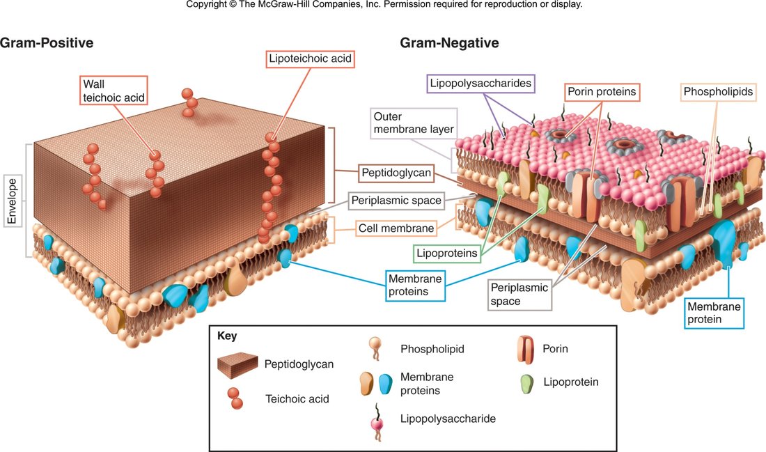
| Gram stain step | Reagent | Time |
|---|---|---|
| 1 | Crystal violet | 1 min |
| 2 | Gram's iodine | 1 min |
| 3 | Acetone alcohol (decolorize) | ~10 sec — the most critical step, and the one most affected by technical variation in timing and reagents |
| 4 | Safranin | 1 min |
| Gram positive | Gram negative | |
|---|---|---|
| Wall | Thick homogenous peptidoglycan, 20–80 nm, with teichoic and lipoteichoic acid and surface proteins | Outer membrane (asymmetric bilayer, outermost layer lipopolysaccharide), periplasmic space, thin peptidoglycan shell, inner cytoplasmic membrane |
| Stain | Retains crystal violet → purple | Loses crystal violet → red from safranin counterstain |
| Strength | Physically strong — resists temperature, pH, osmotic pressure | Chemically strong — resists disinfectants, antibiotics |
Also tested
- Endospore. A dormant, dehydrated form with a tough protective coat that lets a bacterium survive heat, drying and disinfectants for years, conditions that kill the vegetative cell.
- Transpeptidases. They catalyze crosslinking of peptidoglycan and are the target of beta lactam antibiotics, which connects wall synthesis to antibiotic action.
- Lipid A. Lipid A is released from a Gram-negative organism when the cell lyses; it is the toxic portion of the lipopolysaccharide built into the outer membrane, so it reaches the host only when the cell breaks apart.
- Gram-positive versus Gram-negative wall strength. Gram-positive is physically strong, resistant to temperature, pH and osmotic pressure; Gram-negative is chemically strong, resistant to disinfectants and antibiotics. This follows from the thick peptidoglycan sheath versus the outer membrane barrier.
1.5 · Objectives 5 & 6 — Identification methods and culture media
Methods of bacterial identification: microscopic morphology · macroscopic morphology (colony appearance) · physiological and biochemical characteristics · chemical analysis · serological analysis · genetic and molecular analysis (guanine plus cytosine base composition, deoxyribonucleic acid analysis with genetic probes, nucleic acid sequencing and ribosomal ribonucleic acid analysis).
Requirements for growth divide into chemical — water, energy nutrients (carbohydrates, proteins, lipids), vitamins, minerals, oxygen levels — and physical — temperature, pH, osmotic pressure.
| Media type | Definition |
|---|---|
| Synthetic (chemically defined) | Pure organic and inorganic compounds in an exact chemical formula |
| Complex / nonsynthetic | At least one ingredient that is not chemically definable |
| General purpose | Grows a broad range of microbes; usually nonsynthetic |
| Enriched | Complex organic substances — blood, serum, hemoglobin, or special growth factors for fastidious microbes |
| Selective | Contains agents that inhibit some microbes and encourage the desired ones |
| Differential | Allows several types to grow and displays visible differences between them |
The categories combine: blood agar is enriched and differential (gamma, beta and alpha hemolysis); mannitol salt agar is selective and differential.
Also tested
- Synthetic versus complex medium. A synthetic (chemically defined) medium contains pure organic and inorganic compounds in an exact chemical formula; a complex medium contains at least one ingredient that is not chemically definable.
- Selective medium. A selective medium suppresses the growth of unwanted organisms; something in it, such as a high salt concentration, prevents organisms other than the ones sought from growing at all.
- Selective versus differential media. Selective media contain agents that inhibit some microbes and encourage the desired ones; differential media allow several types to grow and display visible differences between them.
1.6 · Objective 7 — The bacterial growth curve
Growth means an increase in number, not size, by binary fission — which is why it is exponential rather than arithmetic.
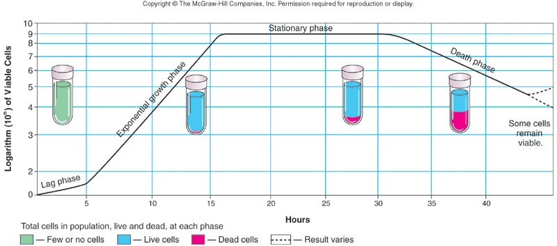
| Phase | What is happening |
|---|---|
| Lag | Flat period of adjustment and enlargement; little growth |
| Exponential growth | Maximum growth, continuing as long as nutrients and environment allow. Most vulnerable to control methods here — the lecture's example is penicillin |
| Stationary | Rate of growth equals rate of death, from depleted nutrients and oxygen and excretion of organic acids and pollutants |
| Death | Limiting factors intensify; cells die exponentially in their own wastes |
Also tested
- Exponential bacterial growth. Each division doubles the population: binary fission turns one cell into two, so the count goes 2, 4, 8, 16 rather than climbing by a fixed amount each generation.
1.7 · Objectives 8 & 9 — Phage and animal virus replication
All viruses have capsids — protein coats enclosing and protecting the nucleic acid — which may be helical, icosahedral or complex. They contain either deoxyribonucleic acid or ribonucleic acid, and may have an envelope or spikes.
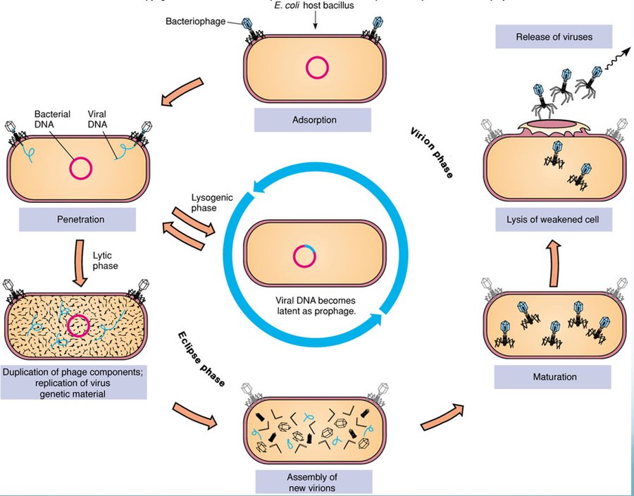
| Step | What happens |
|---|---|
| 1 Adsorption | Binding of virus to a specific molecule on the host cell |
| 2 Penetration | Genome enters the host cell |
| 3 Replication | Viral components produced |
| 4 Assembly | Viral components assembled |
| 5 Maturation | Completion of viral formation |
| 6 Release | Viruses leave the cell to infect others |
| Lytic cycle | Lysogenic cycle |
|---|---|
| Rapid takeover of bacterial metabolism Multiple virus copies produced Destruction of the host cell Generalized transduction — random pieces of host DNA may be transmitted to other bacteria |
Integration of viral genes as prophage May acquire new traits — e.g. the ability to make a toxin Immune to reinfection Viral genes replicated with the host cell Specialized transduction — all cells carry the same host DNA |
Also tested
- Generalized versus specialized transduction. Generalized transduction (lytic cycle) transmits random pieces of host DNA; specialized transduction (lysogenic cycle) means all cells carry the same host DNA.
- Animal virus entry. The virion must be uncoated after entering a host cell, unlike a phage: the whole particle is taken in and the capsid stripped away, whereas a phage injects its genome and leaves the coat outside.
- Phage genome integrated into a bacterial chromosome. The bacterium gains new traits, such as a toxin gene. The prophage is copied along with the chromosome, which is how diphtheria and cholera toxins arise.
- Lytic cycle. Rather than the lysogenic cycle, it features rapid takeover of bacterial metabolism, production of multiple virus copies, and destruction of the host cell. It also produces generalized transduction, in which random pieces of host DNA may be transmitted to other bacteria.
1.8 · Objective 10 — Cytopathic effects of viruses
Virus-induced damage to cells: changes in size and shape · cytoplasmic inclusion bodies · nuclear inclusion bodies · cells fusing to form multinucleated cells · cell lysis · alteration of deoxyribonucleic acid, which may activate oncogenes · transformation of cells into cancerous cells.
Radiation and chemical exposure can activate oncogenes by the same route, which is why this slide links infection to malignant transformation rather than treating them separately.
1.9 · Objective 11 — Health implications of nucleic acid mutations
A mutation is a change in the base sequence of the genome which may impact the phenotype. Mutations occur naturally at a low level but may be induced by radiation, chemicals or viruses, and may be beneficial, neutral or deleterious.
| Route | Mechanism |
|---|---|
| Transformation | Taking up naked DNA from the environment |
| Conjugation | Cell to cell transmission of DNA from a plasmid |
| Transduction | Transmission of genetic material by viral vector |
| Where the mutation is | Health implication |
|---|---|
| Bacteria | New traits — resistance to antibiotics or other control methods, ability to produce toxins or other virulence factors |
| Viruses | High mutation rates change surface antigens and transmission factors. Changing surface properties may weaken or eliminate immunity from vaccination or prior exposure; previously host-specific animal viruses can cross to humans, where immunity is unlikely to exist |
| Human hosts | Chemical or radiation exposure may transform normal cells to cancer cells; oncogenic viruses may do the same; viruses may affect immune cells (human immunodeficiency virus with acquired immunodeficiency syndrome, autoimmune disorders) |
Also tested
- High viral mutation rates. Changing surface properties may weaken or eliminate immunity from vaccination or previous exposure, and animal viruses that were host specific can cross to humans, who are unlikely to have immunity.
- Mutations in human hosts. Health implications include chemical or radiation exposure transforming normal cells to cancer cells, oncogenic viruses causing the same transformation, and viruses affecting immune cells, as with human immunodeficiency virus and acquired immunodeficiency syndrome.
1.10 · Objectives 12 & 13 — Microbial control and the death curve
Aseptic or sterile means removal of all forms of microbial contamination; disinfection means elimination of some. Depending on circumstances, lowering numbers rather than total elimination may be good enough.
Two basic mechanisms: alteration of membrane permeability, and denaturation of proteins and nucleic acids.
| Physical control | Detail |
|---|---|
| Temperature | High can kill; low will NOT — it only slows growth. Dry heat (ovens); wet heat (boiling, autoclave, pasteurization); refrigeration and freezing for preservation |
| pH | Most pathogens prefer neutral, so acid or base inhibits — pickling |
| Osmotic pressure | Increase sugar or salt concentration |
| Filtration | For heat-sensitive solutions — vaccines, sera, beer |
| Radiation | Ionizing (X-ray, gamma) and non-ionizing (ultraviolet) |
| Chemical control | Detail |
|---|---|
| Disinfectants | Used on inanimate objects — Lysol |
| Antiseptics | Used on tissues — Listerine mouthwash |
| True chemical sterilants | Very few. Ethylene oxide (plastics, spices); beta propiolactone (vaccines, tissue grafts, surgical instruments) |
| Adequate for purpose | Halogens such as chlorine or bromine; alcohol |
Antimicrobial chemotherapeutic agents should have selective toxicity — working against the microbial agent without harming the host. Synthetic agents include the sulfa drugs, made from coal tar dyes; antibiotics are substances naturally made by one microorganism against another, such as penicillin from the Penicillium mold, active against Gram-positive bacteria.
Source: Lecture 1 — Review of General Microbiology (Dr. Webster), Slides 1–70, and the PAJ 5200 syllabus instructional objectives.
Also tested
- Constant death rate. With a decimal reduction time of 10 minutes, each interval removes 90% of what remains, so the count falls tenfold each time. Sterilization is therefore defined by probability, not by a moment when the last organism dies.
- Decimal reductions. Each decimal reduction removes ninety percent of whatever remains, so a fall from 10,000,000 to 10,000 organisms, a thousandfold fall, is three decimal reductions.
2 · Antibiotics and Resistance
Instructional Objectives
- Describe major historical events in microbial control.
- Describe criteria for drug selection for prokaryotes and eukaryotes.
- Define therapeutic index including calculations and significance.
- Discuss the effect of drug clearance on dosing schedule.
- Compare and contrast antibiotics and the organisms of major antibiotic drug lines.
- Compare and contrast the basic mechanisms of action of antimicrobial agents and give examples of each.
- Describe mechanisms of drug resistance.
- Describe other potential problems with antimicrobial therapy.
2.1 · Objective 1 — Major historical events in microbial control
| When | Who / what |
|---|---|
| 350–550 AD | Tetracycline traces in Sudanese Nubian skeletal remains — from beer made with fermented grain containing Streptomyces |
| 1600s | Cinchona bark used for malaria and febrile conditions (quinine) |
| 1870 | Burdon-Sanderson: culture fluid covered with mold did not grow bacteria |
| 1877 | Pasteur: anthrax bacilli inhibited by mold growth |
| 1897 | Duchesne healed typhoid-infected guinea pigs with Penicillium |
| 1909 | Ehrlich — drugs as “magic bullets”; developed arsphenamine (salvarsan) against Treponema pallidum |
| 1928 | Fleming — inhibition of Staphylococcus on plates contaminated with Penicillium |
| 1935 | Domagk — sulfonamides from coal tar dyes |
| 1939 | Florey and Chain — produced a stable clinical form of penicillin |
| 1943 | Waksman — Streptomyces produce antibiotics; coined the term, discovered over twenty |
| 1945 | Fleming, Florey and Chain share the Nobel prize in medicine (Waksman's follows in 1952) |
Also tested
- Fleming and penicillin. In 1928 Alexander Fleming observed inhibition of Staphylococcus on plates contaminated with Penicillium mold; Howard Florey and Ernst Chain produced a stable clinical form in 1939, and all three shared the Nobel prize in medicine in 1945.
- Discovery of penicillin. Fleming first observed the inhibition of bacterial growth by a contaminating mold, noticing the zone of inhibition around the mold on a culture plate, which opened the way to penicillin.
2.2 · Objective 2 — Criteria for drug selection
| Term | Definition |
|---|---|
| Chemotherapy | The use of drugs to treat a disease |
| Antimicrobial drug | Interferes with the growth of microbes within a host |
| Antibiotic | A substance produced by a microbe that, in small amounts, inhibits another microbe |
| Selective toxicity | Kills harmful microbes without damaging the host — achieved by targeting processes or structures the pathogen has and the host does not |
| Bactericidal | Bacteriostatic | |
|---|---|---|
| Effect | Kills organisms directly | Inhibits growth |
| Measure | Minimal bactericidal concentration — the minimum level killing 99.9% of test organisms | Minimal inhibitory concentration — the minimum level inhibiting growth |
What makes a good agent — there is no perfect drug, but the lecture lists: selective toxicity · favorable pharmacokinetics (reaching the target site at an effective concentration) · appropriate spectrum of activity (broad = large range of organisms, narrow = small) · lack of side effects · low direct toxicity · low potential for hypersensitivity · good therapeutic index · little resistance development.
Choosing the agent in practice: the disk-diffusion (Kirby Bauer) method reads a zone of inhibition against a chart correcting for concentration, molecular weight and other diffusion factors. The E test (“minimal inhibitory concentration on a stick”) reads that value directly in micrograms per milliliter, where the zone of inhibition meets the strip.
Also tested
- Enzymatic inactivation. This resistance mechanism destroys the antibiotic molecule itself; an enzyme such as beta-lactamase chemically cleaves the drug, so it can no longer bind its target.
- Selective toxicity. Working against the target pathogen without harming the host, normally achieved by interfering with processes or structures found in the pathogen's cells and not the host's; this is why drugs against eukaryotic pathogens are harder to find.
- Broad and narrow spectrum. They refer to the range of organisms the drug will affect: broad meaning a large range, narrow a small one.
- Desirable antimicrobial characteristics. Rapid development of resistance is not desirable; little resistance development is what is wanted.
- Resistance after an antibiotic course. Resistant organisms already present were selected for; the drug removes the susceptible competition rather than creating anything, leaving the few resistant organisms to multiply unopposed.
- Bacteriostatic drugs. They inhibit growth without killing, halting replication and leaving the host's immune system to clear the organisms already there.
- Griseofulvin versus tolnaftate. Griseofulvin inhibits microtubule formation during cell division, interfering with mitosis, and is used for superficial dermatophytes; tolnaftate inhibits squalene epoxidase and therefore ergosterol synthesis, distorting fungal hyphae and stunting mycelial growth.
- Bactericidal versus bacteriostatic drugs. The choice matters most in the neutropenic patient, because a bacteriostatic agent relies on host immunity to finish the job and a patient without functioning neutrophils cannot supply it.
- Efflux pumping. This resistance mechanism keeps an antibiotic from ever reaching a useful concentration inside the cell: the pump exports the drug as fast as it enters, so it never accumulates to a level that would inhibit its target.
2.3 · Objectives 3 & 4 — Therapeutic index and drug clearance
| Therapeutic window | The range of plasma concentrations spanning the minimum concentration for clinical efficacy and toxicity |
| Therapeutic index | A calculation assessing the safety and efficacy of a drug: TI = maximum tolerated dose ÷ minimum inhibitory concentration = TD50 (median toxic dose) ÷ ED50 (median effective dose) |
| Drug clearance | Renal clearance + hepatic clearance + clearance from all other tissues |
Also tested
- Drug clearance and dosage schedule. You must not let the concentration fall below the minimal inhibitory concentration, and it takes time for the drug to enter the system in the first place.
- Drug clearance. It is renal clearance plus hepatic clearance plus clearance from all other tissues, and that total determines how quickly concentration falls, and therefore the dosage schedule.
- Therapeutic index. Expressed as maximum tolerated dose over minimum inhibitory concentration, equivalently median toxic dose (TD50) over median effective dose (ED50); it is a calculation used to assess the safety and efficacy of a drug.
- Antibiotic doses spaced too far apart. Organisms regrow between doses: clearance removes the drug continuously, so once the concentration drops below what inhibits the organism, the suppressed population resumes multiplying.
- Therapeutic index. The index is the toxic dose divided by the effective dose; a toxic dose for half the population of 400 milligrams and an effective dose for half the population of 50 milligrams give an index of 8.
2.4 · Objective 5 — Drug lines and their producing organisms
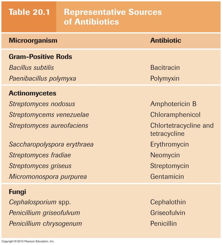
| Drug | Produced by |
|---|---|
| Penicillin | Penicillium chrysogenum (now P. notatum) |
| Cephalosporins | Cephalosporium acremonium |
| Bacitracin | Bacillus subtilis |
| Vancomycin | Amycolatopsis orientalis (formerly Nocardia orientalis) |
| Chloramphenicol | Streptomyces venezuelae |
| Streptomycin | Streptomyces griseus |
| Neomycin | Streptomyces fradiae |
| Gentamycin | Micromonospora purpurea — not a Streptomyces |
| Tetracyclines | Streptomyces aurofaciens |
| Erythromycin | Saccharopolyspora erythraea |
| Polymyxin B | Paenibacillus polymyxa |
| Rifamycin | Amycolatopsis rifamycinica |
| Amphotericin B | Streptomyces nodosus |
| Nystatin | Streptomyces noursei |
| Quinolones, sulfonamides | Artificially produced — sulfonamides from coal tar dyes |
2.5 · Objective 6 — Mechanisms of action
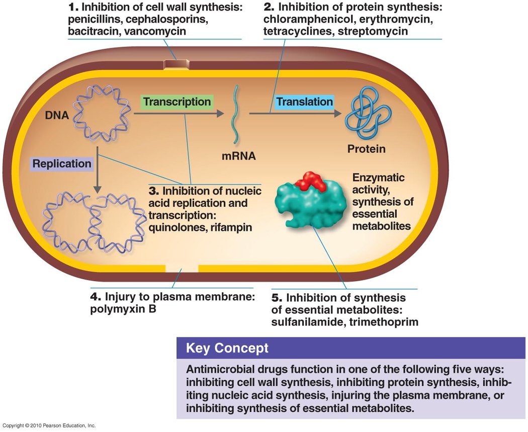
| Target | Agent | Precise mechanism |
|---|---|---|
| Cell wall synthesis | Penicillins | Bind penicillin-binding proteins; inhibit transpeptidase (blocking cross links); produce autolysins (breakdown without synthesis) |
| Cephalosporins | Similar to penicillin's transpeptidase action. 1st gen narrow/Gram-positive → 2nd extended to Gram-negative → 3rd includes pseudomonads, injected → 4th oral | |
| Bacitracin | Interferes with bactoprenol, the transporter carrying peptidoglycan monomers across the membrane, preventing dephosphorylation → osmotic lysis. Topical only, too toxic parenterally | |
| Vancomycin | Glycopeptide binding NAG/NAM subunits, preventing incorporation. “Last line” against resistant Staphylococcus aureus | |
| Isoniazid / ethambutol | Mycobacteria have different walls: isoniazid inhibits mycolic acid synthesis, ethambutol its incorporation | |
| Protein synthesis | Chloramphenicol | Binds 50S; inhibits peptide bond formation. Broad spectrum |
| Aminoglycosides | Change the shape of the 30S subunit → inaccurate reading of messenger RNA codons. Broad spectrum | |
| Tetracyclines | Interfere with transfer RNA attachment. Broad spectrum | |
| Streptogramins | Two components acting together on the 50S subunit. Gram-positives | |
| Erythromycin | Binds 50S; prevents amino acid translocation from transfer RNA to the ribosomal binding site. Gram-positives | |
| Plasma membrane | Polymyxin B | Disrupts the outer membrane of Gram-negatives and the inner membrane by attaching to lipid components. Topical, with bacitracin and neomycin over the counter |
| Nucleic acid synthesis | Rifamycin | Inhibits RNA polymerase. Antituberculosis |
| Quinolones / fluoroquinolones | Inhibit DNA gyrase, preventing unwinding. Urinary tract and hospital acquired infections | |
| Essential metabolites | Sulfonamides | Competitive inhibitors — resemble para-aminobenzoic acid, inhibiting folic acid synthesis. Broad spectrum |
| Antifungal class | Mechanism |
|---|---|
| Polyenes (amphotericin B, nystatin) | High affinity for ergosterol in fungal membranes → pore formation, ergosterol sequestration, reactive oxygen species |
| Echinocandins | Block synthesis of beta-glucan, an essential cell wall component. Intravenous; Candida and Pneumocystis |
| Azoles | Inhibit ergosterol synthesis — membrane |
| Allylamines | For azole-resistant infections |
| Griseofulvin | Inhibits microtubule formation during cell division. Superficial dermatophytes |
| Tolnaftate | Inhibits squalene epoxidase → blocks ergosterol synthesis; distorts hyphae |
| Flucytosine | Cytosine analog — interferes with RNA synthesis and causes miscoding |
| Antiviral strategy | Example / detail |
|---|---|
| Nucleoside and nucleotide analogs | Remdesivir binds viral RNA-dependent RNA polymerase and terminates transcription prematurely. Broad activity against Ebola, respiratory syncytial virus and coronaviruses |
| Protease inhibitors | Block cleavage of viral polypeptide precursors into mature enzymes and structural proteins. Human immunodeficiency virus — indinavir, amprenavir |
| Integrase inhibitors | Prevent viral DNA entering the host chromosome, so it is ultimately degraded |
| Attachment / fusion | Block CCR5 — human immunodeficiency virus |
| Neuraminidase inhibitors | Prevent the virus escaping the host to infect others — influenza |
| Uncoating inhibitors | Amantadine — influenza |
| Interferon | Prevents spread to new cells. Alpha interferon in viral hepatitis induces a transition from the replicating to the nonreplicating form. Adding polyethylene glycol gives a finite treatment duration but greater adverse effects |
Also tested
- Antihelminthic mechanisms. Niclosamide prevents ATP generation (tapeworms) and praziquantel alters membrane permeability (flatworms). Mebendazole inhibits nutrient absorption and ivermectin paralyzes the worm, both for intestinal roundworms.
- Penicillins. They block crosslinking of the cell wall: the beta-lactam ring inactivates the transpeptidase that links peptidoglycan strands, so the wall cannot be completed and the cell bursts.
- Cephalosporins. Originally isolated from Cephalosporium acremonium; first generation narrow spectrum and Gram-positive, second extended to include Gram-negative, third including pseudomonads and injected, fourth oral.
- Aminoglycosides. They act by changing the shape of the 30S subunit so messenger RNA codons are not read accurately; streptomycin, neomycin and gentamycin are broad-spectrum examples.
- Vancomycin. Produced by Amycolatopsis orientalis; a glycopeptide that binds NAG and NAM subunits and prevents their incorporation into the wall, and an important last line against antibiotic-resistant Staphylococcus aureus.
- Echinocandins versus azoles. Echinocandins block synthesis of beta-glucan, an essential component of the fungal cell wall, whereas azoles inhibit ergosterol synthesis, acting on the membrane. Echinocandins are intravenous lipopeptides used against Candida and Pneumocystis.
- Chloramphenicol versus erythromycin. Both bind the 50S ribosomal subunit: chloramphenicol inhibits peptide bond formation and is broad spectrum, while erythromycin prevents amino acid translocation from the transfer RNA to the ribosomal binding site and works against Gram-positives.
- Antimicrobial target absent from human cells. The peptidoglycan cell wall is completely absent from human cells, so drugs that attack it are among the safest antimicrobials available.
2.6 · Objective 7 — Mechanisms of drug resistance
Resistance is the ability of a microorganism to avoid an antibiotic's harmful effects by destroying it, transporting it out of the cell, or undergoing changes that block its effects — three routes that reappear below as the biochemical mechanisms.
| Genetic mechanisms | Cellular and biochemical mechanisms |
|---|---|
| Random genetic mutation Plasmid swapping during conjugation Movement of transposons to plasmids or chromosomes Transduction by bacteriophages Transformation (genes from a recently killed cell) Binary fission shares any of the above |
Drug does not reach the active site: decreased permeability (beta-lactams, quinolones) · decreased transport (aminoglycosides) · increased efflux (tetracyclines, quinolones) Drug inactivation: beta-lactamases, aminoglycoside modifying enzymes Target modification: gyrase modification Bypass of target: vanA, vanB, trimethoprim resistance |
Biofilms enhance resistance three ways: a physical and chemical barrier limiting dissemination and sequestering antibiotics; organisms within are metabolically less active and so less susceptible; and close spatial relationship enhances resistance gene transfer.
Behaviors that drive resistance: misuse or overuse · outdated or weakened antibiotics · using antibiotics for the common cold and other inappropriate conditions · failing to complete the prescribed regimen · using someone else's leftover prescription. Wider contributors include antibiotics in animal feed, environmental warming, and possible correlations with pesticide use and microplastic contamination. Tracking is via the National Antimicrobial Resistance Monitoring System — an interagency partnership monitoring humans (Centers for Disease Control and Prevention), retail meats (Food and Drug Administration) and food animals (United States Department of Agriculture).
Also tested
- Genetic mechanisms of antibiotic resistance. These are random mutation, plasmid swapping during conjugation, movement of transposons, transduction by bacteriophages, transformation, and binary fission sharing any of these. Spontaneous reversion to a wild type state is not listed.
- National Antimicrobial Resistance Monitoring System for Enteric Bacteria. An interagency partnership monitoring resistance of enteric bacteria from humans (Centers for Disease Control and Prevention), retail meats (Food and Drug Administration) and food animals (United States Department of Agriculture), with state and local health departments.
- Fleming's 1945 warning. Self-medication with too small doses does not clear infection; microbes are “educated” to resist penicillin, and penicillin-fast organisms are bred out and passed on until they reach someone with a septicemia or pneumonia penicillin cannot save.
2.7 · Objective 8 — Other problems with antimicrobial therapy
| Problem | Detail |
|---|---|
| Superinfection | Secondary infection following broad spectrum antibiotics, which reduce normal flora and let opportunists take hold. Predisposed by corticosteroids, leukemia, human immunodeficiency virus, systemic lupus, diabetes. Typical organisms: Candida albicans, Clostridioides difficile, hepatitis C virus, human immunodeficiency virus, Aspergillus, resistant staphylococci |
| Hypersensitivity | Most often beta lactams (penicillin, cephalosporins), also quinolones. Immediate or delayed; hives through to anaphylactic shock |
| Resistance | See 2.6 above |
| Direct toxicity — high serum levels of some antibiotics are directly toxic | |
|---|---|
| Aminoglycosides | Renal and auditory toxicity |
| Sulfamethoxazole | Hyperkalemia |
| Ciprofloxacin | Seizures |
| Doxycycline | Esophageal ulcerations |
| Tetracycline | Tooth discoloration — binds calcium during tooth development |
| Chloramphenicol | Aplastic anemia |
| Amphotericin B | Renal and hepatic toxicity |
| Chloroquine | Cardiovascular effects, electrolyte derangements with dysrhythmias |
Drug combinations are one approach to resistant organisms, with three possible results: indifference (no interaction), synergism (combined effect greater than either alone) and antagonism (combined effect less than either alone).
The future: antimicrobial peptides (nisin from lactic acid bacteria, magainin from frogs, cecropin from moths) · targeting biofilm organisms · antisense agents — complementary DNA binding a pathogen's virulence genes to prevent transcription, such as fomivirsen for cytomegalovirus retinitis · phage therapy.
Source: Lecture Number 2 — Antibiotics and Resistance (Dr. Webster), Slides 1–81, and the PAJ 5200 syllabus instructional objectives.
Also tested
- Two-drug interactions. Combining two drugs gives one of three interactions: indifference (no interaction), synergism (the combined effect is greater than either alone) or antagonism (the combined effect is less than either alone).
3 · Microbe-Human Interactions
Instructional Objectives
- Define resident flora
- Summarize the steps in the progression of an infectious disease
- Differentiate between bacterial endo and exo toxins
- Diagram the stages in the course of an infection
- Differentiate between clinical manifestations of an infectious illness
- Describe reservoirs of disease
- Describe the common sources and mitigation methods related to nosocomial infections.
3.1 · Objective 1 — Resident flora
Resident flora includes bacteria, fungi, protozoa, viruses and arthropods. Most areas of the body in contact with the outside environment harbor resident microbes; the large intestine has the highest numbers, mainly strict or facultative anaerobes. Internal organs and tissues are a different matter.
| Region | What lives there |
|---|---|
| Skin | Staphylococci, Corynebacterium, Propionibacterium, yeasts, Mycobacterium smegmatis |
| Mouth | Aerobic streptococci are the most common oral residents |
| Large intestine | The highest bacterial numbers in the body — 10–30% of fecal volume is bacteria |
| Respiratory tract | Staphylococcus aureus, Neisseria meningitidis. The LOWER respiratory tract is essentially sterile — and if it is not, bad things happen |
| Urogenital tract | In females the flora responds to hormonal change; the urinary tract should be sterile except a short portion of the urethra. Same in males |
Normally sterile sites and fluids: heart and blood vessels · kidneys and bladder · brain and spinal column · middle and inner ear · interior of the eyes · blood and cerebrospinal fluid · urine in the kidneys and bladder · amniotic fluid surrounding the fetus.
Also tested
- Protection by resident flora. Resident flora protects the host by occupying the niche first; competition for space and nutrients leaves a newly arrived pathogen nowhere to establish and nothing to consume.
- Benefit of resident microflora. It benefits the host by preventing overgrowth of harmful microbes (competitive exclusion).
- Non-microbial causes of disease. Our own immune responses can cause disease; disease may follow from the host response as much as from the organism.
- Microbial genes in humans. More than a hundred human genes of microbial origin are explained by horizontal gene transfer from bacteria, fungi and plants over time.
- Non-human versus human cells. Non-human cells slightly outnumber human ones, roughly 38 to 39 trillion against about 30 trillion.
- Non-human cells in the body. Bacteria are the most common non-human cell type in the body.
3.2 · Objective 2 — The progression of an infectious disease
The sequence the deck builds, step by step:
| Step | What it means |
|---|---|
| 1. A pathogen with the capacity to cause disease | True (frank) pathogens cause disease in healthy people with normal defenses — influenza and rabies viruses, the plague bacterium, the malarial protozoan. Opportunistic pathogens cause disease when defenses are compromised, or when they grow somewhere unnatural to them — sometimes it is the patient's own flora doing the attacking |
| 2. A weakened host | Age (elderly, young children, premature infants) · genetic or acquired immunological defects · immunosuppressing drugs and organ transplants · stress · chronic conditions such as liver disease or diabetes · inflammation · primary infections already present. Many of these are what you would write as comorbidities |
| 3. A portal of entry | Skin · gastrointestinal tract · respiratory tract · urogenital tract · conjunctiva · pregnancy and birth. Agents are exogenous (from outside) or endogenous (from within) |
| 4. Enough of them | The infectious dose — see below |
| 5. Adhesion | Fimbriae (attachment pili) · flagella · adhesive slimes or capsules (dextran slime, glycocalyx) · suction disks of protozoans · viral spike or capsid proteins · hooks and barbs of helminths and insect larvae |
| 6. Virulence factors | Exoenzymes, toxigenicity, antiphagocytic factors — see 3.3 |
| 7. A pattern of infection | Localized, systemic, mixed, primary, secondary, superinfection — see 3.4 |
| 8. A portal of exit | How the pathogen departs the host, and therefore how it reaches the next one |
Infectious dose (ID) is the minimum number of microbes or viral particles required for infection, usually expressed as ID50 — the dose sufficient to infect 50% of a given population. A SMALL ID50 means GREATER virulence, and therefore a greater degree of pathogenicity.
| Organism | Infectious dose |
|---|---|
| Measles | 1 virus particle |
| Mycobacterium tuberculosis | 10 bacteria |
| Smallpox | 10–100 viral particles |
| Bubonic plague | 100–500 bacterial cells |
| SARS-CoV-1 | about 280 viral particles (animal study) |
| Influenza A | about 790 viral particles |
| Gonorrhea | 1,000 bacterial cells |
| Cholera | 100,000,000 bacterial cells |
Also tested
- True pathogen. A true pathogen sickens a healthy person, carrying enough virulence to overcome intact defenses, which an opportunist cannot do.
- Infectious dose fifty. It describes the dose infecting half of a given population and is the standard expression of infectious dose.
- Opportunistic pathogens. They cause disease with compromised defenses, or growth in an unnatural site, and sometimes it is the patient's own flora attacking.
- Host factors in infectious disease. Immune status is the major host factor.
3.3 · Objective 3 — Endotoxins against exotoxins
| ENDOtoxin | EXOtoxin | |
|---|---|---|
| What it is | Lipid A of the lipopolysaccharide | Proteins |
| Which bacteria | GRAM-NEGATIVE only — it is part of their outer membrane | Certain gram-positive AND gram-negative bacteria |
| Released how | From LYSED or DAMAGED bacteria | SECRETED by LIVING bacteria |
The three virulence factor groups the deck names:
| Group | What it does | Examples |
|---|---|---|
| Exoenzymes | Attack host defenses to allow deeper invasion | Mucinase, hyaluronidase, coagulase, bacterial kinases |
| Toxigenicity | The capacity to produce toxins, grouped by the tissue targeted | Neurotoxins, enterotoxins, hemotoxins, nephrotoxins |
| Antiphagocytic factors | Kill or avoid phagocytes | Leukocidins (“-cidin” = to kill) destroy leukocytes; capsules let the pathogen avoid phagocytosis or resist digestion inside a phagocyte |
Also tested
- Endotoxin release. Endotoxin is released when the bacterial cell lyses, because it is a structural component of the Gram-negative outer membrane.
- Leukocidin. Its function is destroying leukocytes; the suffix means to kill.
3.4 · Objective 4 — The stages in the course of an infection
| Pattern | Definition | Example |
|---|---|---|
| Localized | Microbes enter and remain confined to a specific tissue | Boils, warts |
| Systemic | Spreads to several sites and tissue fluids, usually through the bloodstream | Measles, chicken pox, anthrax, rabies |
| Focal | An agent breaks loose from a local site and spreads | — |
| Mixed | Several microbes grow simultaneously at the infection site | — |
| Primary | The initial infection | — |
| Secondary | Another infection by a different microbe complicating the first | A bacterial lung infection on top of a viral upper respiratory infection |
| Superinfection | A secondary infection resulting from disruption of the natural microflora | Antibiotic therapy for strep throat that results in a vaginal yeast infection |
Also tested
- Superinfection. Unlike an ordinary secondary infection, it follows disruption of the natural microflora, classically after antibiotic therapy.
3.5 · Objective 5 — Clinical manifestations
| Sign | Symptom | |
|---|---|---|
| Whose evidence | OBJECTIVE, as noted by an observer | SUBJECTIVE, as sensed and described by the patient |
| Precision | Often more precise, and may be measured by a clinician | Reported, not measured |
| The deck's example | An inflamed pharynx | A sore throat |
Sequelae are long-term or permanent damage to tissues or organs — paralysis from polio, blindness from gonococcal conjunctivitis, sterility from syphilis or another genital infection, deafness from meningitis, arthritis from Lyme disease.
Also tested
- Sign versus symptom. A sign is observed; a symptom is felt by the patient. The distinction is who perceives it, so a sign can be recorded objectively and a symptom must be reported.
- Sequela. It is lasting damage after the illness: the permanent tissue or organ injury left once the acute illness has resolved, such as blindness following gonococcal conjunctivitis.
- Sign. A sign is observed objectively; it is objective evidence noted by an observer and can often be measured, which separates it from what the patient reports.
3.6 · Objective 6 — Reservoirs of disease
A reservoir is the primary habitat from which a pathogen originates. It may be living or nonliving.
| Type | What the deck says |
|---|---|
| Living — symptomatic | Someone with an obvious, active infection is likely contagious |
| Asymptomatic carriers | May be humans or animals |
| Passive carriers | Medical or dental personnel — carrying the organism without being infected by it |
| Vectors | A live animal that transmits infectious disease — mosquitoes, fleas and ticks; also flies, fruit flies and cockroaches |
| Nonliving | Soil and water |
Zoonosis (plural zoonoses): infections of animals may spread to humans, and vice versa. They can be bacterial, viral, fungal or protozoan in origin — the category is defined by the reservoir, not the organism. A zoonotic infection cannot be completely eliminated without also eradicating the animal reservoir.
| Route | Examples |
|---|---|
| Direct — horizontal | Direct personal contact: kissing or sexual |
| Direct — vertical | Mother to child, transplacental or during vaginal birth |
| Direct — droplet | Close personal contact including saliva, vomit, feces or blood |
| Direct — biological vectors | Mosquitoes, fleas, ticks |
| Indirect | Vehicles and fomites (inanimate objects), and airborne droplet nuclei |
Also tested
- Dormant pathogens in intermediate hosts. Eradication is complicated because spores and cysts persist.
- Nonliving reservoirs. Soil and water are nonliving reservoirs of disease.
3.7 · Objective 7 — Nosocomial infections
Nosocomial infections, also written HAI (hospital-acquired infections), are diseases acquired or developing during a hospital stay. They may prolong the stay, or end in death.
| Facet | Content |
|---|---|
| Most common sites | Urinary tract, respiratory tract, and surgical incisions |
| Most common organisms | Gram-negative: Escherichia coli, Pseudomonas, Klebsiella. Gram-positive: staphylococci and streptococci. Fungi: yeasts. The deck notes the wider list goes by the acronym ESKAPE |
| Mitigation | Universal precautions for sample collection and patient care — based on the assumption that ALL patient specimens are possibly infectious |
Also tested
- Hospital-acquired infection. The most common sites are the urinary tract, airway and surgical wounds; each is routinely instrumented, so catheters, ventilation and surgery breach the barrier that would otherwise exclude organisms.
3.8 · Epidemiology and Koch's postulates
Not separate objectives, but taught in this deck and fair game. Epidemiology is the study of the frequency and distribution of disease and other health-related factors in human populations. Reportable (notifiable) diseases must be reported to public health authorities; in the United States the CDC (Centers for Disease Control and Prevention) publishes the Morbidity and Mortality Weekly Report.
| Measure | Counts | Expressed as |
|---|---|---|
| Prevalence | EXISTING cases, against the entire population | Usually a percentage |
| Incidence | NEW cases over a time period, against the general healthy population | A count over time |
| Mortality rate | DEATHS due to a certain disease | Per 100,000 |
| Morbidity rate | CASES — people afflicted | Per 100,000 |
Over the last hundred years the death rate from infectious disease has dropped while morbidity has remained relatively high. People stopped dying of these illnesses; they did not stop getting them.
| Pattern | Definition |
|---|---|
| Endemic | A relatively steady frequency over a long period in a particular geographic location — often because the reservoir is present |
| Sporadic | Occasional cases at irregular intervals |
| Epidemic | Prevalence increasing beyond what is expected for that population |
| Pandemic | A global epidemic |
Koch's postulates (1880s):
- Find evidence of a particular microbe in every case of a specific disease
- Isolate it from an infected subject and cultivate it artificially in the laboratory
- Inoculate a susceptible healthy subject and observe the resultant disease
- Re-isolate the agent from the newly infected subject
They do not work well for all pathogens. Mycobacterium leprae and Legionella pneumophila are the two the deck names — step 2 is the sticking point when an organism cannot be cultivated artificially. Today they are of limited use, though still applied to bacterial, fungal and protozoan pathogens.
Molecular Koch's postulates (Falkow, 1988) exist because viruses and prions routinely break all the old rules:
- The phenotype or property under investigation should be associated with pathogenic strains or species
- Inactivating the gene or genes associated with the virulence trait should produce a measurable loss of pathogenicity
- Reverting the mutated gene should restore pathogenicity
Also tested
- Prevalence. It measures total existing cases in the population, usually as a percentage.
- Mortality versus morbidity rate. Mortality counts deaths, whereas morbidity counts cases; both are expressed per hundred thousand.
4 · Transmission of Microorganisms
Instructional Objectives
- Describe the chain of infection.
- Describe the relationship between fomite-transmission and nosocomial infections.
- Describe the relationship between fomite-transmission and antibiotic-resistant strains of bacteria and fungi.
Three objectives, and the second and third are both about fomites. That tells you where the weight sits: the chain of infection is the framework, and everything after slide 19 is an argument that non-living surfaces matter more than they look like they should. Roughly half the deck is published studies making that case.
4.1 · The chain of infection
Six links, in order. Break any one and transmission stops — which is the whole point of infection control.
- The infectious agent — and its traits, including whether it is already drug resistant.
- The reservoir, known or unknown.
- The portal of exit from the infected host.
- The mode of transmission to other hosts.
- The portal of entry into the new host.
- A susceptible host — and susceptibility varies between people.
4.2 · Reservoirs, and the four carrier states
Living reservoirs: humans, animals, and — the deck marks both with a question mark — plants and fungi. Non-living reservoirs: soil, water, air, food, and fomites.
Human reservoirs come in four states, and the distinction that matters is whether the person is actually infected:
- Symptomatic and infected — the obvious case.
- Asymptomatic and infected.
- Active carriers — infected, and may or may not show symptoms. Mary Mallon, “Typhoid Mary”, carried Salmonella typhi without symptoms in the early 1900s.
- Passive carriers — NOT infected, but still transmitting. The healthcare or food-service worker with poor hand hygiene.
Active against passive is the distinction to hold. An active carrier has the organism in them; a passive carrier is merely carrying it on them. Both transmit.
4.3 · Zoonoses
Infections of animals that may spread to humans, and the reverse. They can be bacterial, viral, fungal or protozoan. Humans are often the accidental host — plague and anthrax are the examples given — with the lifecycle running through primary and secondary hosts.
Why they cannot be eradicated: you would have to eradicate the animal reservoir too, which is harder still when there are several. And a pathogen that lies dormant in an intermediate host as a fungal spore or protozoan cyst outlasts any campaign against the animal.
Occupational risk is named for veterinary medicine, farm work, landscaping, gardening, zookeeping and slaughterhouse or meat processing work.
| Pathogen | Reservoir | Transmission | Human to human? |
|---|---|---|---|
| Rabies | Mammals | Bite / saliva | Rare — organ transplantation |
| Influenza | Birds, pigs | Respiratory droplet, contact | Yes |
| Ebola | Fruit bats, small primates | Body fluids | Yes |
| Marburg, Nipah | Fruit bats | Body fluids | Yes |
From the recording, not the slides. Rabies is the commonest zoonosis in the United States; malaria is the commonest worldwide — and there is almost no endemic malaria in the United States, though Hawaii has a bird form. Neither ranking appears on any slide, and both are the kind of fact a single question turns on.
Also tested
- Zoonotic infection and eradication. A zoonotic infection cannot be eradicated like smallpox because the animal reservoir persists; eradication requires removing every place the organism can live.
- Hand hygiene. It breaks the mode of transmission link in the chain of infection, because washing removes organisms from the hands that would otherwise carry them from one host to the next.
4.4 · Vectors, and how they differ from reservoirs
A vector is a living thing capable of transmitting disease — mosquitoes, fleas and ticks; flies, fruit flies and cockroaches.
Reservoir and vector are not the same thing, and the difference can be subtle. For malaria the reservoir may be birds while the vector is a particular species of mosquito. The reservoir harbors; the vector delivers.
Also tested
- Vector versus reservoir. The vector carries the organism; the reservoir harbors it. The reservoir is where the organism normally lives and multiplies, while the vector merely transports it to a new host.
4.5 · The modes of transmission
By contact. Direct splits into vertical (mother to child) and horizontal (usually mucous membrane contact — sexual contact, kissing), plus droplets from coughing, sneezing, saliva, blood, sweat and tears. Indirect contact is transmission onto inanimate surfaces first — that is fomite transmission, and it is where the rest of the lecture goes.
By vehicle. Food — gastrointestinal illness is the commonest result. Water — drinking water, and also pools, spas and water parks. Soil — contaminating produce, hands and fingernails; pinworms in children. Air — usually, though not always, an indoor phenomenon, which is why ventilation, filtration and crowding matter.
Also tested
- Indirect contact transmission. A surface comes between the source and the recipient; this is fomite transmission.
- Soil-related transmission. Poorly washed produce is a named route, along with hands and fingernails, and pinworms in children.
4.6 · Fomites and hospital-acquired infection
A fomite is a non-living surface or object that may transmit pathogens. Hospital-acquired infection (HAI) costs longer stays, long-term disability, preventable deaths, expense, and risk to both safety and quality of care. It is most threatening to the immunocompromised, pediatric, geriatric and HIV patients, and those with open wounds, burns or recent surgery.
Clinical fomites named: scalpels and syringes; catheters — a particular problem with biofilm-forming Staphylococcus aureus and S. epidermidis; bed rails and gurneys; telephones, remote controls, keyboards and electronic devices; floors, mops, polishers, brooms and dustpans; pillows, bedding, furniture and curtains; and stethoscopes, neckties, stuffed animals, greeting cards and flowers.
And it is not only hospitals — a public restroom, hotel room, cruise ship cabin or aeroplane tray table is a fomite-filled room too.
Also tested
- Fomite. A fomite is a contaminated non-living object, any inanimate item that carries organisms from one person to another, such as a stethoscope or a keyboard.
4.7 · The studies, and what they did not do
Birthday cake — Dawson and colleagues, 2017
Blowing out the candles put about fifteen times more bacteria on the frosting than the no-blow control. The authors never identified the organisms they cultured, though the discussion named several pathogenic genera — and they said nothing about fungi, yeasts or viruses in saliva. Droplet size is the link to face coverings: the candle test is trying to blow one out while wearing one.
Keyboards — Ide and colleagues, 2019
A systematic review of 75 published studies, swabbing keyboards, mice and pads, and portable tablets. Organisms cultured included coliforms such as E. coli and MRSA.
Viral fomites — Boone and Gerba, 2007
Pre-2007 figures: 1.7 million deaths a year from diarrheal disease and 1.5 million from respiratory infection. Viruses cause about 60% of human infections — and viral disease cannot be cured with antibiotics, so prevention rests on vaccines and antivirals. Crowded indoor environments consistently increase morbidity and mortality: schools, daycare, nursing homes, offices, hotels, cruise ships and hospitals, especially pediatric wards.
Patient-care items — Kanamori and colleagues, 2017
Soap and sanitizer dispensers, humidifiers, nebulizers, pressure transducers, stethoscopes, suction apparatus, thermometers, ultrasound probes and gel, blood pressure monitors, intravenous pumps and poles, telemetry boxes and wires.
From the recording: results are reported as CFU — colony-forming units — because you cannot know whether a visible colony grew from one cell, a hundred or a thousand. The unit is CFU per gram or per milliliter, and serial dilution works for clinical specimens, urine, feces, food, water and milk alike.
Also tested
- Keyboard systematic review. It analyzed seventy-five published studies.
- Viral share of infections. According to the Boone and Gerba review, viruses cause about sixty percent of human infections.
4.8 · Disinfection, coatings and the history
Five best practices for non-critical surfaces, in order: standardize cleaning policy; select EPA-registered hospital disinfectants; educate ALL staff including environmental services; monitor compliance with feedback; implement no-touch decontamination technology.
The history: Semmelweis in the 1840s — handwashing with chlorine solution by obstetricians. Lister in the 1860s — handwashing and wound treatment with carbolic acid.
Chemical agents — bleach, quaternary ammonium salts, phenolics — work only at the correct concentration and for adequate contact time, and they are toxic. Ozone, high-concentration hydrogen peroxide and steam require the room to be vacant, take time facilities may not have, and can damage surfaces and devices.
Coatings borrow from marine anti-fouling paint — copper, arsenic, mercury and tin compounds since the 1960s. Anti-fouling prevents attachment; antimicrobial kills. A hospital coating should be nontoxic, cost effective, commercially available, stable and durable: plastics, copper or silver alloys, photocatalytic coatings, specialized textiles, micro-patterned hydrophobic surfaces.
Also tested
- Persistent pathogens on fomites. They are particularly dangerous because they look like clean surfaces.
- Ozone and hydrogen peroxide disinfection. A room must be vacant before ozone or high-concentration hydrogen peroxide is used, to reduce harm to humans.
- Cleaning and disinfection policy. All staff, without exception, must be educated about it.
- Transmission of hospital-acquired infections. They are primarily transmitted directly, human to human; fomite contact is the secondary route.
- Anti-fouling surface coatings. The idea originated in marine ship coatings, which have used copper, arsenic, mercury and tin compounds since the 1960s.
4.9 · Fomites and antibiotic resistance
Extensive antibiotic use has driven multi-drug resistance, and MDR, XDR and TDR strains — MRSA and carbapenem-resistant Klebsiella pneumoniae among them — are especially problematic in hospitals. Biofilms are polymicrobial networks resistant to cleaning, heat and drugs; Staphylococcus and Pseudomonas form them.
A fomite carrying a persistent pathogen cannot be told apart from a clean surface. That single sentence is the argument for the whole chapter.
Antiseptics in West Africa — Lompo and colleagues, 2023
Two tertiary hospitals in Burkina Faso and Benin. Contamination was highest in liquid soap — 51 of 69 samples. Risk factors: inconsistent preparation, recycled containers including used soft drink bottles, broken pump dispensers, and “topping up”. Counts above 10,000 CFU/mL were Klebsiella pneumoniae, Pseudomonas aeruginosa and Acinetobacter — all gram-negative — from maternity, neonatology, surgery and internal medicine.
How resistance moves between organisms. Conjugation is the big one: donor and recipient need not be the same genus or species, and once a cell gains a resistance plasmid every one of its offspring has it too. Transformation — competent cells taking up free DNA from the environment — matters less. Transduction requires a bacteriophage as the vector.
Put the two halves together and the third objective answers itself: a contaminated surface keeps resistant organisms in circulation, and conjugation lets them hand that resistance to organisms of an entirely different species.
Also tested
- Competent cells. In transformation, competent cells take up DNA fragments from their environment.
- Conjugation and resistance. Horizontal gene transfer by conjugation is significant for resistance because the species need not match, and every daughter cell inherits the plasmid.
- Transduction. This horizontal gene transfer mechanism uses a virus as the carrier: a bacteriophage packages host genetic material and injects it into the next cell, carrying resistance genes between organisms that never touch.
- Hospital fomite contamination. The most heavily contaminated samples came from the maternity and neonatology wards.
5 · Host Defenses: Nonspecific Mechanisms
Instructional Objectives
- Review the lines of host defense.
- Differentiate among the chemical, genetic, and physical defense mechanisms in the first line of defense.
- List the key factors involved in innate immunity.
- Describe the recognition system of host defense.
- Describe the roles of the different classes of leukocytes in the second and third lines of defense.
- Describe the role of a white blood count with differential.
- Describe the characteristic components of inflammation.
- Describe the mechanism of pathogenic fever.
- Explain the beneficial and harmful effects of fever.
- Compare and contrast the origin and functions of interferon.
- Discuss the complement system.
Eleven objectives against 93 slides — this is the biggest lecture of the block. Lectures 1 to 4 carry three or four objectives each. Complement alone runs fourteen slides. The temptation is to revise complement and natural killer cells and let the rest slide, which is exactly how the small objectives — the differential white count, the genetic defense, the effects of fever — get lost.
One idea ties it together. The innate system covers an enormous range of organisms without having a receptor for each one. It manages that by recognizing patterns shared across pathogen families, not individual organisms. Every receptor, plasma protein and cell below is an instance of that trick.
★ HE READ OUT THE EXAM LIST IN THE LAST THREE MINUTES. The closing review of the lecture is a near-verbatim revision plan, and it is reproduced here in his order:
- “You need to know those three lines — first line, second line, third line — and know what belongs in each line. First line, know which things are physical, which are chemical, which are microbiological.”
- “Recognition system. What do we call the receptors that the white blood cells have?”
- “Know the difference between a macrophage and a neutrophil, our two major phagocytes. But how are they different?”
- “White blood cell with differential. What do we use it for? And what can it tell us?”
- “Inflammation — you need to know the major characteristics and how do they come about? How is it that we have the redness, the heat, the pain?”
- “What causes fever? Why is fever good? Why is fever bad?”
- “When do we produce interferon? What kind of invading organism helps us produce interferon?” — and he answered his own question: “remember that it does not kill a virus, but it prevents the viral spread.”
- Complement — “three different pathways, all getting to the important component of C3. Which one’s the first one? The alternative. Then the lectin. And then finally, the classical. And the classical is the one which can be associated with the third line of defense.”
Five things he told the room NOT to memorize, which is just as useful the night before:
- The types of mucins — “I am not asking you to memorize”; the point is only that different surfaces get different ones.
- The chart of toxins defensins can unfold — “a chart I’m not asking you to memorize.”
- The proteins that protect human cells from the membrane attack complex — “you don’t need to fuss about that one.”
- The membrane attack complex assembly chart — “you don’t have to memorize this chart, but just know… it starts putting together pieces to form a pore.”
- The reference ranges for the differential — “which I’m not asking you to memorize … that’s what we have references for.” The PROPORTIONS still matter; the exact numbers do not.
And one framing point he made at the very start: “it is important to understand that the organisms have to hit these barriers IN ORDER. They go through the physical and chemical barriers first, then the processes with the white blood cells. And only after they’ve gone through that can they even start an adaptive immune response.” The three lines are a sequence, not a menu.
5.1 · Objective 1 — The lines of defense
Three categories, and three lines. The categories are barriers (physical and chemical), fixed or hard-wired mechanisms (innate), and the adaptive immune response — the one vaccination exploits, and the only one giving long-term protection.
| Line | What it is | Specific? |
|---|---|---|
| First | Physical, chemical, microbiological and genetic barriers that block invasion at the portal of entry | Nonspecific |
| Second | Protective cells, physiological processes and antimicrobial substances | Nonspecific |
| Third | Acquired on exposure to antigens; produces antibodies or defensive cell lines, and creates immunological memory | Specific |
Two of the three are nonspecific, and that is the whole subject of this lecture. Notice also that the first line includes a genetic barrier — an unusual category that becomes concrete at the defensins in 5.3.
5.2 · Objective 2 — The first line: physical, chemical and genetic
The physical list: intact skin, mucous membranes, the ciliary escalator, the lacrimal apparatus, nasal hairs, saliva, urine, sweat, defecation, the expulsion mechanisms (coughing, sneezing, vomiting), and normal flora.
| Barrier | How it works |
|---|---|
| Epidermis | Stratified squamous epithelium. Rapid desquamation physically removes transient flora. Keratin resists invasion because most pathogens lack keratinase. Keratinocytes make antimicrobial peptides, packaged in lamellar bodies and secreted into a waterproof lipid layer |
| Mucous membranes | Line the gastrointestinal, respiratory and urogenital tracts — a much greater area than skin. Thin, permeable, not keratinized, because gas exchange and absorption demand it |
| Mucus | Glycoproteins, proteoglycans, peptides and enzymes. Mucins are the gigantic glycoproteins that give it its protective properties; the gut goblet cell makes it |
| Lacrimal apparatus | Washes the eye, and carries lysozyme — so it is chemical as well as physical |
| Urine | Mechanical flush, plus high osmolality and an inhibitory pH |
| Saliva and sweat | Mechanical flush plus antimicrobial secretions |
Normal flora is both barriers at once, and that is the point of it. Colonization begins after birth. On the epidermis it is Staphylococcus epidermidis, other coagulase-negative staphylococci, and coryneform bacteria. They compete for living space and nutrients — a physical barrier — and they produce antimicrobial substances — a chemical one.
The atopic dermatitis finding is the cleanest evidence for this. Atopic skin shows dysbiosis with less antimicrobial peptide. When subjects were deliberately colonized with coagulase-negative staphylococci, colonization by Staphylococcus aureus was diminished. Restoring the residents suppressed the pathogen.
Gut commensals do three things beyond blocking colonization: they digest substances we cannot, they provide vitamins, and they help develop gut-associated lymphoid tissue.
The chemical list: sebum, the acid mantle of skin, lysozyme (which acts on peptidoglycan), lactic acid and electrolytes in sweat, digestive secretions, semen (spermine, lysozyme, lactoferrin, phospholipase) and vaginal secretions (lactic acid, beta defensin, hydrogen peroxide). Those last two lists are worth keeping apart — they are easy to swap.
5.3 · Objective 3 — Key factors in innate immunity
Plasma proteins that limit infection: the coagulation system, the kinin system, protease inhibitors, defensins, pentraxins, the acute-phase response, inflammation and its cytokines, and complement.
| System | What it does |
|---|---|
| Coagulation | Clot formation limits pathogen mobility and reduces blood and fluid loss. Platelet degranulation releases cytokines that recruit immune cells and drive repair |
| Kinin | An enzymatic cascade producing bradykinin — dilates vessels and relaxes smooth muscle to enhance elimination of pathogens and tissue repair |
| Protease inhibitors | Block microbial proteases that degrade human tissue. One tenth of all serum proteins; alpha-2 macroglobulin is the example |
Defensins are the predominant family of antimicrobial peptides: 30 to 40 amino acids, amphipathic — hydrophobic and hydrophilic regions — which is how they damage membranes. That mechanism sets their range: bacteria, fungi, and ENVELOPED viruses, because an envelope is a membrane.
They are also the only innate components that can neutralize a broad range of microbial toxins by unfolding them, changing their three-dimensional configuration. The deck calls them antichaperones — chaperones fold proteins, these do the reverse.
This is where the “genetic” defense of objective 2 becomes concrete. Individuals carry 2 to 14 copies of the alpha defensin genes and 2 to 12 of the beta. Copy number determines how much protein is made — so how well defended you are is partly inherited. Four of the six alpha defensins sit in neutrophil granules; the other two are made by intestinal Paneth cells.
Pentraxins bind pathogen surfaces and target them for phagocytosis, bridging pathogen to phagocyte much as antibody does. Short pentraxins come from hepatocytes — serum amyloid P component and C-reactive protein; long pentraxins from myeloid, endothelial and epithelial cells.
The acute-phase response runs on one signal: bacteria induce macrophages to produce interleukin 6, which tells liver cells to raise the defensive plasma proteins and lower others, including albumin. C-reactive protein and serum amyloid A rise more than a hundredfold — which is precisely why C-reactive protein is used to diagnose infection, inflammation and tissue damage. It is a pentraxin opsonin.
Also tested
- Albumin in the acute-phase response. Albumin decreases while defensive plasma proteins increase, as the liver shifts its output.
- C-reactive protein. It is used in diagnosis because it rises over a hundredfold, a large, measurable swing.
- Damage-associated molecular pattern. It signals injury to the host's own cells: molecules normally kept inside a cell appear outside when it is damaged, so the same receptor system provokes inflammation after sterile injury as after infection.
- Acute-phase response cytokine. Interleukin 6, released by macrophages when bacteria induce them, drives the acute-phase response and acts on liver cells.
5.4 · Objective 4 — The recognition system
Recognition has to sort three things, not two: healthy self, non-self, and altered self — tissue damaged or changed by viruses or cancer.
| Term | Meaning |
|---|---|
| PRR | Pattern Recognition Receptor — recognizes structural patterns from types of pathogen |
| PAMP | Pathogen Associated Molecular Pattern — the structure on the microbe that a PRR recognizes. Marks non-self |
| DAMP | Damage Associated Molecular Pattern — signals cells that are damaged, stressed or virus-invaded. Marks altered self |
Each receptor recognizes a pattern shared by a pathogen FAMILY, which is how a limited set of receptors covers a wide range of organisms. Each cell carries a unique combination, which broadens the coverage further. Macrophages are very effective at bacterial and fungal carbohydrates using lectin receptors; natural killer cells at the changed surface proteins of a virus-infected cell.
Where a receptor sits determines what it does, and the Toll-like receptors make the point best:
| Receptor family | Detects | Triggers |
|---|---|---|
| Toll-like — plasma membrane | Many pathogens | Inflammatory cytokines |
| Toll-like — endosomal | Many pathogens | Interferon |
| Scavenger | Microbes — or, with no infection, cellular debris and apoptotic cells | Elimination |
| Retinoic acid inducible gene | Viral RNA | Interferon |
| Cyclic GMP cyclase | Viral DNA | Interferon |
Effector function then runs on cytokines. Some are cached in vesicles and released fast, some must be made on demand, and some are secreted only on cell-to-cell contact. Some are pro-inflammatory and some anti-inflammatory.
Also tested
- Pathogen-associated molecular pattern. It represents a conserved microbial structure; because these structures are shared across whole classes of organism and absent from us, a small set of receptors can detect almost any invader.
- DAMP. A damage-associated molecular pattern signals cells that are damaged, stressed or virus-invaded; PAMPs mark non-self, whereas DAMPs mark altered self.
5.5 · Objectives 7 and 8 — Inflammation and fever
The five cardinal signs, described by Celsus in the first century: rubor (redness), calor (heat), tumor (swelling), dolor (pain), functio laesa (loss of function).
The inflammasome starts it. When macrophages sense infection, pathogen products drive assembly of a protein structure that converts pro interleukin 1 beta into functional interleukin 1 beta in large quantities. A second cascade opens pores in the macrophage membrane to release it — and that kills the macrophage, by pyroptosis. Mutations in inflammasome genes cause autoinflammatory diseases.
| Cytokine | Effect |
|---|---|
| Interleukin 1 beta | Initiates the cytokine release |
| Tumor necrosis factor alpha | Dilates vessels, raising blood volume — producing heat, swelling, redness and pain |
| Interleukin 6 | Increases temperature |
| CXCL8, CCL2, interleukin 12 | Chemokines — attract other white cells |
Getting the leukocytes out of the blood takes two adhesion steps and a squeeze:
- Rolling — L-selectin on the leukocyte binds vascular addressin CD34 on the endothelium. Transient, so the cell rolls along the surface.
- Firm adhesion — integrin LFA-1 binds the immunoglobulin-like ICAM-1.
- Diapedesis — guided by chemokines, the neutrophil squeezes between endothelial cells to reach the infection.
Endothelial cells help by upregulating adhesion molecules and loosening their tight junctions.
Fever — objectives 8 and 9 together. Interleukin 1 beta, interleukin 6 and tumor necrosis factor alpha act both locally and systemically, and the systemic effect is fever. Its benefits are specific and worth memorizing as a list:
- Decreases replication of viral and bacterial pathogens — in bacteria, by starving them of iron
- Increases production and activity of neutrophils
- Enhances T cell proliferation
- Enhances immune signaling
- Enhances tissue resistance to the damaging effects of tumor necrosis factor alpha — protection against the body's own mediator
Tumor necrosis factor alpha is the same molecule doing opposite things at different scales, and this contrast is exam-shaped:
| Released LOCALLY | Released SYSTEMICALLY |
|---|---|
| Increased blood flow and permeability in venules; endothelium becomes adhesive for white cells and platelets; blood in the venules clots, which prevents spread of infection to the blood | Acts on venules in all tissues at once — a state of shock, organ failure, death |
Containment at one site; catastrophe everywhere at once.
Also tested
- Cause of fever. Cytokines reset the set point: interleukin-1, interleukin-6 and tumor necrosis factor act systemically on the hypothalamus, which then defends a higher target temperature.
- Benefit of fever. Fever inhibits pathogen growth; many organisms replicate poorly above normal body temperature, while the host's own enzymatic and cellular responses run faster.
- Danger of high fever. High temperatures damage tissue: above a certain point host proteins begin to denature, so the response that inhibits the organism starts to harm the patient.
- Tumor necrosis factor. This cytokine dilates blood vessels, increasing blood volume in the infected area, to produce heat, swelling, redness and pain.
- Inflammasome. It is a protein complex that activates interleukin 1 beta, and in large quantities.
- Systemic cytokines. Interleukin 1 beta, 6 and TNF alpha have systemic effects including fever; they are the same three that act locally and carry the response beyond the tissue.
- Interleukin 1 beta release. It escapes the macrophage through pores, and the cell dies by pyroptosis; release costs the cell its life.
- Beneficial effect of fever. Fever increases production and activity of neutrophils, alongside enhanced T cell proliferation and immune signaling.
- Four classical signs of inflammation. Redness, heat, swelling and pain (rubor, calor, tumor and dolor), with loss of function sometimes added as a fifth.
- Firm leukocyte adhesion. The integrin pairing LFA-1 to ICAM-1 holds the leukocyte firmly to the endothelium; ICAM-1 is an immunoglobulin-like molecule on the endothelial side.
5.6 · Objective 11 — Complement
More than thirty proteins, ubiquitous in blood and lymph, circulating as inactive zymogens and activated by cleavage in a cascade. Its activity comes from a unique high-energy thioester bond.
The thioester bond is the mechanism. Activation cleaves C3 into C3a and C3b, exposing the bond on C3b. It then has two fates: attacked by water, it becomes soluble and does nothing; or it reacts with a hydroxyl or amino group on a pathogen surface and attaches. That is complement fixation, and it marks the pathogen for destruction. Meanwhile C3a recruits phagocytes.
| Pathway | Order of activation | Trigger |
|---|---|---|
| Alternative | First — quickest | Nonspecific; deposits C3b at the very beginning of infection |
| Lectin | Second | Nonspecific; induced by infection, takes a little time to become effective |
| Classical | Last | C-reactive protein (innate) or antibody (adaptive) — which is why it belongs to both systems |
All three converge on cleaving C3, which is why the deck calls C3 by far the most important molecule in the cascade — the pathways differ mainly in how they reach it. They were discovered in a different order (classical, alternative, lectin), and the system is called “complement” because it was thought to complement the specific immune system.
Regulation is what keeps it off your own cells, and the four regulators split cleanly:
| Regulator | Type | Effect |
|---|---|---|
| Properdin (factor P) | Plasma | Binds C3 convertase on microbial surfaces — INCREASES activation |
| Factor H + factor I | Plasma | H makes C3b susceptible to cleavage by I, giving iC3b, which cannot form a convertase — reduces activation |
| Decay accelerating factor, membrane cofactor protein | Membrane | Rapidly disrupt C3bBb formed on a human cell surface |
C3b is the opsonin — a protein bound to a pathogen that facilitates phagocytosis. Complement receptor 1 binds it to drive phagocytosis, and does double duty protecting human cells by disrupting C3 convertase.
Then the cascade escalates. C3b binding existing C3bBb makes the alternative C5 convertase, which cleaves C5. C5a recruits neutrophils and is the most potent anaphylatoxin; C5b initiates the membrane attack complex. Human cells are protected from that too — S protein, clusterin and factor J stop C5b, C6 and C7 associating with membranes, and homologous restriction factor and CD59 (protectin) stop C9 joining a complex that has already attached.
The evasion story is worth knowing because of its exception. Streptococcus pyogenes and Staphylococcus aureus cover themselves in sialic acid — which factor H has a binding site for, because it is found on human cells. Factor H binds, and their C3b is readily inactivated. But antibody coats the surface and masks the sialic acid before complement binds, so these bacteria resist complement only when no specific antibody is present. Innate immunity alone is beaten here; innate plus adaptive is not.
5.7 · Objective 10 — Interferon
| Interferon | Made by | Does |
|---|---|---|
| Alpha | Lymphocytes and macrophages | Activates natural killer cells |
| Beta | Fibroblasts and epithelial cells | Assists B and T cell maturation and the inflammatory response |
| Gamma | T cells | Inhibits cancer cells, stimulates B cells, activates macrophages and increases their effectiveness |
All are produced in response to viruses, RNA, immune products and various antigens; they bind cell surfaces and induce expression of antiviral proteins, and they inhibit expression of cancer genes and suppress tumors.
The single most misunderstood point in this section, and the deck states it outright: interferon does not kill viruses. It stops spread to surrounding tissue. It protects the neighbors rather than clearing the infection.
Also tested
- Interferon and viruses. Interferon does nothing directly to a virus. It does not kill viruses; it stops spread to surrounding tissue.
- Interferon protection. Interferon protects uninfected cells by inducing antiviral proteins in them, preparing the neighbors.
- Interferon action on viruses. Interferon prevents spread rather than killing the virus. It acts on neighboring uninfected cells, inducing an antiviral state; the already-infected cell is not rescued.
5.8 · Objectives 5 and 6 — Leukocytes and the differential
Learn the percentages — they are what the differential count is read against.
| Cell | % | Appearance | Role |
|---|---|---|---|
| Neutrophils | 55–90 | Lobed nuclei, lavender granules | Phagocytes |
| Eosinophils | 1–3 | Orange granules, bilobed nucleus | Destroy eukaryotic pathogens; minor phagocyte |
| Basophils | 0.5 | Constricted nuclei, dark blue granules | Release potent chemical mediators |
| Monocytes / macrophages | 3–7 | Largest white cells, kidney-shaped nucleus | Phagocytic; housekeeping; antigen presentation; cytokine secretion |
| Lymphocytes | 20–35 | — | B (adaptive humoral), T (adaptive cell-mediated), and non-B non-T including natural killer cells (INNATE) |
Mast cells are related to basophils but nonmotile and bound to connective tissue; whether they share a common progenitor is unsure. A macrophage is the final differentiation of a monocyte, in tissue, and takes special names in particular tissues.
The differential white count totals the number of each type and determines whether they are in normal proportion — proportion, not just total. It is used in diagnosing infection types, inflammation, allergies, immune disorders, leukemia and myelodysplastic syndrome.
Phagocytosis is done principally by macrophages and neutrophils, and the division of labor matters:
| Macrophage | Neutrophil |
|---|---|
| Long-lived, resides in tissue, has other functions, works as infection begins and raises the alarm | Short-lived dedicated killer, circulates in blood, waits for the macrophage alarm to enter tissue |
The respiratory burst: when the engulfed organism meets the neutrophil granules, NADPH oxidase produces superoxide, which picks up hydrogen ions and raises the pH so the digestive granules can break the organism down. Toxic oxygen species can diffuse out and damage host cells, so phagocytes make enzymes such as catalase to inactivate them.
Neutrophils cannot replenish their granules, so they die. Some are phagocytosed by macrophages. Dead organisms, dead neutrophils and dead tissue form pus. Others undergo netosis — bursting so their DNA and defensive proteins form a neutrophil extracellular trap that catches and kills microbes.
Dendritic cells patrol tissue, take up antigen and carry it to the nearest lymph node for T cells. Whether they arrive mature matters: an immature one fails to make a good connection for activation. Plasmacytoid dendritic cells are professional interferon producers — within six hours of activation, 60% of the cell's transcription is making type 1 interferon.
Also tested
- Lymphocytes. They make up 20 to 35 percent; B and T cells are adaptive, while the non-B non-T lymphocytes, including natural killer cells, belong to innate immunity.
- Basophils. They make up 0.5 percent and release potent chemical mediators; they have constricted nuclei with dark blue granules.
- Neutrophils. They make up 55 to 90 percent of white cells and are phagocytes, with lobed nuclei and lavender granules.
- Differential white count. One use is diagnosing infection types; the six uses also include inflammation, allergies, immune disorders, leukemia and myelodysplastic syndrome.
- Monocytes and macrophages. Besides phagocytosis, their roles are cellular housekeeping, antigen presentation to lymphocytes, and cytokine secretion.
- Eosinophils. Their principal role is defense against parasites; their granules carry proteins toxic to organisms too large to phagocytose, particularly helminths.
- Pus. It forms at a site of bacterial infection because spent neutrophils die at the site; each carries one load of granule contents, so the cells accumulate as purulent material.
- Macrophage versus neutrophil. A macrophage can present antigen to lymphocytes, which a neutrophil cannot. This step recruits the specific immune system, so the macrophage bridges innate and adaptive defense while the neutrophil simply kills and dies.
5.9 · Natural killer cells and the innate lymphoid cells
Five innate lymphoid types: natural killer cells, then ILC1 (intracellular pathogens, holding the line until natural killer cells arrive), ILC2 (mucosal, against large extracellular parasites like worms), ILC3 (extracellular bacteria and fungi, abundant in mucosa, containing gut commensals), and lymphoid tissue inducer cells, which build secondary lymphoid tissue.
Natural killer cells are large lymphocytes with cytotoxic granules, giving innate immunity against intracellular infection. Two effector functions split by subpopulation: CD56 dim kills virally infected cells; CD56 bright secretes cytokines to maintain inflammation. A special CD56 bright group in the uterus works with fetal trophoblasts to enlarge the spiral arteries and enhance blood flow to the fetus — and preeclampsia is postulated to involve abnormal killer-cell immunoglobulin-like receptor activity, more inhibitory than activating.
Their cytotoxicity rises 20 to 100 fold on exposure to interferons alpha and beta. Four cytokines activate them early; interferons favor the cytotoxic function while interleukin 12 favors cytokine production.
The one requirement of a natural killer receptor is that it must inhibit the cell from killing healthy self-cells. Killing is a balance: when a cell is infected, malignant or traumatized its protein expression changes, activating signals outweigh inhibitory ones, and it dies. The cell releases cytotoxic granules inducing apoptosis — the target shrinks, chromatin condenses — and a macrophage clears up.
They cooperate in both directions. Macrophages recruit and activate natural killer cells; those return interferon gamma, which enhances macrophage phagocytosis and cytokine secretion. And if natural killer cells cannot contain the infection, they stimulate dendritic cells to migrate to secondary lymphoid tissue and start an adaptive response — control passes to T cells.
Also tested
- Natural killer cells. If they cannot contain an infection, they call in dendritic cells, and control passes to T cells.
5.10 · Where it happens, and how the two systems fit
Encounters occur in the reticuloendothelial system, extracellular fluid, the bloodstream and the lymphatic system. Lymphatic capillaries reach everywhere except the central nervous system, bone, placenta and thymus, and act as the drain-off system for inflammation as well as providing surveillance.
| Primary lymphoid organs | Secondary lymphoid organs and tissues |
|---|---|
| Produce the adaptive cells: thymus (T cells), bone marrow (B cells) | Where lymphoid cells engage pathogens: spleen, lymph nodes, SALT (skin), MALT (mucosal), GALT (gut — Peyer's patches and appendix), BALT (bronchial) |
The induced innate response fills the gap from four hours to four days. It is still nonspecific — it does not differ by invader — but uses more extensive pattern recognition by macrophages, neutrophils and dendritic cells.
The closing contrast is the one to take into the exam.
- Normal: infection cleared by innate and adaptive together.
- No innate immunity: infections are uncontrolled, and the adaptive response cannot be deployed at all.
- No adaptive immunity: infection is initially controlled by innate immunity but cannot be cleared from the body.
The asymmetry is the point: innate immunity is the prerequisite, not the backup. The deck's evidence that it works is simple — we carry vast populations of resident microbes and are well most of the time — and rare inheritable innate defects cause a substantial reduction in protection.
6 · The Acquisition of Specific Immunity
Instructional Objectives
- Relate innate and specific immunity.
- Review the role of leukocytes in the inflammatory process.
- Describe the interaction between antigens, antibodies, and leukocytes.
- Compare and contrast antibody structure, class, and function in specific immunity.
- Compare and contrast laboratory tests implemented in immunology.
- Compare and contrast genetic basis for specificity, diversity and self/non-self discrimination.
- Compare and contrast the mechanisms of humoral and cellular immune responses.
- Compare and contrast classical and alternative complement pathways in specific immunity.
This is the second half of a pair. Lecture 5 covered the two nonspecific lines; this one covers the third, and the two are examined together. The hinge between them is worth stating: organisms must hit the barriers in order — physical and chemical first, then the white cell processes, and only after those can an adaptive response begin.
Where the deck is thin, this guide says so. Objective 5 (laboratory tests) appears only as antibody titres and the BAU unit. Objective 8 (classical against alternative complement) was covered in Lecture 5 and survives here only as complement fixation by antibody. Nothing has been invented to fill either.
6.1 · Objective 1 — The four kinds of acquired immunity
Specific immunity is adaptive and highly specific, acquired through contact with infectious agents — against innate immunity, which we are born with. A special type of innate immunity is species immunity: illnesses we cannot contract because we are human.
Acquired immunity sorts on two axes at once — active or passive, and natural or artificial. Four boxes, and the exam will want you to place a scenario in one:
| NATURAL | ARTIFICIAL | |
|---|---|---|
| ACTIVE you make it | Recovery from infection — including subclinical or asymptomatic infection | Vaccination. Degree and duration vary with each disease |
| PASSIVE you receive it | Maternal antibody — across the placenta, then milk-borne through nursing | Immunotherapy — pooled serum (gamma globulin) or donor antibody. Used for hepatitis A, rabies, tetanus |
Two numbers from this section. Children acquire 99% of natural passive immunity in utero — nursing adds beneficial microbes and maternal antibody the placenta cannot pass, but the bulk arrives before birth. And antibody titres are what tell you whether a vaccine on your record is still measurably effective, or whether a booster is needed before starting a hospital or laboratory post.
Also tested
- Vaccination. It produces artificial active immunity: the antigen is administered deliberately, so it is artificial, and the recipient mounts their own response and forms memory, so it is active.
- Vaccination immunity type. Vaccination confers artificial active immunity; the degree and duration of protection vary with each disease.
- Pooled gamma globulin. Given after a hepatitis A exposure, it is artificial passive immunity: immunotherapy with donated antibody, also used for rabies and tetanus.
6.2 · Objective 2 — The leukocytes, in order
All five originate in the bone marrow, and are counted by manual examination of a stained blood smear or by automated hematology.
“Never let monkeys eat bananas” — highest to lowest percentage: Neutrophils, lymphocytes, monocytes, eosinophils, basophils.
Lecture 5 gives the actual figures, and they are worth carrying together: neutrophils 55–90%, lymphocytes 20–35%, monocytes 3–7%, eosinophils 1–3%, basophils 0.5%.
6.3 · Objectives 3 and 6 — MHC, and the genetics of self
The Major Histocompatibility Complex is also called human leukocyte antigen. Its receptors are on all cells except red blood cells, and its genes sit on chromosome 6 in a multi-gene complex of classes I, II and III. It does two jobs: recognition of self, and rejection of foreign tissue — the same system, read two ways.
| Class I | Class II |
|---|---|
| Displays unique self molecules and regulates immune reactions. Some T cells must engage it before reacting to foreign cells — the cytotoxic (Tc) cells | The immune regulatory receptors. Found on macrophages, antigen-presenting cells and B lymphocytes, and REQUIRED for an APC to bring antigen to a T helper (CD4) cell |
Each person inherits a unique MHC profile, but it can be close enough to another's to allow transfusion or transplantation — which is also the requirement for marrow donation in 6.9.
Clonal selection is the genetic answer to objective 6, and the order of events is the part that catches people:
- Lymphocytes use more than 500 genes to build their receptors.
- Undifferentiated lymphocytes divide and mutate continuously in the embryo and fetus, generating millions to billions of cell types, each with a unique receptor.
- Any clone with a specificity for self is eliminated before the fetus is harmed. A defect here gives an inherited autoimmune disorder such as severe combined immunodeficiency.
- The surviving naive pool holds 1014 to 1018 possible variations — up to a quintillion — waiting in lymphatic tissue.
The point to hold: specificity exists BEFORE the antigen does. It is pre-programmed in the genome. Antigen entry does not instruct a lymphocyte what to recognize — it selects the clone that already carries the matching receptor. That is what “clonal selection” names, and getting it backwards is the classic error.
6.4 · The two receptors
| B-cell receptor (immunoglobulin) | T-cell receptor | |
|---|---|---|
| Structure | Four polypeptide chains — two identical heavy, two identical light. Y-shaped | Two parallel chains. Relatively small — equivalent to one fork of the Y |
| Regions | Variable and constant | Variable and constant, formed by genetic recombination |
| Secreted? | Yes — as antibody | NEVER |
| Recognizes | Free antigen | Antigen only when presented with MHC |
The variable-region genes are locked in for the life of the cell and its progeny, including its memory cells. The first receptor on a young B cell is a small version of IgM; mature B cells carry IgD.
Maturation splits by organ. B cells are directed by bone marrow stromal cells and migrate to lymph nodes, spleen and gut-associated lymphoid tissue. T cells are directed by the thymus and its hormones — which is why the thymus unites the immune and endocrine systems. T-cell receptors are the CD markers (cluster of differentiation): CD4 on helper cells, CD8 on cytotoxic cells.
Also tested
- Class two major histocompatibility complex molecules. They are carried by antigen-presenting cells and display engulfed material to the helper T cell, which bears the matching cluster of differentiation 4 marker.
6.5 · Antigens and their special categories
An antigen is anything provoking a response in specific lymphocytes — it must be perceived as foreign and be big enough to attract attention. The antigenic determinant or epitope is the small molecular group actually recognized, and one antigen may carry many.
Size decides antigenicity. Foreign cells and complex molecules over 100,000 molecular weight are the most antigenic, and usually large proteins. Molecules under 1,000 — haptens — are generally not antigenic unless attached to a larger carrier. Drugs, metals and industrial chemicals behave this way, which is how an occupational allergy to latex or cleaning chemicals arises.
| Category | What it is | Consequence |
|---|---|---|
| Autoantigen | Self tissue for which tolerance is inadequate | Accounts for some autoimmune disorders |
| Alloantigen | A surface marker of one individual that is antigenic to another of the same species | Gives the blood groups and the MHC profile; incompatibility causes transfusion reaction or graft-versus-host disease |
| Heterophilic | Molecules from unrelated species with similar determinants | Mammalian heart muscle and group A streptococcal cell wall may cross react |
| Superantigen | A potent T-cell stimulator | Cytokine storm — staphylococcal toxic shock toxin, enterotoxin |
| Allergen | Any antigen provoking allergy — Type I hypersensitivity | Classified by portal of entry: inhaled, ingested, injected, contact |
Also tested
- Hapten. A hapten is a molecule under 1,000 molecular weight that is antigenic only when attached to a larger carrier. Drugs, metals and industrial chemicals behave this way, which is how occupational allergies to latex or cleaning chemicals arise.
- Allergen categories. Bee or wasp venom belongs in the injected category, alongside some medications.
6.6 · Objective 7 — Presentation, and the two interleukins
T-cell dependent antigens must be processed by an antigen-presenting cell, which alters the antigen and attaches it to its class II MHC receptor. Presentation is a three-way collaboration: APC, T helper cell, and an antigen-specific B or T cell.
Two interleukins, two sources, two jobs — and they are easy to swap.
- Interleukin 1 is secreted by the APC to activate the T helper cell. It is also an endogenous pyrogen, stimulating fever and inflammation.
- Interleukin 2 is produced by the activated T helper cell to activate B cells and other T cells, especially enhancing helper function. Its dysregulation can contribute to lupus and rheumatoid arthritis.
Once B cells process antigen, interact with helper cells and receive growth and differentiation factors, they undergo clonal expansion into plasma cells, which secrete antibody, and memory cells — which pause partway through mitosis. That pause is why the secondary response is so much faster.
The naming. B-cell responses are antibody-mediated immunity (or humoral-mediated in older texts); T-cell responses are cell-mediated immunity.
Also tested
- Extracellular versus intracellular pathogens. The defense needed varies accordingly. The same organism may need both kinds over its life cycle, since some organisms are intracellular at one stage and extracellular at another.
- Interleukin 2. Once the T helper cell is activated, interleukin 2 activates B cells and other T cells and enhances T helper function. Dysregulation of it can contribute to lupus and rheumatoid arthritis.
- Activating a helper T cell. An antigen-presenting cell must use class two major histocompatibility complex, the immune regulatory receptors it carries, to bring bound antigen to a helper T cell.
- Cell-mediated immunity. Responses that stimulate T cells are called cell-mediated immunity.
- MHC class II. Class II is the MHC class found on macrophages, antigen-presenting cells and B lymphocytes, part of the immune regulatory receptors.
6.7 · Objective 4 — The five immunoglobulins
Structure first. A large Y-shaped protein of four chains. The two identical Fab ends (antigen-binding fragment) bind antigen; the Fc end (crystallizable fragment) binds cells of the immune system and allows the molecule to swivel.
| Class | Form | The thing that separates it |
|---|---|---|
| IgG | Monomer | MOST prevalent. The ONLY class crossing the placenta. Responsible for long-term immunity. Made in the primary response and in very large titre in secondary responses |
| IgA | Monomer or dimer | SECOND most prevalent. Lines mucosal epithelium; free or secretory form in saliva, tears, colostrum and mucus. Local immunity against enteric, respiratory and genitourinary pathogens |
| IgM | PENTAMER | By far the LARGEST, so too big to cross the placenta. First responder of the primary response. Important complement fixer; binds B cells |
| IgD | Monomer | High titre in NEWBORNS, very low in adults and children. Main function is binding B cells, probably triggering activation and regulation |
| IgE | Monomer | LEAST common in serum and the SHORTEST-LIVED. Against allergens and parasitic worms. Binds mast cells and basophils; mediates asthma and anaphylaxis through histamine |
Four things antibody does once bound — and each has a mechanism worth separating:
| Reaction | How |
|---|---|
| Opsonization | Coating the organism so phagocytes can engulf it. Matters most for slippery envelopes, waxy capsules and slime layers. The word is Greek — something cooked with food, a condiment or coating |
| Neutralization | Blocking a virus's attachment spike proteins so it cannot enter a host cell — which also makes it easier to phagocytose |
| Agglutination | Cross-linking adjacent cells. The IgM pentamer, with ten binding sites, is particularly effective |
| Complement fixation | Antibody binds, leaving sites for complement proteins, which use perforins to lyse the envelope. This is objective 8's content in this lecture |
Also tested
- Antibody functions. Once bound to antigen, antibodies perform four functions: agglutination, opsonization, fixation and neutralization.
- Antibody and complement-mediated lysis. Antibody leads to it by binding the bacterium, which is complement fixation in the specific immune response; it leaves sites for complement proteins to bind and use perforins.
- Antibody neutralization of viruses. Antibody neutralizes a virus by blocking its attachment spikes, and it also makes the virus easier to phagocytose.
- Immunoglobulins that bind B cells. IgM and IgD bind to B cells; this is IgD's main function, and one IgM shares.
6.8 · Objective 5 — Primary against secondary response
| PRIMARY | SECONDARY |
|---|---|
| After first exposure. Produces IgM and IgG, with a gradual rise in titre. B cells make plasma cells for each, plus memory B cells | After a second or later contact. Rapid and stronger, because of the memory cells. A much higher titre of IgG, followed by gradual IgM |
The secondary response is also called the anamnestic response. Titre is measured in binding antibody units (BAU), and in a healthy person with robust responses it does not fall to zero between exposures.
T cells and cell-mediated immunity. T cells act directly against antigen and foreign cells but require MHC activation. All of them produce cytokines; sensitized T cells become long-lasting memory T cells. Four types:
| Type | Marker | Role |
|---|---|---|
| T helper (TH) | CD4 | Assists other T and B cells — the conductor of the response |
| Cytotoxic (TC) | CD8 | Secretes enzymes that lyse cells — especially virally infected cells, cancer cells, and cells from other humans and animals |
| Delayed hypersensitivity (TD) | — | Allergy appearing hours or days after contact, or after accumulated contact |
| T suppressor (TS) | — | Limits the actions of other T and B cells |
Also tested
- Antibody titre over time. In a healthy individual with robust immune responses, it typically does not fall to zero.
6.9 · The applications
Passive immunization (immunotherapy) gives short-term protection — on the order of two to three months. Early use transfused horse serum antitoxins for tetanus and diphtheria; horse sera are still sometimes used for diphtheria, botulism and spider or snake bites, with serum sickness or anaphylaxis as the risks. Current pooled gamma globulin is used for hepatitis A and B, HIV, measles and generally immunodeficient patients. Convalescent plasma and monoclonal antibodies have a role in viral infection.
The monoclonal antibody endings are free marks, because the name tells you the source.
- -mab — monoclonal antibody (a protein)
- -omab — mouse
- -ximab — chimeric
- -zumab — humanized
- -umab — entirely human
Two examples: adalimumab for rheumatoid and psoriatic arthritis, Crohn disease and plaque psoriasis; pembrolizumab for melanoma and lung cancer.
Vaccination is deliberate exposure to material that is antigenic but NOT pathogenic. The practice reached England through Lady Montagu; the first effective human vaccination was Jenner's, using cowpox against smallpox; Pasteur later developed one against rabies.
An effective vaccine should have low toxicity, protect against exposure, stimulate BOTH antibody-mediated and cell-mediated responses, produce lasting memory B and T cells, generally not need numerous doses or boosters, and be inexpensive, easy to give and long-lived on the shelf. A sterilizing vaccine — one leaving the target non-functioning and unable to mutate — would be best of all.
Most vaccines contain killed whole cells or inactivated viruses; live attenuated organisms; acellular or subunit antigens; toxoids from purified antigenic components; or genetically engineered organisms or antigens.
Marrow and stem cell donation requires a close class I MHC match plus further DNA compatibility testing. Marrow is harvested from the sternum, femur or iliac crest; circulating peripheral stem cells are collected by apheresis after a mobilizing drug. The recipient receives drug and radiation therapy beforehand to reduce the risk of rejecting the donor cells.
Also tested
- Opsonization. It coats the organism so phagocytes can engulf it; the coating marks the target and gives the phagocyte a surface to bind, greatly increasing the rate of engulfment.
- Second antigen exposure. The response is faster and produces far more immunoglobulin G. Memory cells from the first exposure are already present and pre-committed, so the response begins sooner, climbs higher and switches class.
- Donated antibodies. Protection lasts only a few months because no memory cells are produced; the recipient never mounts a response, so nothing remains to reproduce the protection once it decays.
- Chimeric monoclonal antibody ending. The ending -ximab indicates a chimeric monoclonal antibody.